Introduction
Aplasia cutis congenita (ACC) is a disease that is
characterized by a localized or widespread, complete or partial
absence or scarcity of skin at birth. The condition is believed to
result from the disrupted development or degeneration of skin in
utero and was first described by Cordon in 1767 (1). Frieden (2) classified ACC into nine groups according
to the pattern and location, underlying causes and anomalies of the
condition. Clinically, ACC lesions generally appear as
well-demarcated, translucent, ulcerated membranes, through which it
is possible to visualize the underlying structures; however the
defect may also heal in utero (3). Numerous factors have been considered as
possible causes of ACC, including placental infarcts, genetics,
teratogenic substances, intrauterine infections and trauma,
vascular compromise, amniogenesis, adhesions of the amniotic
membrane to the fetal skin, amniotic rupture sequence, ectodermal
dysplasia, imperfect neural tube closure and maternal intrapartum
drug use (4). Treatment for ACC
varies depending on the condition of the infant (5), although conservative treatment is the
most popular regimen. Although there have been reports of the
surgical treatment of ACC, the use of the scalp as a donor site has
rarely been reported (6). The
present study describes a case of ACC with the lesion on the right
lower extremity, which was healed by skin grafting with the head as
the donor site.
Case report
A 1-week-old infant was transferred to the
Department of Burns and Plastic Surgery at the 175th Hospital of
PLA (Zhangzhou, China) with skin defects on the right hand and
lower extremity, which had been present since birth. The
28-year-old mother was healthy, without a history of drug intake,
trauma or infectious diseases during pregnancy. The infant was born
at term by spontaneous vaginal delivery from non-consanguineous
parents. There were no cases of ACC or congenital anomalies of any
other organ in the family history of the infant. Weekly ultrasounds
of the fetus were not performed during pregnancy. No fetus
papyraceus (FP) accompanied the delivery.
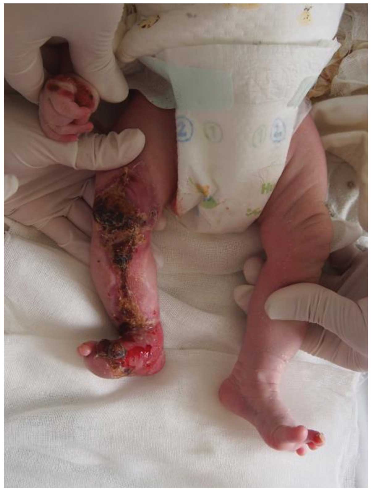
The infant presented with irregular linear and large
skin defects on the right lower extremity with eschar and small
lesions on the right hand (Fig. 1).
When the patient first visited the department, the wounds were
covered by a simple dressing. Physical examination showed no
evidence of other abnormalities. All urine and blood tests, as well
as laboratory tests examining kidney and liver function, were
normal. Conservative treatment was employed for the first few days
following admission to the hospital; however, this did not
alleviate the condition. In order to accelerate the healing process
and prevent wound infection, skin grafting was performed. The
surgical procedure was conducted as follows: i) The patient
received a general anesthetic (3 mg/kg propofol); ii) razor-thin
skin was attained from the right parietal region of the scalp using
an electrical dermatome (Zimmer, Inc., Warsaw, IN, USA), and used
as a dressing to bind up the donor site; iii) the razor-thin skin
should be cleaned of remaining hair prior to grafting; iv) the
surface granulation tissue of skin defect was resected, the lesion
cleaned was three times, the donor skin was transplanted onto the
lesion and the lesion area was binded; and v) operation was
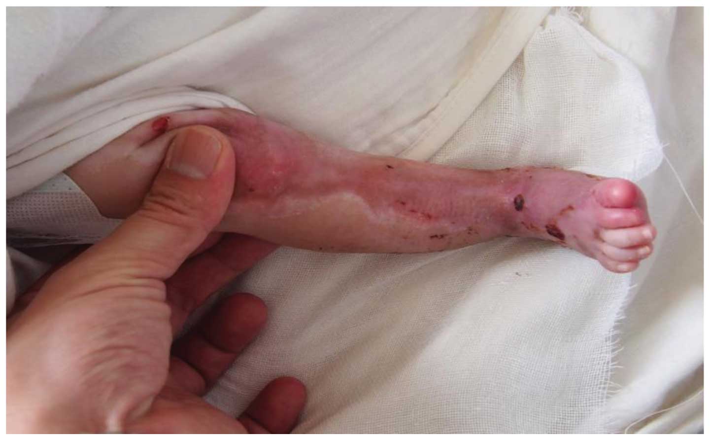
completed successfully. The wounds healed within 14 days of
admission, with only a little hyperpigmentation (Fig. 2).
Discussion
ACC is a rare condition with an underlying mechanism
that remains to be fully elucidated. To date, only 500 cases have
been reported in the literature (5).
This congenital skin disorder can occur as an isolated condition,
such as the case reported in the present study, or can coexist with
other genetic syndromes. One of the factors found to facilitate the
development of ACC is maternal cigarette smoking (5). In addition, the association of
methimazole, diclofenac sodium, valproic acid, marijuana and
cocaine with ACC has already been reported (6).
FP has been found to be closely associated with ACC.
The recent increase in the use of in vitro fertilization
techniques has caused a marked rise in the incidence of multiple
gestation pregnancies (5). FP, or
the mortality of the co-twin, which occurs as a result of
multi-gestational pregnancies, has been shown to give rise to ACC.
Forty-nine cases of FP associated with ACC have been reported in
the literature (3). ACC associated
with FP (group V of Frieden's classification) (2), is characterized by classic large and
symmetrical lesions that are most widely distributed on the trunk
and extremities (7). The severity
and size of ACC lesions may be determined by the time of mortality
of the fetus. When the mortality occurs in the late-first to
early-second trimester and prior to 14 weeks' gestation, the
lesions tend to be smaller, linear, actuate, triangular and
well-demarcated (7).
Several theories have been proposed in order to
explain the phenomenon of ACC occurring in the setting of FP
(5,6); however, based on the recent evidence,
transient hypovolemia may be the primary cause (3). Evidence from Doppler ultrasound has
demonstrated acute transfusion from the surviving to the dying
twin. The ischemia of the skin and other organs of the viable twin
may result from hypotension and hypovolemia.
Individuals affected by ACC most commonly exhibit a
unique circular defect on the scalp. Skin defects are the primary
symptom but can also be accompanied by congenital pulmonary
(8) and other malformations, such as
lumbosacral sinus tract (3), faun
tail nevus and spinal cord malformation (9,10). An
ACC scalp defect can be an indicator of internal organ involvement
and increase the risk of complications (6). The occurrence of complications can
increase the likelihood of a poor outcome. Attention should be paid
to the prevention and treatment of complications such as infection,
bleeding, electrolyte imbalance from increased epidermal water
loss, nutritional deficiency from chronic blood loss and pain at
wound dressing (6).
The selection of treatment depends on the condition
of the infant and the complications that occur. Multiple treatment
regimens have been reported for cases of ACC (6); however, most cases of ACC associated
with FP can be healed under conservative treatment, in accordance
with the literature (4,6,11).
Cerebral damage, bleeding and infections should be carefully
monitored when the patient is being conservatively treated with
dressing changes. A multidisciplinary approach can be useful.
Treatment for ACC is currently controversial and may be
conservative, including dressings, surgery or a combination of
both. There are a range of conservative dressing options, such as
moist dressing, burn cream, dressing with silver sulfadiazine cream
and dressings containing epidermal growth factor. Skin grafting and
local flaps are the predominant surgical treatment option for ACC.
In the literature, a number of cases have been reported in which
ACC has been treated using autologous dermis grafts, allogenic
dermal grafts and cultured epithelial autografts (12). Skin graft is recommended for large
facial defects that are at risk of sagittal sinus thrombosis and
cerebral hemorrhage (13). Surgical
management should be selected for large and deep lesions that are
not healed through conservative treatment.
The present study reported a case of a large lesion
on the right lower extremity that bled during dressing changes. In
the past, autologous dermal and allogenic dermal grafts, as well as
cultured epithelial autografts, have been used as treatment for the
disease (14). In the present case,
an autologous graft was selected. Using the head as the donor site,
razor-thin skin was transplanted on the lesion, which healed within
a 7-day period without leaving any scars. The lesions are
predominantly located on the midline vertex of the scalp, with
occasional skull defects, thus the scalp is rarely selected as the
donor site. In addition, the fontanelle of the infant is not fully
developed, which results in a high risk of cerebral infection.
However, in patients without a scalp defect, using the scalp as the
donor site may result in fewer additional scars compared with other
donor sites. In conclusion, the use of the scalp as a donor site
for skin grafting appears to be an effective treatment for large
and deep ACC lesions.
References
|
1
|
Cordon M: Extract from a letter describing
three children from the same mother born with parts of the
extremities devoid of skin. J Med Chir Pharm. 26:556–557. 1767.(In
French).
|
|
2
|
Frieden IJ: Aplasia cutis congenita: A
clinical review and proposal for classification. J Am Acad
Dermatol. 14:646–660. 1986. View Article : Google Scholar : PubMed/NCBI
|
|
3
|
Mazza JM, Klein JF, Christopher K and
Silverberg NB: Aplasia cutis congenita in a setting of fetus
papyraceus associated with small fetal abdominal circumference and
high alpha-fetoprotein and amniotic acetylcholinesterase. Pediatr
Dermatol. 32:138–140. 2013. View Article : Google Scholar : PubMed/NCBI
|
|
4
|
Blouin MM, Bernard J, Caron F and Auger I:
Aplasia cutis congenita of the trunk and scalp associated with
fetus papyraceus. Int J Dermatol. 50:733–735. 2011. View Article : Google Scholar : PubMed/NCBI
|
|
5
|
Ustuner P, Dilek N, Saral Y and Ustüner I:
Coexistence of aplasia cutis congenita, faun tail nevus and fetus
papyraceus. J Dermatol Case Rep. 7:93–96. 2013. View Article : Google Scholar : PubMed/NCBI
|
|
6
|
Tempark T and Shwayder TA: Aplasia cutis
congenita with fetus papyraceus: Report and review of the
literature. Int J Dermatol. 51:1419–1426. 2012. View Article : Google Scholar : PubMed/NCBI
|
|
7
|
Klein RQ, Robinson DM, Lieber CD and
Antaya RJ: Symmetric aplasia cutis congenita associated with fetus
papyraceus: Report of two cases. Pediatr Dermatol. 28:467–469.
2011. View Article : Google Scholar : PubMed/NCBI
|
|
8
|
Schaffer JV, Popiolek DA and Orlow SJ:
Symmetric truncal aplasia cutis congenita following multifetal
reduction of a sextuplet pregnancy. J Pediatr. 153:860–863. 2008.
View Article : Google Scholar : PubMed/NCBI
|
|
9
|
Louise L, Annabel M, Hubert L, Isabelle G
and Gerard L: Fetus papyraceus: Congenital pulmonary anomalies
associated with congenital aplasia cutis on the surviving twin.
Pediatr Dermatol. 30:e143–e145. 2013. View Article : Google Scholar : PubMed/NCBI
|
|
10
|
Cho AY, Lee SS, Lee Y, Kim CD, Lee JH and
Seo YJ: Aplasia cutis congenita with hair collar sign and dermal
melanocytosis. Int J Dermatol. 51:745–747. 2012. View Article : Google Scholar : PubMed/NCBI
|
|
11
|
Kelly BJ, Samolitis NJ, Xie DL and
Skidmore RA: Aplasia cutis congenita of the trunk with fetus
papyraceus. Pediatr Dermatol. 19:326–329. 2002. View Article : Google Scholar : PubMed/NCBI
|
|
12
|
Wexler A, Harris M and Lesavoy M:
Conservative treatment of cutis aplasia. Plast Reconstr Surg.
86:1066–1071. 1990. View Article : Google Scholar : PubMed/NCBI
|
|
13
|
Skoufi G, Lialios G, Plachouras N,
Kutsogiannis D and Mperis A: Aplasia cutis congenita: Successful
conservative treatment. Pediatr Int. 48:507–509. 2006. View Article : Google Scholar : PubMed/NCBI
|
|
14
|
Anderl H, Frisch H and Hussl H: Successful
treatment of an extensive form of aplasia cutis congenita by dermis
grafts. Chir Plast (Berl). 5:183–190. 1980.
|