Introduction
Intraosseous ganglia within the carpal bones are
relatively rare, with only a limited number of cases previously
reported (1–3). They are benign, non-neoplastic bone
lesions that have similar histological characteristics to those of
soft tissue ganglion cysts (4,5). The
most common clinical symptom is wrist pain. Carpal bone cysts are
one of the causes of chronic pain in the wrist joint; however, the
etiology of cyst formation remains unknown (6–9). Single
wrist intraosseous thecal cysts are most common in the lunate and
scaphoid bones (9). In clinical
treatment, tumor excision is the best strategy for painful ganglia,
and is usually followed by autogenous bone grafting in the
traditional treatment (10,11). However, the anatomical structure of
the wrist joint is complicated, and thus the surgical incision may
damage this structure, leading to multiple complications, including
joint stiffness and disturbances of the fragile vascular system of
the lunate bone (12).
Various artificial substances have been developed
and used as bone grafting materials. For instance, bone cement is a
grafting material with several advantages, including ease of
handling, biological safety and sufficient compressive strength
(6). Therefore, the present study
used bone cement injection to treat 4 patients with intraosseous
ganglia of the carpal bones and evaluated the therapeutic effects
of this treatment.
Patients and methods
Patient description
The present study was approved by the Ethics
Committee of the Third Hospital of Hebei Medical University
(Shijiazhuang, China). Written informed consent was obtained from
all patients. Between January 2012 and December 2013, 4 patients (3
men and 1 woman) presented at the Third Hospital of Hebei Medical
University with wrist pain and activity limitation, and were
diagnosed with intraosseous ganglion of the carpal bone by
radiography. Briefly, X-rays and computed tomography (CT) scans
showed that there were well-defined, round or oval bright defects
surrounded by a circular sclerotic bone in the lunate bone,
adjacent to the joint. In addition, magnetic resonance imaging
(MRI) and Technetium-99m (T99) bone scans showed no cortical
expansion, periosteal reaction or internal calcification. All these
patients were treated by resection of the cyst, followed by
injection of bone cement. Patients with secondary inflammation or
degenerative joint disease were excluded from the study. Patient
characteristics are shown in Table
I.
 | Table I.General information of the case
series. |
Table I.
General information of the case
series.
| Patient no. | Age (years) | Gender | Lesion side | Pain duration
(months) | Surgery duration
(min) | Intraoperative blood
loss (ml) | Follow-up
(months) |
|---|
| 1 | 43 | Female | Left | 12 | 20 | 5 | 18 |
| 2 | 33 | Male | Right | 8 | 30 | 10 | 15 |
| 3 | 60 | Male | Right | 10 | 15 | 3 | 12 |
| 4 | 42 | Male | Right | 18 | 16 | 4 | 22 |
Surgical procedure and postoperative
care
Prior to surgery, patients received conservative
treatment, including diclofenac sodium sustained-release tablets
(75 mg daily; Beijing Novartis Pharma Co., Ltd., Beijing, China) or
celecoxib capsules (0.2 g daily; Pfizer Inc., New York, NY, USA),
for 1 month. All patients were operated in the supine position, and
local anesthesia or nerve tissue anesthesia was used. Anesthetic
consisted of 2–4 ml Lidocaine (Shanghai Fosun Pharmaceutical Group
Co., Ltd., Shanghai, China). The lesions were located in the lunate
bone and thus the dorsal approach was adopted (13,14).
Guided by C-arm fluoroscopy, a guide pin was used to position the
bone cyst location. Subsequent to performing a 2-mm skin incision,
the trocar was inserted into the skin until it reached the center
of the cyst. Next, the trocar core with jelly-like liquid was
removed from the cyst. A small drill was passed through the trocar
to reach the cyst wall. Tissue was collected and used for
histological analyses. Normal saline was used to flush the surgical
position with a syringe, and 0.3 ml bone cement (Mendec Spine;
Tecres Medical, Verona, Italy) was then injected into the cavity.
When the bone cement was completely solidified, the sleeve needle
was pulled out, with no bone cement leakage observed. The skin
wound was immediately bound, and the patients began flexion
exercises from the next day following the surgery.
Histological analysis of tissues
specimens
The collected tissues specimens were fixed for 24 h
in 10% formalin and paraffin-embedded. Subsequently, serial
sections of 4-µm thickness were cut, dewaxed and stained with
hematoxylin and eosin, prior to analysis under a microscope. These
sections were used to confirm the pathology.
Postoperative management
At 1 day after the surgery, patients were able to
perform finger flexion exercises, and 1 week later, patients were
able to perform wrist flexion and extension functional exercises.
Postoperative X-ray examination was performed in the outpatient
service at 1, 3, 6, 12 and 18 months after surgery. CT scanning was
also performed in the last follow-up. Patients were free to return
to work when their wrist was able to bear the work intensity.
Evaluation
The following preoperative and postoperative results
were recorded by independent observers: Pain value (1 to 10) was
measured using the visual analog scale (VAS) system (15); hand strength was determined based on
the lateral force (percentage of the contralateral force); wrist
flexion extension was measured with a protractor; Mayo Wrist Score
(MWS) (16); and the Disabilities of
the Arm, Shoulder and Hand (DASH) score (17).
Results
Patient details
The patients participating in the present study
included 3 males and 1 female, aged between 33 and 60 years (mean
age, 44.5 years; Table I). In 3 of
the patients, the lesion was in the right side, while in 1 case the
lesion was located in the left side, and no patient presented a
bilateral lesion. All the patients were subjected to X-ray
examination due to the effect of wrist joint pain on daily
activities. Patients with a history of trauma, local infection or
neurovascular lesions were excluded from the current study. The
pain duration was between 8 and 18 months before admission, with a
mean duration of 12 months. Conservative treatments, including
plaster fixation and nonsteroidal drugs, did not relieve the
symptoms. The ranges of motion of wrist flexion and extension were
limited in all patients, and their grip strength was reduced
compared with that of the healthy wrist. Histological analyses to
confirm the pathology were performed prior to surgery for 3
patients; however, the tissue sample was too small for a
pathological diagnosis in 1 patient. For the 3 cases, destruction
in the internal capsule wall of the carpal bone was significant
such that the integrated capsule wall structure could not be
observed.
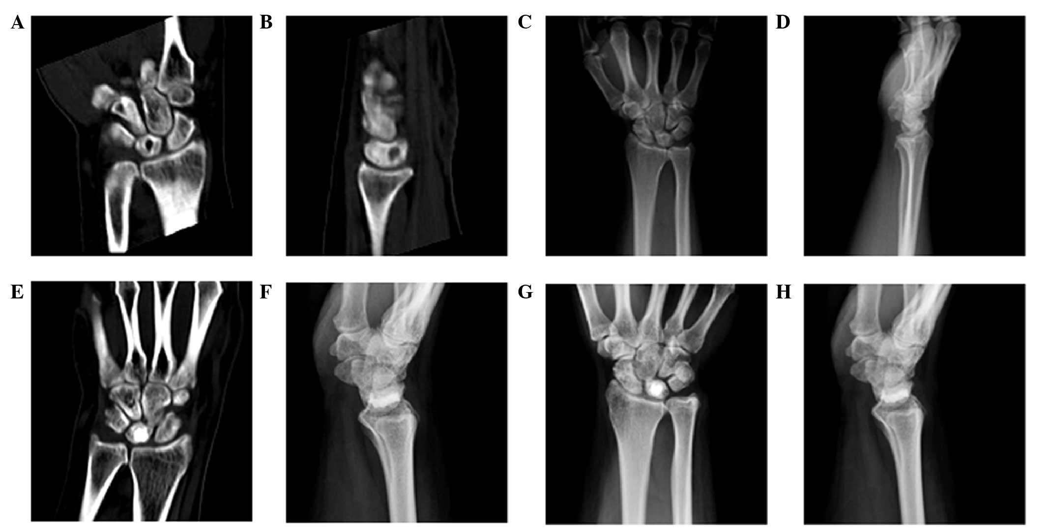
Preoperative imaging
In the four patients included in the present study,
preoperative CT scans demonstrated that the cysts were located
within the bone analogous central region, and no cortical bone
destruction was observed (Fig. 1A and
B) Furthermore, preoperative X-ray examinations revealed that
the lesions in all patients had a well-defined, round and
osteolytic low density area with sclerosing edge; however, the
degree of sclerosis was not the same for all patients (Fig. 1C and D). All lesions were located at
the lunate bone.
Surgical treatment and follow-up
The mean surgery duration for all 4 patients was 20
min (range, 15–30 min), with a mean blood loss amount of 5.5 ml
(range, 3–10 ml) during the surgery. Postoperatively, the wounds
healed well in all patients. The mean follow-up time was 16.8
months (range, 12–22 months). Wrist pain symptoms disappeared
following surgery, and 3 of the patients presented increased grip
strength at the 24-month follow-up. In all cases, increased wrist
flexion and extension range were reported in the wrist joint, which
are presented as mobility in Table
II. Bone cement absorption was not observed by CT examination
during the follow-up period (Fig. 1E and
F), although X-ray scans suggested that bone absorption had
occurred (Fig. 1G and H). In
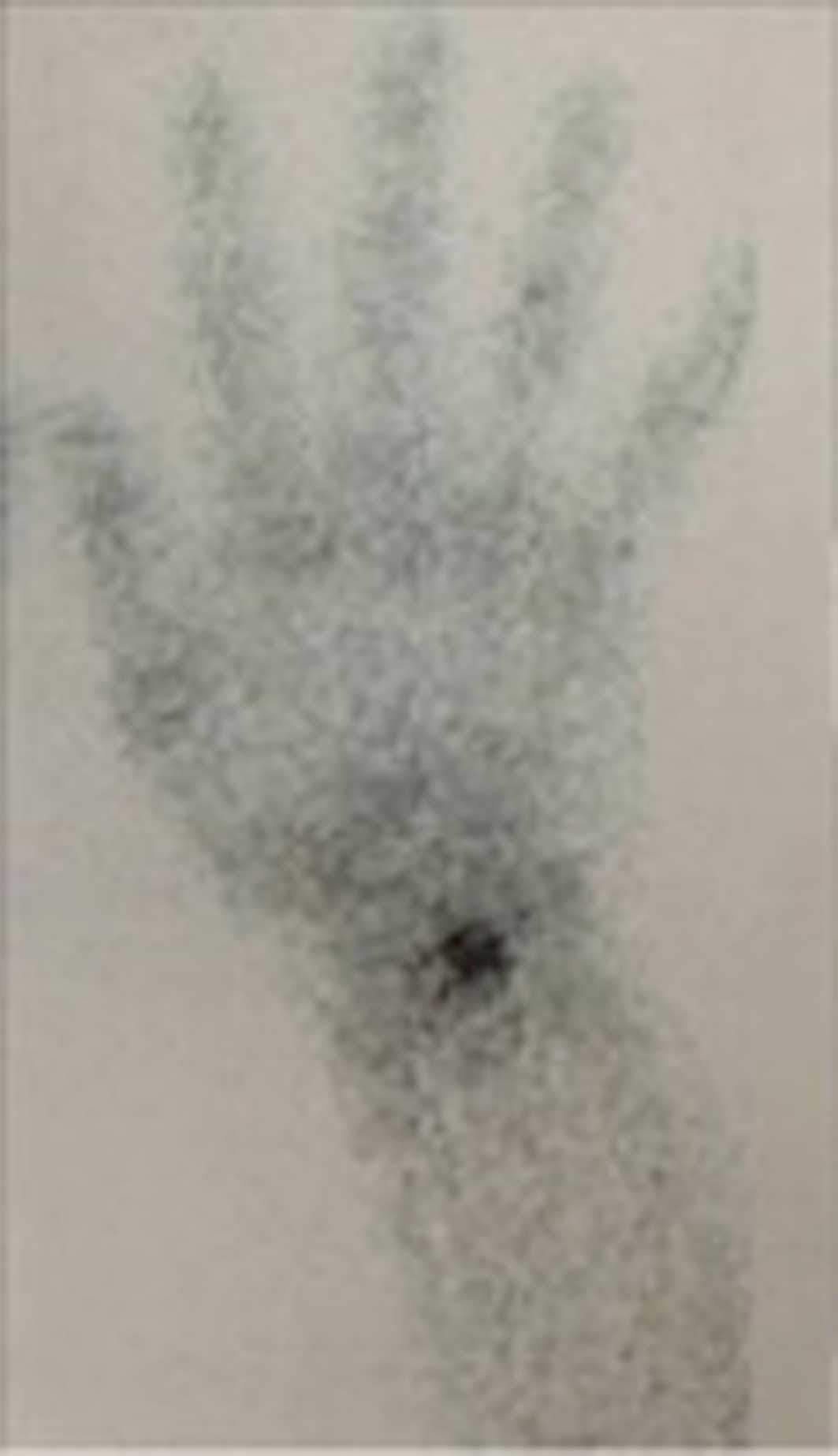
addition, T99 bone scans showed that partial absorption of bone
cement in the lunate bone was increased, without absorption of bone
cement in other parts of the carpal bone (Fig. 2). Furthermore, no tumor recurrence,
rejection reaction or other complications were reported during the
follow-up, and all patients returned to work within 4 weeks.
| Table II.Preoperative and postoperative
evaluation of the patients. |
Table II.
Preoperative and postoperative
evaluation of the patients.
|
| Pain on VAS | Grip strength (% of
healthy wrist) | Mobility (% of
healthy wrist) |
|
|
|
|---|
|
|
|
|
|
|
|
|
|---|
| Patients | Pre | Post | Pre | Post | Pre | Post | MWS | DASH score | Patient
satisfaction |
|---|
| 1 | 8 | 0 | 37 | 91 | 40 | 70 | 80 | 7.7 | Satisfied |
| 2 | 7 | 1 | 42 | 87 | 38 | 66 | 75 | 12.8 | Satisfied |
| 3 | 8 | 0 | 32 | 90 | 50 | 75 | 80 | 8.5 | Satisfied |
| 4 | 8 | 0 | 46 | 82 | 45 | 80 | 80 | 15.0 | Satisfied |
| Mean | 7.8 | 0.3 | 39.3 | 87.5 | 43.3 | 72.8 | 78.8 | 11.0 |
|
VAS score, MWS and DASH score
results
The present study evaluated the pain that patients
experienced based on their VAS scores, as well as the grip strength
of all patients (Table II). At 12
months postoperatively, the mean VAS score was 0.3 (range, 0–1),
indicating that the pain experienced by patients was reduced, when
compared with the preoperative pain, with a mean VAS score of 7.8
(range, 7–8). Similarly, the mean strength score improved from 39.3
(range, 32–46) preoperatively to 87.5 (range, 82–91)
postoperatively. Following treatment, the mean MWS and DASH score
reached 78.8 (range, 75–80) and 11 (range, 7.7–15.0), respectively
(Table II). All the patients were
satisfied with the treatment, and the aforementioned results
suggested a good recovery in all cases.
Discussion
Ganglia can be observed in various anatomic
locations, including the metaphysis adjacent to the growth plate of
the tubular bones; in particular, the humerus and femur. Ganglia
may be classified as intraosseous, subperiosteal, intraarticular or
soft tissue ganglia. Soft tissue ganglia are commonly observed,
occasionally presenting with superficial erosion and close
proximity to the bone; however, their intraosseous counterparts
have been rarely reported (18,19). In
the majority of patient cohorts, intraosseous ganglia are most
commonly detected in the lower extremities, particularly in the
tibia and femur, and predominantly occur in the
epiphyseal-metaphyseal area of the long tubular bones (20). Benign or malignant tumors are rare in
carpal bones, with intraosseous ganglion being the most common type
at this site (7).
The cause of the intraosseous ganglia development
remains unclear. Two main types have been suggested, including the
idiopathic or primary type (lesion possibly formed due to a
degenerative bone process) and the secondary type (lesion caused by
the cortical penetration of a previously existing soft tissue
ganglion) (2,21). Kambolis et al (22) have previously suggested that
ganglionic cysts of the bone developed by invasion of the
ganglion-like connective tissue into the bone from the local soft
parts. In addition, Schajowicz et al (23) stated that ~85% of the reported cases
are of intraosseous origin associated with altered mechanical
stress leading to intramedullary vascular disturbance and aseptic
necrosis.
Intraosseous ganglion is one of the most frequent
lytic defect of the wrist. Its location in the lunate bone is
frequently discovered by chance on an X-ray scan performed for
another condition, or due to wrist pain, and very rarely due to a
lunate fracture.
X-ray films, CT and MRI examinations help reach the
diagnosis of intraosseous ganglia. Radiographs and CT scans
typically show eccentrically located, well-defined, rounded or
ovoid lucent defects with a surrounding zone of sclerosis adjacent
to the articular surface (24,25). No
expansion of the cortex, periosteal reaction or internal
calcification are observed, and these features can help to
distinguish between the intraosseous ganglion and numerous other
lesions, such as degenerative cysts, giant cell tumors and osteoid
osteomas (26). The joint space is
normal in all intraosseous ganglia, while certain lesions present a
communication in particular with the scapholunate joint (24,25). CT
and MRI assist in the clarification of the extent of the
abnormality and allow for more accurate preoperative planning. In
specific lesions, an intraarticular extension may be visible in
association with the defect in the surface of the lunate upon MRI
examination (24,25).
The lunate bone has a central position in the wrist.
Surgery on the lunate bone can be performed in either the dorsal or
palmar position. The palmar anatomical structure is relatively
complex and its location is relatively deep. Furthermore, the
median nerve that is found in the carpal tunnel may be disrupted
when performing in the palmar position (27). Conversely, the dorsal approach is
performed in a superficial location, in which only an extensor
tendon is located (27). Therefore,
in order to reduce the occurrence of complications during the
treatment of intraosseous ganglia, we recommend a dorsal approach
surgery.
Following resection, the ganglion cyst cavity within
the lunate bone should be filled to avoid bone fractures caused by
the axial pressure (28). Bone
cement is a type of biological bone substitute materials that has
been applied in clinical treatment for years, and is particularly
suitable for the repair of bone defects subsequent to resection of
bone tumor (29,30). The traditional treatment strategy for
carpal bone cyst with persistent symptoms is treatment by open
lesion scraping, followed by autogenous bone transplantation, which
severely limits the daily activities of patients (2,3).
Although this treatment can improve the patients' clinical
symptoms, several studies have reported that, after surgery, ~40%
of patients still present symptoms that influence their daily life
or have a limited flexibility of flexion and extension activities
(7,9). Certain studies (31,32) have
applied resection of the lunate bone cyst and bone graft under
wrist arthroscopy-assisted minimally invasive surgery for reducing
the complications caused by incision surgery. However, the surgical
technology requirements for the wrist joint are relative high,
since the corresponding equipment is necessary, which limits its
application. In our previous study, balloon dilatation-based bone
cement was successfully applied to the treatment of ischemic lunate
bone necrosis using vertebroplasty and bone cement conveying
equipment (33). Bone cement
injection has certain limitations, since it is not suitable for the
removal of a damaged cyst, with the exception of the dorsal cortex
and outer residual cortex, since it may cause bone cement leakage
to the wrist joint cavity. Therefore, CT preoperative evaluation is
essential.
The incision made during percutaneous surgery is
small, thus it is not possible to fully reveal and scrape the
lunate bone cyst, cystic wall and its contents, which results in
difficulties obtaining material for pathologic examination. In the
present study, pathological examination was performed in 3
patients, but it was not possible in 1 case since the sample was
too small for pathological diagnosis. Upon pathologic examination
in the 3 cases, the destruction in the internal capsule wall of
carpal bone was large, thus the integrated capsule wall structure
could not be obtained.
The recurrence of ganglion cysts in the foot inner
bone following surgical resection rate has been reported to be
~6.1%; however, no recurrence has been previously reported within
the carpal bones ganglion cyst following surgery (34). It has been suggested that recurrences
do not necessarily develop due to inadequate excision or curettage,
but may occur in conjunction with the connective tissue metaplasia
at the operative site or in tissue immediately adjacent to it
(2,35). In the present study, no recurrence
was detected during follow-up.
A balloon kyphoplasty instrument is typically used
for cyst curetting and injection of bone cement; however, it is not
suitable for lunate bone cysts. Based on the early curative effect,
there is a need to further improve the surgical instruments. Due to
the small number of cases and the short period of follow-up in the
present study, the long-term efficacy of this procedure requires
further assessment. Since the present study used the technique to
treat a carpal lunate bone cyst, it is unclear whether this
technique would be suitable for other wrist cysts, and this
requires further clinical research.
In conclusion, the results of the present study
suggested that bone cement injection is an effective and safe
therapeutic strategy for the treatment of intraosseous ganglion of
the carpal bone.
Acknowledgements
The present study was supported by a grant from the
Key Project of Medical Research of Hebei Province (no.
ZL20140209).
References
|
1
|
Dumas P, Georgiou C, Chignon-Sicard B,
Balaguer T, Lebreton E and Dumontier C: Intra-osseous ganglion cyst
of the carpal bones. A review of the literature underlining the
importance of systematic computed tomography. Chir Main. 32:3–7.
2013.(In French). View Article : Google Scholar : PubMed/NCBI
|
|
2
|
Logan SE, Gilula LA and Kyriakos M:
Bilateral scaphoid ganglion cysts in an adolescent. J Hand Surg Am.
17:490–495. 1992. View Article : Google Scholar : PubMed/NCBI
|
|
3
|
Arabori M, Kitazawa H, Akisue T, Kuroda R,
Fujioka H, Doita M and Kurosaka M: Intraosseous ganglion of the
phalanx. Clin Imaging. 32:73–76. 2008. View Article : Google Scholar : PubMed/NCBI
|
|
4
|
Warren NP and Harris NH: Juxta-articular
aneurysmal bone cyst. J R Soc Med. 81:291–292. 1988.PubMed/NCBI
|
|
5
|
Barth E and Hagen R: Juxta-articular bone
cyst. Acta Orthop Scand. 53:215–217. 1982. View Article : Google Scholar : PubMed/NCBI
|
|
6
|
Yajima H, Murata K, Kawamura K, Kawate K
and Takakura Y: Treatment of intraosseous ganglia and bone cysts of
the carpal bones with injectable calcium phosphate bone cement.
Hand Surg. 13:167–173. 2008. View Article : Google Scholar : PubMed/NCBI
|
|
7
|
Waizenegger M: Intraosseous ganglia of
carpal bones. J Hand Surg Br. 18:350–355. 1993. View Article : Google Scholar : PubMed/NCBI
|
|
8
|
Schacherer TG and Aulicino PL:
Intraosseous ganglia of the carpal bones. Orthop Rev. 20:889–892.
1991.PubMed/NCBI
|
|
9
|
Tham S and Ireland DC: Intraosseous
ganglion cyst of the lunate: Diagnosis and management. J Hand Surg
Br. 17:429–432. 1992. View Article : Google Scholar : PubMed/NCBI
|
|
10
|
Kligman M and Roffman M: Bilateral
intraosseous ganglia of the scaphoid and lunate bones. J Hand Surg
Eur Vol. 22:820–821. 1997. View Article : Google Scholar
|
|
11
|
Matsumine A, Kusuzaki K, Matsubara T,
Okamura A, Okuyama N, Miyazaki S, Shintani K and Uchida A: Calcium
phosphate cement in musculoskeletal tumour surgery. J Surg Oncol.
93:212–220. 2006. View Article : Google Scholar : PubMed/NCBI
|
|
12
|
Daly PJ, Sim FH, Beabout JW and Unni KK:
Intraosseous Ganglion Cysts. Orthopedics. 11:1715–1719.
1988.PubMed/NCBI
|
|
13
|
Trail IA, Murali R, Stanley JK, Hayton MJ,
Talwalkar S, Sreekumar R and Birch A: The long-term outcome of
four-corner fusion. J Wrist Surg. 4:128–133. 2015. View Article : Google Scholar : PubMed/NCBI
|
|
14
|
Bhatia DN: Direct “Cystoscopic” Approach
for Arthroscopic Decompression of an Intraosseous Ganglion of the
Lunate. Arthrosc Tech. 4:e223–e229. 2015. View Article : Google Scholar : PubMed/NCBI
|
|
15
|
Huskisson EC: Measurement of pain. Lancet.
2:1127–1131. 1974. View Article : Google Scholar : PubMed/NCBI
|
|
16
|
Amadio PC, Berquist TH, Smith DK, Ilstrup
DM, Cooney WP 3rd and Linscheid RL: Scaphoid malunion. J Hand Surg
Am. 14:679–687. 1989. View Article : Google Scholar : PubMed/NCBI
|
|
17
|
Hudak PL, Amadio PC and Bombardier C:
Development of an upper extremity outcome measure: The DASH
(disabilities of the arm, shoulder and hand)[corrected]. The Upper
Extremity Collaborative Group (UECG). Am J Ind Med. 29:602–608.
1996. View Article : Google Scholar : PubMed/NCBI
|
|
18
|
Williams HJ, Davies AM, Allen G, Evans N
and Mangham DC: Imaging features of intraosseous ganglia: A report
of 45 cases. Eur Radiol. 14:1761–7169. 2004. View Article : Google Scholar : PubMed/NCBI
|
|
19
|
Angelides AC: Ganglions of the hand and
wrist. Green's Operative Hand Surgery (3rd). Green DP: Churchill
Livingstone. (New York). 2159–2162. 1993.
|
|
20
|
Helwig U, Lang S, Baczynski M and
Windhager R: The intraosseous ganglion. A clinical-pathological
report on 42 cases. Arch Orthop Trauma Surg. 114:14–17. 1994.
View Article : Google Scholar : PubMed/NCBI
|
|
21
|
Lorente R, Moreno M and Quiles M:
Bilateral intraosseous ganglia of the lunate: A case report. J Hand
Surg Am. 17:1084–1085. 1992. View Article : Google Scholar : PubMed/NCBI
|
|
22
|
Kambolis C, Bullough PG and Jaffe HI:
Ganglionic cystic defects of bone. J Bone Joint Surg Am.
55:496–505. 1973.PubMed/NCBI
|
|
23
|
Schajowicz F, Clavel Sainz M and Slullitel
JA: Juxta-articular bone cysts (intra-osseous ganglia): A
clinicopathological study of eighty-eight cases. J Bone Joint Surg
Br. 61:107–116. 1979.PubMed/NCBI
|
|
24
|
Haller J, Resnick D, Greenway G, Chevrot
A, Murray W, Haghighi P, Sartoris DJ and Chen CK: Juxtaacetabular
ganglionic (or synovial) cysts: CT and MR features. J Comput Assist
Tomogr. 13:976–983. 1989. View Article : Google Scholar : PubMed/NCBI
|
|
25
|
Benis J and Turpin F: The role of imaging
in the assessment of vascularity at hand and wrist. Chir Main.
29(Suppl 1): S21–S27. 2010.(In French). View Article : Google Scholar : PubMed/NCBI
|
|
26
|
Ferkel RD, Field J, Scherer WP, Bernstein
ML and Kasimian D: Intraosseous ganglion cysts of the ankle: A
report of three cases with long-term follow-up. Foot Ankle Int.
20:384–388. 1999. View Article : Google Scholar : PubMed/NCBI
|
|
27
|
Van den Dungen S, Marchesi S, Ezzedine R,
Bindou D and Lorea P: Relationship between dorsal ganglion cysts of
the wrist and intraosseous ganglion cysts of the carpal bones. Acta
Orthop Belg. 71:535–539. 2005.PubMed/NCBI
|
|
28
|
Uriburu IJ and Levy VD: Intraosseous
ganglia of the scaphoid and lunate bones: Report of 15 cases in 13
patients. J Hand Surg Am. 24:508–515. 1999. View Article : Google Scholar : PubMed/NCBI
|
|
29
|
Guo H, Wei J and Liu CS: Development of a
degradable cement of calcium phosphate and calcium sulfate
composite for bone reconstruction. Biomed Mater. 1:193–197. 2006.
View Article : Google Scholar : PubMed/NCBI
|
|
30
|
Stubbs D, Deakin M, Chapman-Sheath P,
Bruce W, Debes J, Gillies RM and Walsh WR: In vivo evaluation of
resorbable bone graft substitutes in a rabbit tibial defect model.
Biomaterials. 25:5037–5044. 2004. View Article : Google Scholar : PubMed/NCBI
|
|
31
|
Ashwood N and Bain GI: Arthroscopically
assisted treatment of intraosseous ganglions of the lunate: A new
technique. J Hand Surg Am. 28:62–68. 2003. View Article : Google Scholar : PubMed/NCBI
|
|
32
|
Bain GI, Turner PC and Ashwood N:
Arthroscopically assisted treatment of intraosseous ganglions of
the lunate. Tech Hand Up Extrem Surg. 12:202–207. 2008. View Article : Google Scholar : PubMed/NCBI
|
|
33
|
Chen W, Wang J, Pan J, Zhang Q, Shao X and
Zhang Y: Primary results of Kienböck's disease treated using
balloon kyphoplasty system. Arch Orthop Trauma Surg. 132:677–683.
2012. View Article : Google Scholar : PubMed/NCBI
|
|
34
|
Murff R and Ashry HR: Intraosseous ganglia
of the foot. J Foot Ankle Surg. 33:396–401. 1993.
|
|
35
|
Pellegrino EA and Olson JR: Bilateral
carpal lunate ganglia. Clin Orthop Relat Res. 87:225–227. 1972.
View Article : Google Scholar : PubMed/NCBI
|