Introduction
Osteoarthritis (OA) is a chronic, progressive and
degenerative joint disease, involving single or multiple joints. OA
of the knee (KOA) is the most common disabling disease,
characterized by the degeneration and degradation of cartilage,
subchondral bone remodeling, osteophyte formation and synovial
inflammation, which affects the patient's quality of life and
constitutes a heavy financial burden (1–3). With
the exception of oral and intra-article injection medications that
relieve the symptoms and improve joint function, there is no
approved medical treatment that halts disease progression and joint
destruction (1,4).
Various surgical methods, including microfracture
(5,6)
and subchondral drilling (7), have
been proposed to regenerate articular cartilage. However, due to
the complications and inferior quality of the regenerative
fibrocartilage, risky and cost-effective joint replacement surgery
is often ultimately required (8).
Previous studies have investigated tissue engineering and cellular
therapies for treating early stage OA, and autologous chondrocyte
implantation has demonstrated positive clinical outcomes (9,10).
Nevertheless, due to the poor self-renewal and regeneration
potential of chondrocytes, it is a slow process that may lead to
fibrocartilage rather than hyaline cartilage (11,12).
Furthermore, this two-stage surgical procedure and is predominantly
used to treat cartilage defects caused by injury rather than
OA.
Therefore, research attention in this field has
shifted to the more promising treatment of mesenchymal stem cells
(MSCs). MSCs, which can be derived from blood, bone marrow,
skeletal muscle, adipose, skin and synovial membrane (13), have the capacity to differentiate
into osteocytes, adipocytes, chondrocytes, myoblasts, tenocytes
(14,15), secrete bioactive molecules that
stimulate angiogenesis and tissue repair, and reduce the response
of T cells and inflammation (16,17).
Previous clinical trials have reported that mild/moderate OA or
advanced OA can be treated efficiently using autologous or
allogenic MSCs through implantation, micro fracture or
intra-articular injections (18–20).
However, so far, no meta-analytic research has evaluated the
efficacy and safety of MSCs in treating patients with KOA.
Therefore, the present meta-analysis was conducted
to analyze the clinical outcomes of MSC treatment on patients with
KOA patients by analyzing pain and functional changes, compared
with their pretreatment condition, or placebo controls.
Materials and methods
Search strategy and eligibility
criteria
Electronic databases: including PubMed (ncbi.nlm.nih.gov/pubmed), EMBASE (embase.com) and Web of Science (webofknowledge.com), were used to comprehensively
search for all relevant studies published in English from the
earliest record to December 2014. The following keywords were used:
‘cartilage defect’, ‘cartilage repair’, ‘osteoarthritis’, ‘knee
osteoarthritis’, ‘stem cells’, ‘mesenchymal stem cells’ (MSCs),
‘bone marrow concentrate’, ‘adipose-derived mesenchymal stem cells’
(ADMSCs), ‘synovial-derived mesenchymal stem cells’ and ‘peripheral
blood-derived mesenchymal stem cells’, as medical subject headings
or text words. In addition, Cochrane Systematic Reviews (cochrane.org/evidence) and ClinicalTrials.gov were manually searched for
additional references. Articles were considered eligible if they
met the following criteria: i) Patients were ≥18 years-old and had
KOA symptom or diagnosed with KOA by clinical and imaging
examination; ii) MSCs administered to at least one treatment group;
iii) ≥3-month follow-up; iv) ≥1 valid outcome measurement before
and after the administration of MSCs, such as the visual analogue
scale (VAS), International Knee Documentation Committee (IKDC)
Subjective Knee Form, Lysholm scale, and Western Ontario and
McMaster Universities Osteoarthritis Index (WOMAC); and v) outcomes
were presented as continuous data [mean ± standard deviation (SD)].
Studies that lacked an intervention plan or pain and functional
measurements were excluded.
Data extraction and study quality
assessment
Two independent reviewers searched the electronic
databases and evaluated the eligibility of the searched articles
and subsequently extracted data using a standardized form,
including data on the study type, number of patients enrolled,
patient characteristics, disease duration, dosage of MSCs, outcome
measurements, follow-up time and adverse events. If additional data
was necessary, the authors were contacted for further information.
The Jadad scoring system was used to assess the methodological
quality of the randomized controlled trials (RCTs) (21). The quality of the included RCTs
ranged from 0–5 points, with a score of <3 indicating a
low-quality study. The Newcastle-Ottawa Scale (NOS) was used to
assess the quality of other studies according to selection,
comparability, exposure, and outcome, including single-arm
prospective and quasi-experimental studies (22). NOS was scored out of 9 points, with
total scores <4 points defined as low quality. Discrepancies
between the two independent evaluations of potential articles were
resolved by discussion and consensus.
Data synthesis and analysis
Data were extracted from four time points at or
closest to the 3rd, 6th, 12th and 24th months after MSCs treatment.
Effect size (ES) was calculated from knee joint pain and functional
changes and the results were compared with the pretreatment
baseline or between different treatment arms. VAS was extracted
from the included articles. If >1 functional measurement was
included in an article, only one functional scale in line with the
order of IKDC, Lysholm and WOMAC was chosen. As multiple treatment
groups wew included in some articles, each group was selected as a
separate status set to analysis. Mean ± SD between the pretreatment
baseline condition and functional scores after treatment was used
to evaluate the effectiveness of MSCs therapy. Positive ES values
demonstrated a pain or functional improvement, and vice versa. For
studies in which the measurement score and SD was deficient, the
value was calculated from the P-value of the corresponding
hypothesis test. If the measurement scores and SD could not be
extracted in some articles, a correlation of 0.5 was used to
estimate the dispersion. A random effect model was used to pool the
ESs with a 95% confidence interval (95%CI) on the basis of
heterogeneity. A positive pooled ES with a 95%CI >0 indicated an
advantage of MSCs compared with the pretreatment condition or
reference treatments.
Assessment of heterogeneity and
sensitivity
Statistical heterogeneity was assessed via the
I-square and Cochran's Q tests. A P-value of <0.10 for
χ2 test or an I-square >50% was indicative of the
existence of substantial heterogeneity (21). Subgroup analysis was performed
according to variables of the study design, different dosages,
arthroscopic debridement (AD), activation agent, as well as the
severity of Kellgren-Lawrence (K-L) grades. Sensitivity analysis
was performed by excluding some articles with extreme ES values to
assess whether the movement resulted in serious changes in the
total result. Funnel plots were used to assess the potential
publication bias. All analyses were conducted using Review Manager
Version 5.2 (The Cochrane Collaboration, Oxford, UK).
Results
Study characteristics
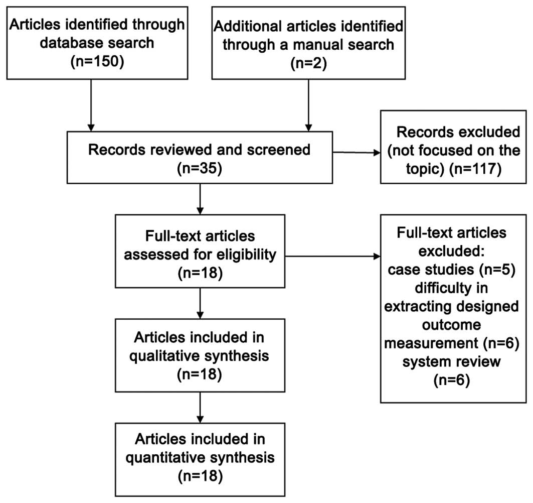
A total of 152 studies were initially searched, of
which 117 were removed after title and abstract screening. Of the
35 citations, 18 clinical studies which met the inclusion criteria
were identified for eligibility (Fig.
1); five case studies (17,22–26) were
excluded and nine studies (24,27–34) were
removed due to difficulties in extracting the outcome measurements.
Four systematic reviews (35–38) were
also excluded. An assessment of the remaining 18 studies revealed
that 10 used a single-arm prospective design (18–20,39–45),
four used quasi-experimental trials (46–49) and
four used RCT (50–53) (Table
I). A total of 565 participants (226 males and 339 females)
were included from the 18 studies. The duration from the onset of
knee pain to registration in each study was 3 months to ≥7 years.
The follow-up period was 3–24 months. The majority of studies
recruited patients with KOA with a severity grade of 1–4 on the K-L
scale. K-L grade s 1–2, and grades 3–4 were defined as early OA and
advanced OA, respectively (Table
II).
 | Table I.Summary of studies using MSCs to
treat KOA patients. |
Table I.
Summary of studies using MSCs to
treat KOA patients.
| Author, year | Number of
patients | Mean age
(year) | BMI | Disease
duration | Double blind | ITT | Outcome
measure | Follow-up time
(month) | Adverse events | Quality
assessment | Ref. |
|---|
| Single-arm,
prospective follow-up studies |
| Buda
et al, 2010 | 20 (12M, 8F) | NM | NM | ≥12 months | No | Yes | IKDC | 6, 12, 24 | None | 4a | (39) |
| Gobbi
et al, 2011 | 15 (10M, 5F) | 48 (32–58) | 24.5±2.53 | NM | No | Yes | VAS, IKDC | 6, 12, 24 | None | 4a | (40) |
|
Davatchi et al,
2011 | 4 (2M, 2F) | 57.7±5.0 | 30.25±4.86 | ≥7 years | No | Yes | VAS | 6 | None | 4a | (18) |
|
Emadedin et al,
2012 | 6 (6F) | 53.8±8.9 | 31.6±4.2 | NM | No | Yes | VAS, WOMAC | 2, 6, 12 | None | 4a | (19) |
| Koh
et al, 2013 | 18 (6M, 12F) | 54.6±7.8 | NM | ≥6 months | No | No | VAS, Lysholm | 24 | Marked pain in 1
patient | 4a | (20) |
|
Turajane et al,
2013 | 5 (1M, 4F) | 57.2±1.92 | 25.36±4.46 | ≥3 months | No | Yes | VAS, WOMAC | 1,6 | None | 4a | (41) |
| Orozco
et al, 2013 | 12 (6M, 6F) | 49±17.3 | NM | ≥6 months | No | Yes | VAS, WOMAC | 3, 6, 12, 24 | Local pain with
discomfort in 6 patients | 4a | (42) |
|
| Author, year | Number of
patients | Average age
(year) | BMI | Disease
duration | Double blind | ITT | Outcome
measure | Follow-up time
(month) | Adverse event | Quality
assessment | Ref. |
|
| Kim
et al, 2014 | 41 (17M, 24F) | 60.7 (53–80) | <30 | ≥12 months | No | Yes | VAS, IKDC | 3, 6, 12 | Joint swelling in
69 knees, pain in 31 knees | 4a | (43) |
| Koh
et al, 2013 | 30 (5M, 25F) | 70.3 (65–80) | NM | ≥12 months | No | Yes | VAS, Lysholm | 3, 12, 24 | Slight pain in 3
patients | 4a | (44) |
| Gobbi
et al, 2014 | 25 (16M, 5F) | 46.5±8.55 | 24.4±3.0 | ≥3 years | No | Yes | VAS, IKDC | 12, 24 | None | 4* | (45) |
| Quasi-experimental
studies |
| Koh and
Choi, 2012 | 50 (MSCs + PRP
group: 8M, 17F; PRP group: 8M; 17F) | MSC group:
54.2±9.3; placebo group: 54.4±11.3 | NM | ≥12 months | No | Yes | VAS, Lysholm | 3, 12 | Marked pain with
swelling in 1 patient | 5a | (46) |
| Koh
et al, 2014 | 56 (Group 1: 8M,
13F; Group 2: 14M, 21F) | Group 1: 55.3±4.1;
Group 2: 57.4±5.7 | Group 1: 26.7±3.1;
Group 2: 26.3±3.0 | ≥12 months | No | No | IKDS | 12, 24 | None | 5a | (47) |
| Jo
et al, 2014 | 18 (LDG: 1M, 2F;
MDG: 0M, 3F; HDG: 2M, 10F) | LDG: 63±8.6 MDG:
65±6.6 HDG: 61±6.2 | LDG: 26±1.0 MDG:
28±2.1 HDG: 26±2.1 | ≥4 months | No | Yes | VAS, WOMAC | 3, 6 | Mild (LDG:3; MDG:
2; HDG:5) | 5a | (48) |
| Kim
et al, 2014 | 54 (MSCs group:
14M, 23F; MSC + fibrin glue group: 8M, 9F) | MSCs group:
57.5±5.9; MSC + fibrin glue group: 57.7±5.8 | MSCs group:
26.3±3.2; MSC + fibrin glue group: 27.3±2.9 | ≥18 months | No | No | IKDC | 12, 24 | None | 5a | (49) |
| Randomized
controlled trials |
| Varma
et al, 2010 | 50 (AD: 25; AD +
MSC: 25) | AD group:
48.20±5.13; AD + MSC: 50.67±5.38 | NM | NM | No | Yes | VAS | 3, 6 | None | 3b | (50) |
| Saw
et al, 2013 | 50 (HAG: 7M, 17F;
HA + PBSC group 10M, 15F) | HAG: 42±5.91 HA +
PBSC: 38±7.33 | HA: 24.83±4.04 HA +
PBSC: 24.91±4.15 | ≥12 months | No | No | IKDS | 6, 12, 24 | None | 3b | (51) |
| Wong
et al, 2013 | 56 (HTO + MSC: 15M,
13F HTO: 14M, 14F) | HTO + MSC: 53
(36–54) HTO: 49 (24–54) | HTO + MSC:
23.81±2.17 HTO: 23.89±3.20 | NM | No | Yes | IKDS, Tegner,
Lysholm | 6, 12, 24 | None | 3b | (52) |
|
Vangsness et al,
2014 | 55 (LDG: 11M, 7F;
HDG: 14M, 4F HAG: 13M, 6F) | LDG: 44.6±9.82 HDG:
45.6±12.42 HAG: 47.8±8 | LDG: 29.86±7.94;
HDG: 29.09±5.91; HAG: 26.89± 4.05 | NM | Yes | No | VAS, Lysholm | 6, 12, 24 | Mild (LDG:18; HDG:
17; HAG: 17) | 5b | (53) |
| Table II.Summary of the preparations and
injection details of MSCs in the retrieved trials. |
Table II.
Summary of the preparations and
injection details of MSCs in the retrieved trials.
| Author, year | MSC origin | Number of
cells | Delivery
system | Method of
implementation | Activation
agent | K-L grade | Comparison | Ref. |
|---|
| Single-arm,
prospective follow-up studies |
| Buda
et al, 2010 | Autologous
BMAC | NM | AD | Implantation | HA, membrane
scaffold | NM | None | (39) |
| Gobbi
et al, 2011 | Autologous
BMAC | NM |
Mini-arthrotomy | Implantation | Collagen
matrix | NM | None | (40) |
|
Davatchi et al,
2011 | Autologous
BMSCs |
8–9×10⁶ | None | Intra-articular
injection | None | 3–4 | None | (18) |
|
Emadedin et al,
2012 | Autologous
BMSCs |
2.0–2.4×10⁷ | None | Intra-articular
injection | None | 4 | None | (19) |
| Koh
et al, 2013 | Autologous
AMSCs |
1.18×10⁶ | AD,
synovectomy | Intra-articular
injection | PRP | 3–4 | None | (20) |
|
Turajane et al,
2013 | Autologous BSC | NM | Microfracture | Intra-articular
injection | GFAP, HA | 2 | None | (41) |
| Orozco
et al, 2013 | Autologous
BMSCs |
40×106 | None | Intra-articular
injection | None | 2–4 | None | (42) |
| Kim
et al, 2014 | Autologous
BMSCs |
2.4×10⁵ | Microfracture and
AD | Intra-articular
injection | Adipose
tissues | 1–4 | None | (43) |
| Koh
et al, 2013 | Autologous
AMSCs |
4.04×10⁶ | Arthroscopic
lavage | Intra-articular
injection | None | 2–4 | None | (44) |
| Gobbi
et al, 2014 | Autologous
BMAC | NM | AD | Implantation | Collagen cell
sheets | NM | None | (45) |
| Quasi-experimental
studies |
| Koh and
Choi, 2012 | Autologous
AMSCs |
1.89×10⁶ | AD,
synovectomy | Intra-articular
injection | PRP | 3 | MSCs + PRP vs.
PRP | (46) |
| Koh
et al, 2014 | Autologous
AMSCs |
3.8×10⁶ | AD | Implantation | None | 1–2 | None | (47) |
| Jo
et al, 2014 | Autologous
AMSCs | LDG:
1×10⁷ MDG: 5×10⁷ HDG:10×107 | None | Intra-articular
injection | None | 3–4 | None | (48) |
| Kim
et al, 2014 | Autologous
AMSCs |
3.9×10⁶ | AD | Implantation | Fibrin glue | 1–2 | MSCs vs. MSCs +
fibrin glue | (49) |
| Randomized
controlled trials |
| Varma
et al, 2010 | Buffy coat | Not mentioned | None | Intra-articular
injection | None | 1–2 | None | (50) |
|
| Author, year | Number Origin of
MSCs | of cells | Implementation
Delivery system | method | Activation
agent | K-L grade | Comparison | Ref. |
|
| Saw
et al, 2013 | Autologous
PBSCs | NM | Microfracture
injection |
Intra-articular | None HA + PBSC
group | NM | HA group vs. | (51) |
| Wang
et al, 2013 | Autologous
BMAC |
1.46×10⁷ | HTO injection |
Intra-articular | HA vs. HTO
group | NM | HTO + MSCs
group | (52) |
|
Vangsness et al,
2014 | Allogenic
BMSCs |
5.0×107,15×107
meniscectomy | Partial medial | Intra-articular
injection | None | NM | Group A: LD MSCs +
HA Group B: HD MSCs + HA; control group: HA | (53) |
Effects of MSCs
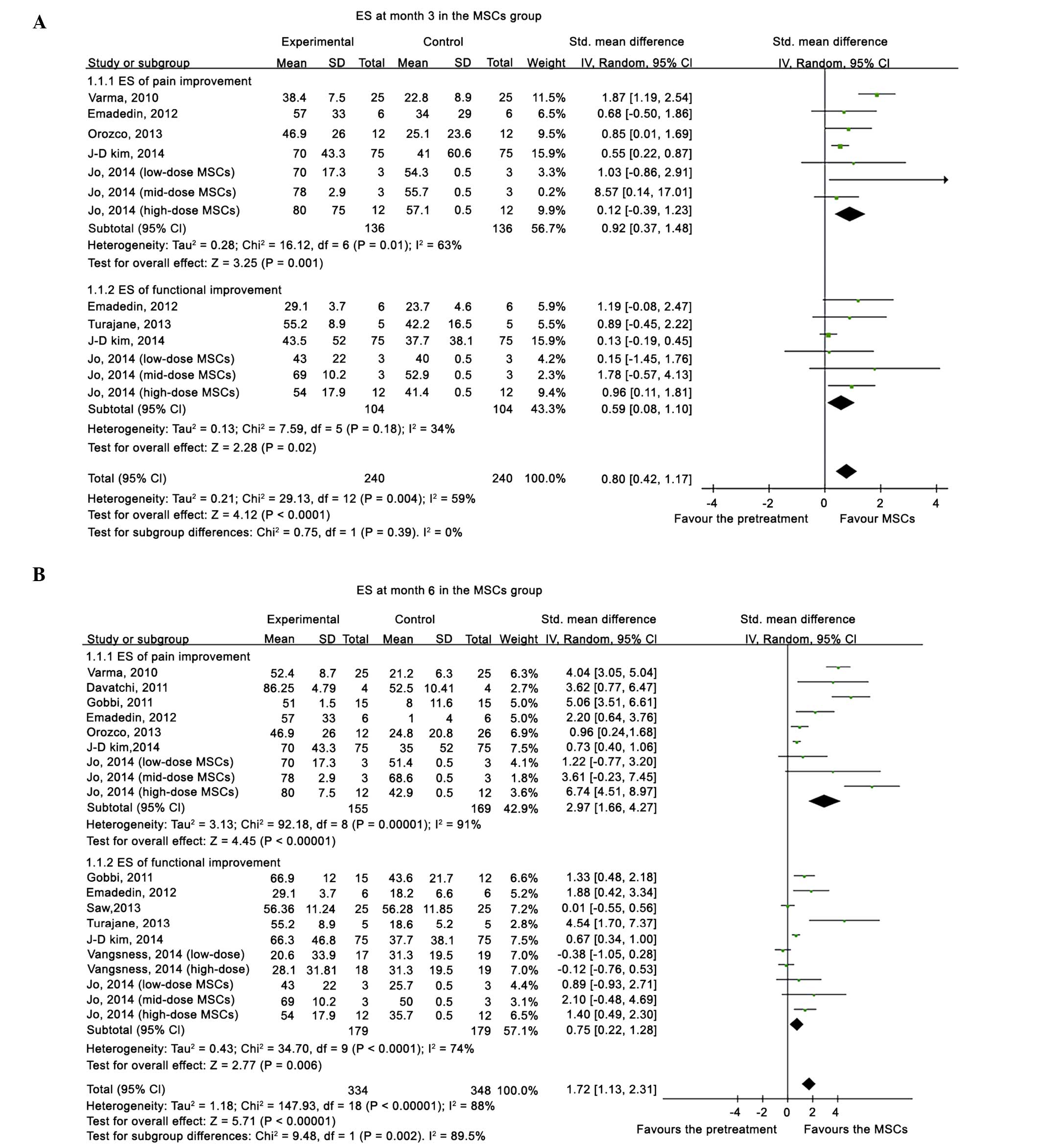
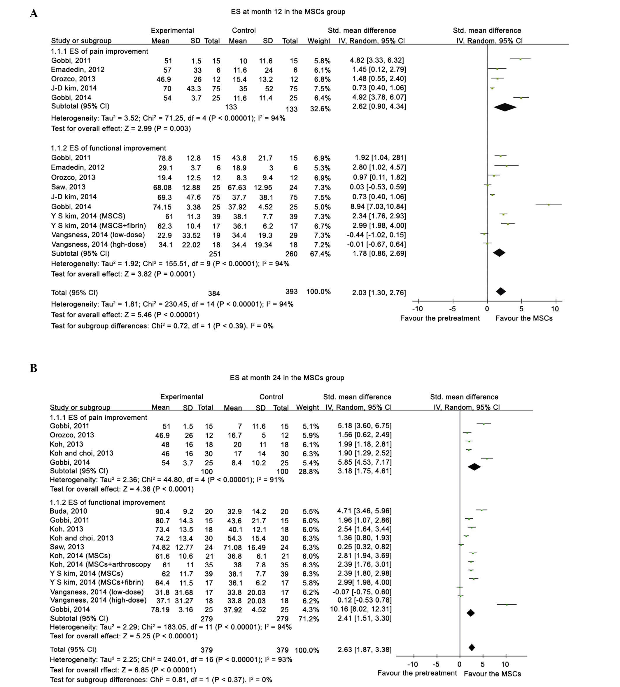
Compared with the pretreatment condition, a pooled
ES of 0.80 (95%CI, 0.42–1.17) was determined at 3 months, 1.72
(95%CI, 1.13–2.31) at 6 months, 2.03 (95%CI, 1.30–2.76) at 12
months (Fig.2), and 1.81 (95%CI,
1.62–2.00) at 24 months (Fig. 3),
which all favored the status after MSCs treatment. Following the
exclusion of an outlier with an extremely high ES, the beneficial
effects from MSCs treatment remained, with an ES of 0.77 (95%CI,
0.41–1.13) at 3 months, 1.49 (95%CI, 0.93–2.04) at 6 months, 1.63
(95%CI, 0.99–2.27) at 12 months, and 1.74 (95%CI, 1.55–1.93) at 24
months. A significant superiority of MSCs intervention was
demonstrated by a high summed ES at 12 and 24 months without an
overlap of the 95%CI of ES at 3 months, which indicated that the
treatment effect of MSCs on KOA patients improved significantly
over time. However, after excluding the data from quai-experimental
and single-arm prospective studies and only using the data from
RCTs, the treatment of MSCs did not demonstrate superiority.
Relative to the baseline, patients improved in the pain and
functional scale scores at all time points.
Stratified analysis
Participants receiving MSC treatment were stratified
according to the study design, administration dosage, AD,
activation agents and K-L grades. Point estimates of the pooled ES
in the single-arm prospective studies and quasi-experimental trials
were higher than those in the RCTs, and an uncertainty in the
treatment effectiveness emerged regarding participants in the RCTs
at 6, 12 and 24 months, since the 95%CI of the summed ES crossed
the value of 0. Stratified analysis failed to demonstrate a
dose-responsiveness association in the MSC numbers. However, the
treatment effectiveness in the MSC groups with AD or activation
agents was superior to the MSC groups without AD and activation
agents, particularly at 12 months in the activation agents group
(ES, 3.13; 95%CI, 1.55–4.71) compared with the group without
activation agents (ES, 0.67; 95% CI, 0.01–1.34). And the early OA
group exhibited a higher ES point estimate at all time points than
the advanced OA group (Table
III).
| Table III.Analysis of the effect sizes of MSC
treatment stratified by the indicated subgroups. |
Table III.
Analysis of the effect sizes of MSC
treatment stratified by the indicated subgroups.
| Subgroup | Pooled effect size
at month 3 | Pooled effect size
at month 6 | Pooled effect size
at month 12 | Pooled effect size
at month 24 |
|---|
| Study design |
|
|
|
|
|
Single-arm follow-up
study | 0.48
(0.18–0.77) | 1.48
(0.51–2.44) | 2.66
(1.69–3.62) | 2.87
(1.99–3.75) |
|
Quasi-experimental study | 0.75
(0.17–1.32) | 1.37
(0.59–2.14) | 2.53
(1.96–3.10) | 2.53
(2.18–2.89) |
|
Randomized controlled
trial | 1.87
(1.19–2.54) | 1.09
(−0.35–2.53) | 0.14
(0.49–0.20) | 0.12
(0.24–0.48) |
| MSCs doses
administered |
|
|
|
|
|
<5×10⁶ | 0.34
(−0.08–0.75) | 0.70
(0.46–0.93) | 1.60
(0.73–2.46) | 2.25
(1.54–2.97) |
|
5×10⁶-5×10⁷ | 0.89
(0.36–1.42) | 1.39
(0.80–1.99) | 1.60
(0.55–2.65) | −0.07
(−0.75–0.60) |
|
>1×10⁷ | 0.67
(0.09–1.26) | 1.91
(0.58–3.23) | −0.01
(−0.67–0.64) | 0.12
(−0.53–0.78) |
|
Arthroscopic debridement |
|
|
|
|
|
Yes | 0.37
(0.01–0.74) | 0.45
(−0.16–1.06) | 2.20
(1.30–3.09) | 2.32
(1.61–3.03) |
| No | 1.02
(0.58–1.47) | 1.48
(0.80–2.16) | 1.41
(0.83–2.00) | 1.56
(0.62–2.49) |
| Activation
agent |
|
|
|
|
|
Yes | 0.37
(0.01–0.74) | 1.40
(0.26–2.54) | 3.13
(1.55–4.71) | 2.82
(2.07–3.56) |
| No | 1.02
(0.58–1.47) | 1.29
(0.53–2.05) | 0.67
(0.01–1.34) | 0.84
(0.16–1.52) |
| Severity of
degeneration |
|
|
|
|
| Early
OA | 1.55
(0.66–2.45) | 4.10
(3.16–5.04) | 2.53
(1.96–3.10) | 2.53
(2.18–2.89) |
|
Advanced OA | 0.78
(0.34–1.22) | 2.40
(1.34–3.46) | 1.99
(0.70–3.28) | 2.54
(1.64–3.44) |
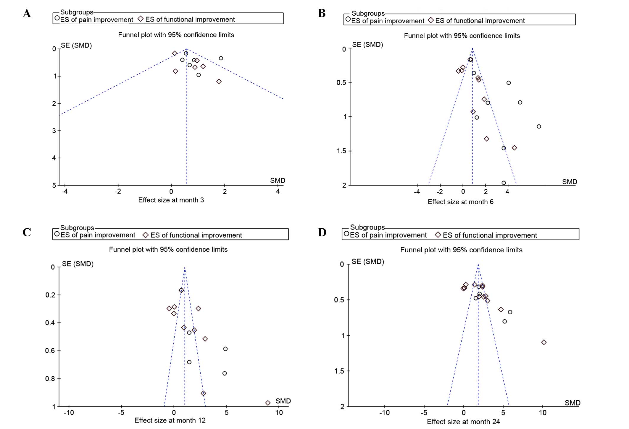
Adverse effects and publication
bias
Seven of the 18 trials reported adverse events after
MSC treatment, in which the predominant symptoms were local
swelling and transient regional pain. All of the adverse events
reported by patients were self-limited or were remedied with
therapeutic measures. None of the patients included in the present
study were diagnosed with cancer that was associated with MSC
therapy. Asymmetry was observed in the funnel plots based on the
ESs of changes in the pain and functional scales from baseline
(Fig. 4).
Discussion
The present meta-analysis comparing the conditions
of patients with KOA before and after treatment with MSCs
demonstrated a continual efficacy for at least 24 months. Following
analysis of the pooled ESs at 12 and 24 months, these values were
higher than the summed ESs at 3 months, which indicated that the
treatment effect of MSCs did not decrease in a time-dependent
manner. However, a dose-responsiveness association was not
demonstrated in the MSC numbers. The treatment effectiveness in the
MSC groups treated with AD or activation agents was superior to the
MSCs groups alone. Notably, the early OA group exhibited a higher
ES point estimate at all time points, as compared with the advanced
OA group.
To the best of our knowledge, no previous
meta-analytic research has quantified the effectiveness of MSC
treatment and analyzed the factors and modified the outcomes.
Several reviews of the literature (35–38) have
analyzed the role of MSCs therapy in KOA. Barry and Murphy
(37) stressed that paracrine factor
must be used as a measure to evaluate the potential treatment of
MSCs in order to replace traditional measures based on
differentiation and cell-surface markers. They also outlined that
early-stage clinical trials are underway for test the method of
intra-articular injection of MSCs into the knee. However, the
optimal dose and vehicle have not been established. Filardo et
al (38) reported that, due to
the prevalence of low-quality preclinical studies and clinical
trials, knowledge on the treatment of MSCs for cartilage
regeneration remains preliminary, despite the growing interest in
the biological approach. Rodriguez-Merchan (35) highlighted the efficacy of utilizing
intra-articular injections of MSCs to treat KOA; however, the
results of the treatment are simply encouraging. Kristjansson and
Honsawek (36) discussed and
assessed three ways in which MSCs may be used to treat OA patients
by intra-articular injections and implantation as well as micro
fracture. They reported that with higher numbers of MSCs injected
superior results would be obtained. However, in order to facilitate
the treatment, a single injection of MSCs alone or in combination
of growth factors would be the ultimate solution.
The present meta-analysis suggested that MSC
treatment significantly improved pain and functional status,
relative to the basal evaluations in KOA, and the beneficial effect
was maintained for two years after treatment. Furthermore, the
treatment effectiveness did not reduce over time. Several factors
mentioned by anecdotal research may modify the ESs of MSC
treatment. In terms of the study design, the pooled ESs in
single-arm and quasi-experimental studies were likely to be higher
than those in RCTs. However, the results of these RCT studies
suggested that MSCs also reduce pain and improve function in
patients with KOA. Regarding the number of MSCs used in treatment,
a dose-responsiveness relationship remained unclear. Jo et
al (48) enrolled 18 patients
who were injected with ADMSCs into the knee. The study consisted of
three groups, the low-dose (1.0×10⁷ cells), mid-dose
(5.0×10⁷), and high-dose (1.0×10⁸) groups.
However, a significant improvement in joint function and reduction
in pain was observed in the low and mid-dose groups. Conversely, in
previous studies, an increased number of cells yielded superior
results. Therefore, the optimal dose and vehicle are yet to be
established. One potential modifier is the AD. The present
stratified analysis suggested that AD potentially contributed to an
increase in treatment effectiveness. Another issue is the addition
of activation agents, particularly at 12 months in the activation
agents group (ES, 3.13; 95% CI, 1.55–4.71) compared with the group
without activation agents (ES, 0.67; 95%CI, 0.01–1.34). The present
subgroup analysis showed that the efficacy varied according to the
degenerative severity, which was associated with the regenerative
potential of damaged cartilage. These results are compatible with
the findings of the majority of previous trials, and the early OA
group exhibited a higher ES point estimated at all time points than
the advanced OA group.
Acknowledgements
The study was supported by grants from the National
Science Foundation of China (grant nos. 81160225, 81260453 and
81360451) and the Xinjiang Bingtuan Special Program of Medical
Science (grant nos. 2014CC002, 2013BA020 and 2012BC002).
References
|
1
|
Findlay DM: If good things come from
above, do bad things come from below? Arthritis Res Ther.
12:1192010. View
Article : Google Scholar : PubMed/NCBI
|
|
2
|
Goldring MB and Goldring SR: Articular
cartilage and subchondral bone in the pathogenesis of
osteoarthritis. Ann NY Acad Sci. 1192:230–237. 2010. View Article : Google Scholar : PubMed/NCBI
|
|
3
|
Gross JB, Guillaume C, Gégout-Pottie P,
Mainard D and Presle N: Synovial fluid levels of adipokines in
osteoarthritis: Association with local factors of inflammation and
cartilage maintenance. Biomed Mater Eng. 24(Suppl 1): S17–S25.
2014.
|
|
4
|
Hawker GA, Mian S, Bednis K and Stanaitis
I: Osteoarthritis year 2010 in review: Non-pharmacologic therapy.
Osteoarthritis Cartilage. 19:366–374. 2011. View Article : Google Scholar : PubMed/NCBI
|
|
5
|
Sakata K, Furumatsu T, Abe N, Miyazawa S,
Sakoma Y and Ozaki T: Histological analysis of failed cartilage
repair after marrow stimulation for the treatment of large
cartilage defect in medial compartmental osteoarthritis of the
knee. Acta Med Okayama. 67:65–74. 2013.PubMed/NCBI
|
|
6
|
Lee GW, Son JH, Kim JD and Jung GH: Is
platelet-rich plasma able to enhance the results of arthroscopic
microfracture in early osteoarthritis and cartilage lesion over 40
years of age? Eur J Orthop Surg Traumatol. 23:581–587. 2013.
View Article : Google Scholar : PubMed/NCBI
|
|
7
|
Eldracher M, Orth P, Cucchiarini M, Pape D
and Madry H: Small subchondral drill holes improve marrow
stimulation of articular cartilage defects. Am J Sports Med.
42:2741–2750. 2014. View Article : Google Scholar : PubMed/NCBI
|
|
8
|
Dowsey MM, Gunn J and Choong PF: Selecting
those to refer for joint replacement: Who will likely benefit and
who will not? Best Pract Res Clin Rheumatol. 28:157–171. 2014.
View Article : Google Scholar : PubMed/NCBI
|
|
9
|
Knutsen G, Drogset JO, Engebretsen L,
Grøntvedt T, Isaksen V, Ludvigsen TC, Roberts S, Solheim E, Strand
T and Johansen O: A randomized trial comparing autologous
chondrocyte implantation with microfracture. Findings at five
years. J Bone Joint Surg Am. 89:2105–2112. 2007. View Article : Google Scholar : PubMed/NCBI
|
|
10
|
Lee CR, Grodzinsky AJ, Hsu HP, Martin SD
and Spector M: Effects of harvest and selected cartilage repair
procedures on the physical and biochemical properties of articular
cartilage in the canine knee. J Orthop Res. 18:790–799. 2000.
View Article : Google Scholar : PubMed/NCBI
|
|
11
|
Vasiliadis HS and Wasiak J: Autologous
chondrocyte implantation for full thickness articular cartilage
defects of the knee. Cochrane Database Syst Rev.
10:CD0033232010.PubMed/NCBI
|
|
12
|
Mandelbaum B, Browne JE, Fu F, Micheli LJ,
Moseley JB Jr, Erggelet C and Anderson AF: Treatment outcomes of
autologous chondrocyte implantation for full-thickness articular
cartilage defects of the trochlea. Am J Sports Med. 35:915–921.
2007. View Article : Google Scholar : PubMed/NCBI
|
|
13
|
Phinney DG and Prockop DJ: Concise review:
Mesenchymal stem/multipotent stromal cells: The state of
transdifferentiation and modes of tissue repair-current views. Stem
cells. 25:2896–2902. 2007. View Article : Google Scholar : PubMed/NCBI
|
|
14
|
Delorme B, Ringe J, Pontikoglou C,
Gaillard J, Langonné A, Sensebé L, Noël D, Jorgensen C, Häupl T and
Charbord P: Specific lineage-priming of bone marrow mesenchymal
stem cells provides the molecular framework for their plasticity.
Stem Cells. 27:1142–1151. 2009. View
Article : Google Scholar : PubMed/NCBI
|
|
15
|
Oreffo RO, Cooper C, Mason C and Clements
M: Mesenchymal stem cells: Lineage, plasticity and skeletal
therapeutic potential. Stem Cell Rev. 1:169–178. 2005. View Article : Google Scholar : PubMed/NCBI
|
|
16
|
Robey PG and Bianco P: The use of adult
stem cells in rebuilding the human face. J Am Dent Assoc.
137:961–972. 2006. View Article : Google Scholar : PubMed/NCBI
|
|
17
|
Caplan AI: Why are MSCs therapeutic? New
data: New insight. J Pathol. 217:318–324. 2009. View Article : Google Scholar : PubMed/NCBI
|
|
18
|
Davatchi F, Abdollahi B Sadeghi, Mohyeddin
M and Nikbin B: Mesenchymal stem cell therapy for knee
osteoarthritis: 5 years follow-up of three patients. Int J Rheum
Dis. 2015.
|
|
19
|
Emadedin M, Aghdami N, Taghiyar L, Fazeli
R, Moghadasali R, Jahangir S, Farjad R and Eslaminejad M Baghaban:
Intra-articular injection of autologous mesenchymal stem cells in
six patients with knee osteoarthritis. Arch Iran Med. 15:422–428.
2012.PubMed/NCBI
|
|
20
|
Koh YG, Jo SB, Kwon OR, Suh DS, Lee SW,
Park SH and Choi YJ: Mesenchymal stem cell injections improve
symptoms of knee osteoarthritis. Arthroscopy. 29:748–755. 2013.
View Article : Google Scholar : PubMed/NCBI
|
|
21
|
Higgins JP, Thompson SG, Deeks JJ and
Altman DG: Measuring inconsistency in meta-analyses. BMJ.
327:557–560. 2003. View Article : Google Scholar : PubMed/NCBI
|
|
22
|
Kuroda R, Ishida K, Matsumoto T, Akisue T,
Fujioka H, Mizuno K, Ohgushi H, Wakitani S and Kurosaka M:
Treatment of a full-thickness articular cartilage defect in the
femoral condyle of an athlete with autologous bone-marrow stromal
cells. Osteoarthritis Cartilage. 15:226–231. 2007. View Article : Google Scholar : PubMed/NCBI
|
|
23
|
Wakitani S, Nawata M, Tensho K, Okabe T,
Machida H and Ohgushi H: Repair of articular cartilage defects in
the patello-femoral joint with autologous bone marrow mesenchymal
cell transplantation: Three case reports involving nine defects in
five knees. J Tissue Eng Regen Med. 1:74–79. 2007. View Article : Google Scholar : PubMed/NCBI
|
|
24
|
Centeno CJ, Busse D, Kisiday J, Keohan C,
Freeman M and Karli D: Increased knee cartilage volume in
degenerative joint disease using percutaneously implanted,
autologous mesenchymal stem cells. Pain Physician. 11:343–353.
2008.PubMed/NCBI
|
|
25
|
Centeno CJ, Busse D, Kisiday J, Keohan C,
Freeman M and Karli D: Regeneration of meniscus cartilage in a knee
treated with percutaneously implanted autologous mesenchymal stem
cells. Med Hypotheses. 71:900–908. 2008. View Article : Google Scholar : PubMed/NCBI
|
|
26
|
Wakitani S, Mitsuoka T, Nakamura N,
Toritsuka Y, Nakamura Y and Horibe S: Autologous bone marrow
stromal cell transplantation for repair of full-thickness articular
cartilage defects in human patellae: Two case reports. Cell
Transplant. 13:595–600. 2004. View Article : Google Scholar : PubMed/NCBI
|
|
27
|
Wakitani S, Imoto K, Yamamoto T, Saito M,
Murata N and Yoneda M: Human autologous culture expanded bone
marrow mesenchymal cell transplantation for repair of cartilage
defects in osteoarthritic knees. Osteoarthritis Cartilage.
10:199–206. 2002. View Article : Google Scholar : PubMed/NCBI
|
|
28
|
Nejadnik H, Hui JH, Choong EP Feng, Tai BC
and Lee EH: Autologous bone marrow-derived mesenchymal stem cells
versus autologous chondrocyte implantation: An observational cohort
study. Am J Sports Med. 38:1110–1116. 2010. View Article : Google Scholar : PubMed/NCBI
|
|
29
|
Wakitani S, Okabe T, Horibe S, Mitsuoka T,
Saito M, Koyama T, Nawata M, Tensho K, Kato H and Uematsu K: Safety
of autologous bone marrow-derived mesenchymal stem cell
transplantation for cartilage repair in 41 patients with 45 joints
followed for up to 11 years and 5 months. J Tissue Eng Regen Med.
5:146–150. 2011. View
Article : Google Scholar : PubMed/NCBI
|
|
30
|
Saw KY, Anz A, Merican S, Tay YG,
Ragavanaidu K, Jee CS and McGuire DA: Articular cartilage
regeneration with autologous peripheral blood progenitor cells and
hyaluronic acid after arthroscopic subchondral drilling: A report
of 5 cases with histology. Arthroscopy. 27:493–506. 2011.
View Article : Google Scholar : PubMed/NCBI
|
|
31
|
Hauser RA and Orlofsky A: Regenerative
injection therapy with whole bone marrow aspirate for degenerative
joint disease: A case series. Clin Med Insights Arthritis
Musculoskelet Disord. 6:65–72. 2013.PubMed/NCBI
|
|
32
|
Orozco L, Munar A, Soler R, Alberca M,
Soler F, Huguet M, Sentís J, Sánchez A and García-Sancho J:
Treatment of knee osteoarthritis with autologous mesenchymal stem
cells: Two-year follow-up results. Transplantation. 97:e66–e68.
2014. View Article : Google Scholar : PubMed/NCBI
|
|
33
|
Centeno CJ, Schultz JR, Cheever M, Freeman
M, Faulkner S, Robinson B and Hanson R: Safety and complications
reporting update on the re-implantation of culture-expanded
mesenchymal stem cells using autologous platelet lysate technique.
Curr Stem Cell Res Ther. 6:368–378. 2011. View Article : Google Scholar : PubMed/NCBI
|
|
34
|
Centeno CJ, Schultz JR, Cheever M,
Robinson B, Freeman M and Marasco W: Safety and complications
reporting on the re-implantation of culture-expanded mesenchymal
stem cells using autologous platelet lysate technique. Curr Stem
Cell Res Ther. 5:81–93. 2010. View Article : Google Scholar : PubMed/NCBI
|
|
35
|
Rodriguez-Merchán EC: Intra-articular
injections of mesenchymal stem cells for knee osteoarthritis. Am J
Orthop (Belle Mead NJ). 43:E282–E291. 2014.PubMed/NCBI
|
|
36
|
Kristjnsson B and Honsawek S: Current
perspectives in mesenchymal stem cell therapies for osteoarthritis.
Stem Cells Int. 2014:1943182014.PubMed/NCBI
|
|
37
|
Barry F and Murphy M: Mesenchymal stem
cells in joint disease and repair. Nat Rev Rheumatol. 9:584–594.
2013. View Article : Google Scholar : PubMed/NCBI
|
|
38
|
Filardo G, Madry H, Jelic M, Roffi A,
Cucchiarini M and Kon E: Mesenchymal stem cells for the treatment
of cartilage lesions: From preclinical findings to clinical
application in orthopaedics. Knee Surg Sports Traumatol Arthrosc.
21:1717–1729. 2013. View Article : Google Scholar : PubMed/NCBI
|
|
39
|
Buda R, Vannini F, Cavallo M, Grigolo B,
Cenacchi A and Giannini S: Osteochondral lesions of the knee: A new
one-step repair technique with bone-marrow-derived cells. J Bone
Joint Surg Am. 92(Suppl 2): S2–S11. 2010. View Article : Google Scholar
|
|
40
|
Gobbi A, Karnatzikos G, Scotti C, Mahajan
V, Mazzucco L and Grigolo B: One-step cartilage repair with bone
marrow aspirate concentrated cells and collagen matrix in
full-thickness knee cartilage lesions: Results at 2 year follow-up.
Cartilage. 2:286–299. 2011. View Article : Google Scholar : PubMed/NCBI
|
|
41
|
Turajane T, Chaweewannakorn U,
Larbpaiboonpong V, Aojanepong J, Thitiset T, Honsawek S, Fongsarun
J and Papadopoulos KI: Combination of intra-articular autologous
activated peripheral blood stem cells with growth factor addition/
preservation and hyaluronic acid in conjunction with arthroscopic
microdrilling mesenchymal cell stimulation improves quality of life
and regenerates articular cartilage in early osteoarthritic knee
disease. J Med Assoc Thai. 96:580–588. 2013.PubMed/NCBI
|
|
42
|
Orozco L, Munar A, Soler R, Alberca M,
Soler F, Huguet M, Sentís J, Sánchez A and García-Sancho J:
Treatment of knee osteoarthritis with autologous mesenchymal stem
cells: A pilot study. Transplantation. 95:1535–1541. 2013.
View Article : Google Scholar : PubMed/NCBI
|
|
43
|
Kim JD, Lee GW, Jung GH, Kim CK, Kim T,
Park JH, Cha SS and You YB: Clinical outcome of autologous bone
marrow aspirates concentrate (BMAC) injection in degenerative
arthritis of the knee. Eur J Orthop Surg Traumatol. 24:1505–1511.
2014. View Article : Google Scholar : PubMed/NCBI
|
|
44
|
Koh YG, Choi YJ, Kwon SK, Kim YS and Yeo
JE: Clinical results and second-look arthroscopic findings after
treatment with adipose-derived stem cells for knee osteoarthritis.
Knee Surg Sports Traumatol Arthrosc. 23:1308–1316. 2015. View Article : Google Scholar : PubMed/NCBI
|
|
45
|
Gobbi A, Karnatzikos G and Sankineani SR:
One-step surgery with multipotent stem cells for the treatment of
large full-thickness chondral defects of the knee. Am J Sports Med.
42:648–657. 2014. View Article : Google Scholar : PubMed/NCBI
|
|
46
|
Koh YG and Choi YJ: Infrapatellar fat
pad-derived mesenchymal stem cell therapy for knee osteoarthritis.
Knee. 19:902–907. 2012. View Article : Google Scholar : PubMed/NCBI
|
|
47
|
Koh YG, Choi YJ, Kwon OR and Kim YS:
Second-look arthroscopic evaluation of cartilage lesions after
mesenchymal stem cell implantation in osteoarthritic knees. Am J
Sports Med. 42:1628–1637. 2014. View Article : Google Scholar : PubMed/NCBI
|
|
48
|
Jo CH, Lee YG, Shin WH, Kim H, Chai JW,
Jeong EC, Kim JE, Shim H, Shin JS, Shin IS, et al: Intra-articular
injection of mesenchymal stem cells for the treatment of
osteoarthritis of the knee: A proof-of-concept clinical trial. Stem
Cells. 32:1254–1266. 2014. View Article : Google Scholar : PubMed/NCBI
|
|
49
|
Kim YS, Choi YJ, Suh DS, Heo DB, Kim YI,
Ryu JS and Koh YG: Mesenchymal stem cell implantation in
osteoarthritic knees: Is fibrin glue effective as a scaffold? Am J
Sports Med. 43:176–185. 2015. View Article : Google Scholar : PubMed/NCBI
|
|
50
|
Varma HS, Dadarya B and Vidyarthi A: The
new avenues in the management of osteo-arthritis of knee-stem
cells. J Indian Med Assoc. 108:583–585. 2010.PubMed/NCBI
|
|
51
|
Saw KY, Anz A, Siew-Yoke JC, Merican S,
Ching-Soong Ng R, Roohi SA and Ragavanaidu K: Articular cartilage
regeneration with autologous peripheral blood stem cells versus
hyaluronic acid: A randomized controlled trial. Arthroscopy.
29:684–694. 2013. View Article : Google Scholar : PubMed/NCBI
|
|
52
|
Wong KL, Lee KB, Tai BC, Law P, Lee EH and
Hui JH: Injectable cultured bone marrow-derived mesenchymal stem
cells in varus knees with cartilage defects undergoing high tibial
osteotomy: A prospective, randomized controlled clinical trial with
2 years' follow-up. Arthroscopy. 29:2020–2028. 2013. View Article : Google Scholar : PubMed/NCBI
|
|
53
|
Vangsness CT Jr, Farr J II, Boyd J,
Dellaero DT, Mills CR and LeRoux-Williams M: Adult human
mesenchymal stem cells delivered via intra-articular injection to
the knee following partial medial meniscectomy: A randomized,
double-blind, controlled study. J Bone Joint Surg Am. 96:90–98.
2014. View Article : Google Scholar : PubMed/NCBI
|