Introduction
Gliomas occur in ectodermal tissue and are
characterized by aggressive proliferation and expansion into
surrounding brain tissue. They reportedly account for ~50% of the
incidence rate of neuroepithelial tumors (1). Malignant gliomas are invasive and
rapidly growing tumors, which are refractory to available
treatments, so they continue to be a major therapeutic challenge.
Long-term survivors are rare and the median survival of patients
with malignant gliomas is ~1 year, which has not changed notably in
the last 30 years (2). In recent
years, glioma therapy has advanced substantially, especially due to
molecular biology research. Gene and viral therapies are gradually
being developed, with several previous studies and a small number
of pre-clinical research reports focused on this area (3–5).
Myxoma virus (MYXV) is a type of poxvirus and has a
large double-stranded DNA genome that allows for the potential
insertion of large (25 kb), therapeutically relevant, eukaryotic
genes (6). MYXV is a rabbit-specific
virus and brings about a lethal disease in the European rabbit
(7,8). However, it is an oncolytic virus that
is non-pathogenic for all other vertebrate species tested including
humans. Despite the extremely narrow species host range, MYXV can
effectively infect certain non-rabbit cells in vitro,
including primary murine cells with genetic defects of the
interferon response, and a variety of human tumor cells in
vitro experiments (9–12). It has been shown in previous studies
that MYXV exerts an effective and oncolytic potential against human
malignant gliomas (8,9). The mechanism underlying the progress of
this oncolysis is not yet fully understood. However, there is
increasing evidence that the activity of AKT plays a central role
in modulating MYXV-mediated oncolysis of diverse tumor cells
(13,14).
In this study, MYXV was shown to affect cell
viability and induce cell apoptosis, resulting in a dose- and
time-dependent cytotoxic effect on neuroglioma cells. The
phosphorylation of AKT in the neuroglioma cell lines was induced by
MYXV, indicating that MYXV induced oncolysis of malignant gliomas
through regulating the activation of AKT. These results suggest
that MYXV could have therapeutic value in the treatment of
malignant gliomas.
Materials and methods
Cell cultures
The human neuroglioma cells lines U251 (TCHu 58) and
A172 (TCHu171) obtained from the Type Culture Collection of the
Chinese Academy of Sciences (Shanghai, China) and were grown in
DMEM/F12 (Hyclone, Beijing, China) containing 10% fetal bovine
serum (FBS; Hyclone) at 37°C in a humidified 5% CO2 incubator. All
cells were passaged until they reached 80% confluence, harvested by
trypsin treatment, and replaced in the medium. Each cell line was
tested routinely for mycoplasma contamination.
Virus and cell infection
Parental myxoma virus (Lausanne strain) was obtained
from Grant McFadden at the University of Florida (Gainsville, FL,
USA) and was amplified in BGMK cells as previously described
(15). In brief, BGMK cells were
infected with myxoma virus and then harvested following 72 h. These
cells were lysed through filtration, and then the supernatant was
clarified via a 36% sucrose pad. Pellets in the supernatant were
eliminated by discontinuous (40/36/32/28/24) percent sucrose
gradient and viral virions were extracted from the 40/36%
interface. A derivative of myxoma virus, designated vMyxGFP
(16), was prepared and titrated on
BGMK cells as described previously (15). Inactive myxoma virus was prepared by
irradiating vMyxGFP with UV light for 3 h. U251 and A172 cells were
infected with vMyxGFP or inactive myxoma virus at multiplicities of
infection (MOIs) of 0, 0.1, 0.5, 1, 5 and 10 for 1 h at 37°C, and
then the cells were washed with phosphate-buffered saline (PBS) and
cultured with fresh medium for subsequent experiments.
MTT assay
MTT assay was used to detect the viability of cells
infected with 6 gradients (0, 0.1, 0.5, 1, 5 and 10) of
multiplicity of infection (MOI) of MYXV in U251 and A172 cells. The
MOI 5, with 40–60% cell lethality was then used in the subsequent
experiments. In brief, cells were plated by using fresh DMEM/F12
medium (Hyclone) supplemented with 10% FBS in 96-well plates at a
density of 5,000 cells per well. Following overnight cultivation at
37°C, cells were infected with MYXV for 1, 2 or 3 days. After
incubating with an additional 25 µl MTT solution (5 mg/ml) for 4 h
under 5% CO2 at 37°C, 100 µl dimethyl sulfoxide (DMSO) was added
and the container was agitated for 5 min. The precipitated formazan
was dissolved, and detected using a microplate reader at 595
nm.
Cell death assay
Apoptotic and necrotic cell populations were
evaluated using Annexin V-FITC/propidium iodide (PI). U251 and A172
cells were seeded by a density of 5×10⁴ cells in flasks
and incubated under 5% CO2 at 37°C in an incubator until they
reached 80% confluence. Cells treated with MYXV at MOIs of 0, 1 and
5 were harvested at 0, 1 and 2 days post-infection, then washed
with PBS twice, centrifuged at 805 × g for 5 min and
resuspended in 1 ml binding buffer. Following this, cells were
stained with 5 µl PI and examined using an Annexin V-FITC apoptosis
detection kit (Cell Signaling Technology, Inc., Beverly, MA, USA).
Cells were incubated at room temperature in a dark environment for
15 min. The percentage of cell death was assessed by flow cytometry
(FCM) using CellQuest software (BD Biosciences, San Jose, CA,
USA).
Western blot analysis
To study the effect of MYXV on phosphorylated AKT
(p-AKT) in neuroglioma cells, total AKT and p-AKT expression was
evaluated in U251 and A172 cells by western blot analysis.
Neuroglioma cells were infected with 5 MOI of MYXV, and untreated
cells were set as a negative control. After being infected for 0, 1
or 2 days, whole proteins were extracted from cell lysates with
RIPA lysis buffer (Beyotime Institute of Biotechnology, Shanghai,
China; 50 nM Tris, pH 7.4, 150 mM NaCl, 1% Triton X-100, 1% sodium
deoxycholate, 0.1% SDS, 0.05 mM EDTA), separated by 10% SDS-PAGE
and transferred onto nitrocellulose membranes (Hoffman-La Roche,
AG, Basel, Switzerland). The membranes were blocked with 5% skimmed
milk in Tris-buffered saline, then incubated with primary
polyclonal antibodies anti-AKT (pan; catalogue no. C67E7) at a
dilution of 1:1,000 and monoclonal anti-p-AKT (Ser-473; catalogue
no. D9E) XP at a dilution of 1:1,000 at 4°C overnight, both
purchased from Cell Signaling Technology, Inc (Beverly, MA, USA).
Afterwards, the membranes were incubated for 1 h at room
temperature with horseradish peroxidase-conjugated secondary
antibody (1:10,000; catalogue no. HAF017; R&D Systems;
Bio-Techne, Minneapolis, MN, USA). The bands were visualized using
an ECL Western Blotting Kit (BioVision, Inc., Milpitas, CA, USA)
and quantified by Quantity One version 4.6.2 software (Bio-Rad
Laboratories, Inc., Hercules, CA, USA).
Statistical analysis
All experiments were repeated a minimum of three
times. All data are presented as mean ± standard deviation wherever
applicable. GraphPad Prism version 5 software was used to perform
statistical analysis. We used a Student's t-test and two-way
analysis of variance (ANOVA) to determine significance. P<0.05
was considered to represent statistically significant
differences.
Results
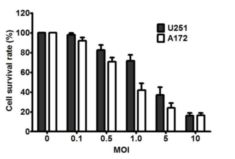
MYXV affected cell viability in a
dose-dependent manner
Different infective doses of cell lethality
were investigated and similar results were found in two human
malignant glioma cell lines (U251 and A172). As shown in Fig. 1, both U251 and A172 cells were
susceptible to infection by MYXV, which was consistent with a
previous study (16). Furthermore,
>80% of the U251 and A172 cells were killed by MYXV at an MOI of
10. When the MOI was 5, both U251 and A172 cell lethality was
between 40 and 60%, which was determined to be the most suitable
infection status for the present study. In addition, the cell
viabilities of the U251 and A172 cells that did not receive myxoma
virus were used as negative control for all experiments in the
present study. Based on this result, an MOI of 5 was used in the
subsequent experiments.
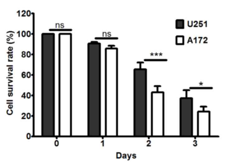
MYXV affected cell viability in a
time-dependent manner
The effect of different incubation periods of MYXV
on cell death was determined by MTT assay. Neuroglioma cells were
infected with MYXV at MOI of 5. After incubation periods of 0, 1, 2
and 3 days, the survival rates of U251 and A172 were detected by
MTT. Cell survival rates were reduced as the length of the
incubation period increased, and A172 cells were more sensitive to
MYXV than U251 cells (P<0.05; Fig.
2).
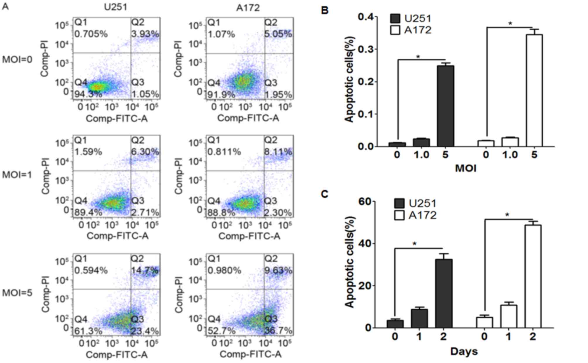
MYXV induced cell apoptosis in a time-
and dose-dependent manner
FCM was conducted to detect the effect of MYXV on
cell apoptosis. Neuroglioma cells were infected with MYXV at
different MOIs (0, 1 and 5). As shown in Fig. 3A and B, compared with MOI 0, the
proportion of apoptotic cells in U251 and A172 cells increased at
MOI 5, which indicated that both U251 and A174 cells were
susceptible to being killed by MYXV in a dose-dependent manner
(P<0.05; Fig. 3A and B).
Furthermore, the percentage of apoptotic cells in U251 and A172
after treatment with 5 MOI MYXV for 0, 1 and 2 days were calculated
(Fig. 3C). With the increase of time
post-infection, cell apoptosis rate of U251 and A172 cells also
increased compared to day 0 (P<0.05; Fig. 3C), which suggested that MYXV induced
cell apoptosis in a time-dependent manner.
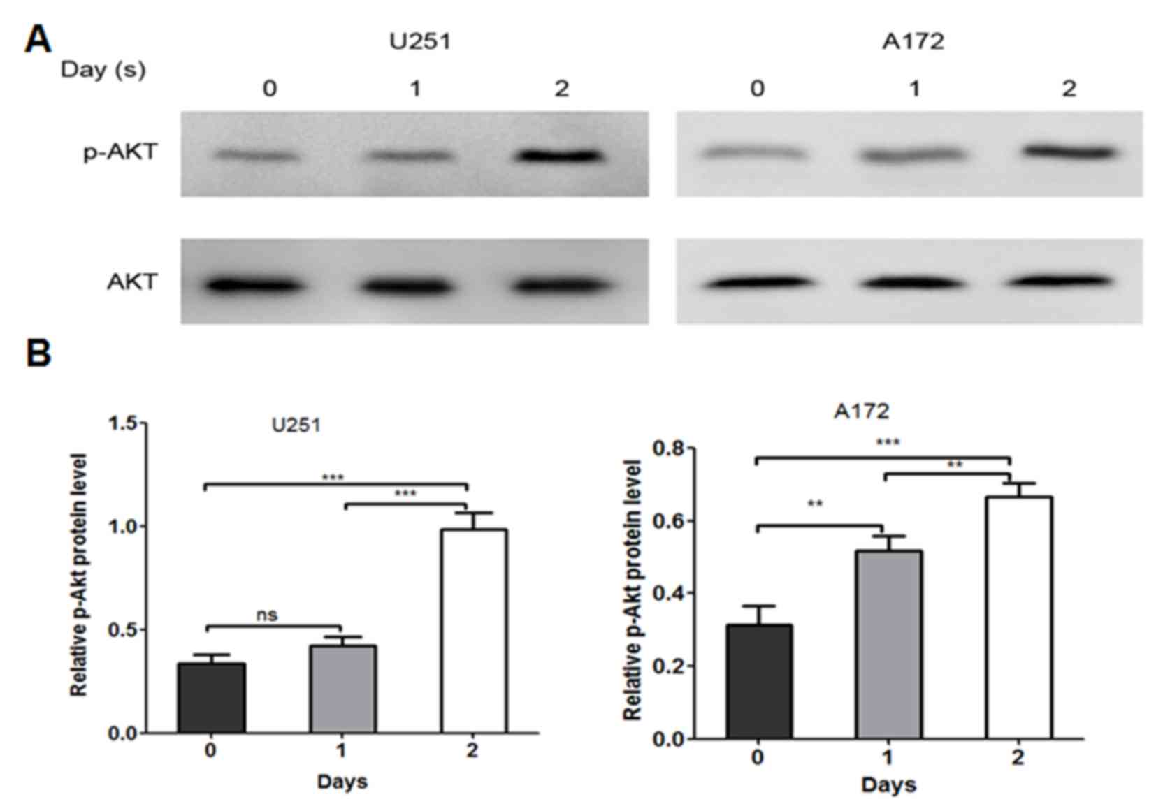
Effect of MYXV on p-AKT activity
The levels of p-AKT are directly involved in the
susceptibility of tumor cells to infection with MYXV (9,17). To
examine whether similar effects are observed in U251 and A172 cell
lines, western blot analysis was used to detect the expression
levels of total AKT and p-AKT in U251 and A172 cells that had been
infected by MYXV at MOI 5 for 0, 1 and 2 days. Consistent with
previous studies, MYXV induced the phosphorylation of AKT in both
U251 and A172 cell lines in a time-dependent manner (Fig. 4). Then a densitometric analysis was
performed on these results by Quantity One version 4.6.2 software.
According to the densitometric analysis, in U251 cells, there was a
significant increase in the expression levels of p-AKT on day 2
compared with day 0 (P<0.05; Fig.
4B) and in A172 cells, there was a significant increase in
p-AKT expression levels on day 1 and 2 compared to day 0
(P<0.05; Fig. 4B).
Discussion
Malignant gliomas are the most common and aggressive
primary central nervous system tumor in humans. Numerous types of
conventional pro-apoptotic therapies have been applied to resistant
malignant gliomas, such as radiotherapy, chemotherapy and adjuvant
therapies, but these have not been effective in killing glioma
cells (18,19). It is therefore crucial to develop an
original approach for malignant glioma therapy. Oncolytic
virotherapy, a novel and promising cancer therapeutic strategy, is
reported to be more effective and have fewer side effects than
conventional cancer therapies (20–22), and
has previously been investigated as a treatment for gliomas
(23). Candidate oncolytic viruses
should produce few side-effects, be non-pathogenic and exhibit
selective anti-tumor activities.
MYXV belongs to the poxviridae family. It is a
rabbit-specific virus and exhibits restricted pathogenicity for all
other vertebrate species, including humans (24,25).
Despite its limited host range specificity, MYXV has been shown to
selectively infect diverse forms of human tumor cells, including
glioma cells (8). In the present
study, MYXV acted as a vital factor that affected cell survival
rates. In addition, MYXV exerted such effects in a dose and
time-dependent manner. The results also indicated that MYXV
contributed to apoptosis in human neuroglioma cell lines U251 and
A172 in a dose and time-dependent manner.
Notably, MYXV exerts a selective tropism for tumor
cells with elevated levels of p-AKT (8). AKT, also known as protein kinase B, is
a serine/threonine protein kinase which plays an important role in
cell survival and apoptosis (17).
AKT phosphorylates a series of proteins and inhibits apoptosis via
a number of mechanisms (26). It has
been confirmed that the majority of both malignant and recurrent
glioma cells exhibit PTEN gene inactivation or deletion, which
increases AKT activity and results in cell proliferation and
inhibition of apoptosis. Data from the current study indicated that
the expression levels of p-AKT significantly increased in
MYXV-infected U251 and A172 cell lines compared with cell lines
receiving an inactive virus.
Furthermore, the present results verified the stated
association between the activation of AKT and MYXV-mediated
oncolysis in vitro. This highlights the potential oncolytic
function of MYXV on human glioma cells and provides a promising
therapeutic target for human malignant glioma tumors. Further
studies are required to verify whether MYXV can promote oncolysis
through modulating the levels of activated AKT within other types
of tumor cell in vitro, which could make MYXV a key factor
in improving the outcome of treatment for various cancer types.
Acknowledgements
This work was supported by Shenzhen International
Cooperation Research Funding (grant no. GJHZ20120614154914623) and
Shenzhen Key Laboratory of Neurosurgery (grant no.
ZDSYS20140509173142601).
References
|
1
|
Laws ER: Management of (malignant)
intracranial gliomas. Practical Handbook of Neurosurgery. 549–558.
2009. View Article : Google Scholar
|
|
2
|
Scott JN, Rewcastle NB, Brasher PM, Fulton
D, MacKinnon JA, Hamilton M, Cairncross JG and Forsyth P: Which
glioblastoma multiforme patient will become a long-term survivor? A
population-based study. Ann Neurol. 46:183–188. 1999. View Article : Google Scholar : PubMed/NCBI
|
|
3
|
Wilcox ME, Yang W, Senger D, Rewcastle NB,
Morris DG, Brasher PM, Shi ZQ, Johnston RN, Nishikawa S, Lee PW and
Forsyth PA: Reovirus as an oncolytic agent against experimental
human malignant gliomas. J Natl Cancer Inst. 93:903–912. 2001.
View Article : Google Scholar : PubMed/NCBI
|
|
4
|
Csatary LK, Gosztonyi G, Szeberenyi J,
Fabian Z, Liszka V, Bodey B and Csatary CM: MTH-68/H oncolytic
viral treatment in human high-grade gliomas. J Neurooncol.
67:83–93. 2004. View Article : Google Scholar : PubMed/NCBI
|
|
5
|
Freeman AI, Gomori JM, Linetsky E,
Zakay-Rones Z, Panet A, Libson E, Irving CS, Galun E and Siegal T:
Phase I/II trial of intravenous OV001 oncolytic virus in resistant
glioblastoma multiforme (GBM). J Clin Oncol. 22:15152004.
View Article : Google Scholar
|
|
6
|
Stanford MM and McFadden G: Myxoma virus
and oncolytic virotherapy: A new biologic weapon in the war against
cancer. Expert Opin Biol Ther. 7:1415–1425. 2007. View Article : Google Scholar : PubMed/NCBI
|
|
7
|
Kirn DH and Thorne SH: Targeted and armed
oncolytic poxviruses: A novel multi-mechanistic therapeutic class
for cancer. Nat Rev Cancer. 9:64–71. 2009. View Article : Google Scholar : PubMed/NCBI
|
|
8
|
Lun X, Yang W, Alain T, Shi ZQ, Muzik H,
Barrett JW, McFadden G, Bell J, Hamilton MG, Senger DL and Forsyth
PA: Myxoma virus is a novel oncolytic virus with significant
antitumor activity against experimental human gliomas. Cancer Res.
65:9982–9990. 2005. View Article : Google Scholar : PubMed/NCBI
|
|
9
|
Lun XQ, Zhou H, Alain T, Sun B, Wang L,
Barrett JW, Stanford MM, McFadden G, Bell J, Senger DL and Forsyth
PA: Targeting human medulloblastoma: Oncolytic virotherapy with
myxoma virus is enhanced by rapamycin. Cancer Res. 67:8818–8827.
2007. View Article : Google Scholar : PubMed/NCBI
|
|
10
|
Almansour NM, Pirogova E, Coloe PJ, Cosic
I and Istivan TS: A bioactive peptide analogue for myxoma virus
protein with a targeted cytotoxicity for human skin cancer in
vitro. J Biomed Sci. 19:652012. View Article : Google Scholar : PubMed/NCBI
|
|
11
|
Coffey MC, Strong JE, Forsyth PA and Lee
PW: Reovirus therapy of tumors with activated ras pathway. Science.
282:1332–1334. 1998. View Article : Google Scholar : PubMed/NCBI
|
|
12
|
Norman KL, Hirasawa K, Yang AD, Shields MA
and Lee PW: Reovirus oncolysis: The Ras/RalGEF/p38 pathway dictates
host cell permissiveness to reovirus infection. Proc Natl Acad Sci
USA. 101:pp. 11099–11104. 2004; View Article : Google Scholar : PubMed/NCBI
|
|
13
|
Werden SJ and McFadden G: Pharmacological
manipulation of the Akt signaling pathway regulates myxoma virus
replication and tropism in human cancer cells. J Virol.
84:3287–3302. 2010. View Article : Google Scholar : PubMed/NCBI
|
|
14
|
Correa RJ, Komar M, Tong JG, Sivapragasam
M, Rahman MM, McFadden G, Dimattia GE and Shepherd TG: Myxoma
virus-mediated oncolysis of ascites-derived human ovarian cancer
cells and spheroids is impacted by differential AKT activity.
Gynecol Oncol. 125:441–450. 2012. View Article : Google Scholar : PubMed/NCBI
|
|
15
|
Smallwood SE, Rahman MM, Smith DW and
McFadden G: Myxoma virus: Propagation, purification, quantification
and storage. Curr Protoc Microbiol Chapter. 14:Unit: 14A.
12010.
|
|
16
|
Johnston JB, Barrett JW, Chang W, Chung
CS, Zeng W, Masters J, Mann M, Wang F, Cao J and McFadden G: Role
of the serine-threonine kinase PAK-1 in myxoma virus replication. J
Virol. 77:5877–5888. 2003. View Article : Google Scholar : PubMed/NCBI
|
|
17
|
Wang G, Barrett JW, Stanford M, Werden SJ,
Johnston JB, Gao X, Sun M, Cheng JQ and McFadden G: Infection of
human cancer cells with myxoma virus requires Akt activation via
interaction with a viral ankyrin-repeat host range factor. Proc
Natl Acad Sci USA. 103:pp. 4640–4645. 2006; View Article : Google Scholar : PubMed/NCBI
|
|
18
|
Hentschel SJ and Lang FF: Current surgical
management of glioblastoma. Cancer J. 9:113–125. 2003. View Article : Google Scholar : PubMed/NCBI
|
|
19
|
Stupp R, Mason WP, van den Bent MJ, Weller
M, Fisher B, Taphoorn MJ, Belanger K, Brandes AA, Marosi C, Bogdahn
U, et al: Radiotherapy plus concomitant and adjuvant temozolomide
for glioblastoma. N Engl J Med. 352:987–996. 2005. View Article : Google Scholar : PubMed/NCBI
|
|
20
|
Thorne SH and Kirn DH: Future directions
for the field of oncolytic virotherapy: A perspective on the use of
vaccinia virus. Expert Opin Biol Ther. 4:1307–1321. 2004.
View Article : Google Scholar : PubMed/NCBI
|
|
21
|
Shah AC, Benos D, Gillespie GY and Markert
JM: Oncolytic viruses: Clinical applications as vectors for the
treatment of malignant gliomas. J Neurooncol. 65:203–226. 2003.
View Article : Google Scholar : PubMed/NCBI
|
|
22
|
Norman KL, Farassati F and Lee PW:
Oncolytic viruses and cancer therapy. Cytokine Growth Factor Rev.
12:271–282. 2001. View Article : Google Scholar : PubMed/NCBI
|
|
23
|
Hu CW, Yin GF, Wang XR, Ren BW, Zhang WG,
Bai QL, Lv YM, Li WL and Zhao WQ: IL-24 induces apoptosis via
upregulation of RNA-activated protein kinase and enhances
temozolomide-induced apoptosis in glioma cells. Oncol Res.
22:159–165. 2014. View Article : Google Scholar : PubMed/NCBI
|
|
24
|
Górski J, Mizak B and Chrobocińska M:
Control of rabbit myxomatosis in poland. Rev Sci Tech. 13:869–879.
1994. View Article : Google Scholar : PubMed/NCBI
|
|
25
|
Rivers TM and Ward SM: Infectious
myxomatosis of rabbits: Preparation of elementary bodies and
studies of serologically active materials associated with the
disease. J Exp Med. 66:1–14. 1937. View Article : Google Scholar : PubMed/NCBI
|
|
26
|
Barrett JW, Alston LR, Wang F, Stanford
MM, Gilbert PA, Gao X, Jimenez J, Villeneuve D, Forsyth P and
McFadden G: Identification of host range mutants of myxoma virus
with altered oncolytic potential in human glioma cells. J
Neurovirol. 13:549–560. 2007. View Article : Google Scholar : PubMed/NCBI
|