Introduction
Large skin defects, which are often observed in the
clinic following trauma, burn, high-tension wounds, tumorectomy and
large flap donor zones, have become a global problem that numerous
surgeons encounter. It is often difficult to achieve primary
closure on large skin defects on the trunk and limbs of patients
due to a paucity of skin. Furthermore, a variety of methods have
been used for closing large skin defects, including the use of skin
grafts, local flaps, tissue stretching, skin expansion, free flaps
and closure by secondary surgery. Limited by their considerable
failure rate, risk of complications, complexity, delayed healing
and poor appearance; these techniques have not been very widely
used.
Biomechanical characteristics that define skin
include creep, extensibility, viscoelasticity and stress relaxation
(1–3). Creep describes the ability of the skin
to extend under stretching at continuous tension (4). Viscoelasticity includes viscosity and
elasticity, where elasticity describes the characteristic that the
deformation due to stress loads can be temporary if the stress is
rapidly relieved, whereas viscosity describes the permanent
deformation that results when stress is maintained (3). As the corollary of creep, stress
relaxation indicates that if skin is stretched over a constant
distance, the amount of tension that is required to keep the skin
stretched will slowly decrease over time (1,2,5).
Based on these principles, skin-stretching systems
have been designed to exploit the potential afforded by these
biomechanical characteristics to realize the primary closure of
large skin defects within a short time. By applying a mechanical
load to skin stretchers, one edge of the wound can be gradually
pulled to the opposite side, achieving complete wound closure. This
method is effective in theory. However, understanding how to
achieve optimum efficiency in stretching skin in practice remains
under investigation.
In 1987, Bashir (6)
was the first to attempt a stretching technique to achieve skin
expansion by traction with Kirschner and silver wires.
Subsequently, several types of skin-stretching systems have
emerged. In 1992, a specific piece of equipment termed a
skin-stretching device was invented by Hirshowitz et al
(7); this device attempted to close
a wound by harnessing the viscoelastic property of the skin. This
new device and improved versions accomplished the effective primary
closure of large skin defects by markedly reducing the size of the
defects in cases involving tumors, ulcers, scars, tattoos, burns,
nevi and donor-site defects (6–12).
Nevertheless, notable complications following skin stretching,
including wound dehiscence, hypertrophic scar, infection, marginal
necrosis and delayed healing arose, and this device was unable to
prevent them (6–9).
Using a different form that grasped the skin edge
without Kirschner wires, Wilhelm Fleischmann designed the
EASApprox® skin-stretching system, which uses special
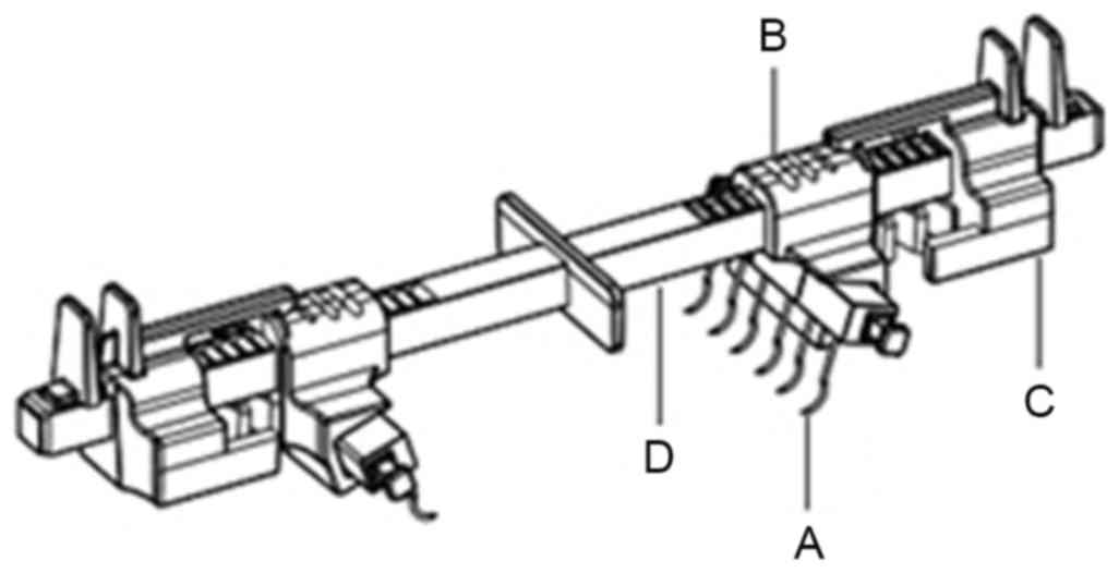
hooked needles and a tension indicator (Fig. 1). This device was used to improve the
potential of skin biomechanical properties. Three makers on the
indicator included with the EASApprox® skin-stretching
device represent 0.5 (5 N), 1.5 (15 N) and 3.0 kg (30 N) of
stretching tension. This innovation has several advantages,
including simple operation, increased healing and reduced
complications. In order to evaluate the effectiveness of the new
device at closing skin defects, the present study was performed
using Bama miniature pigs; the new EASApprox® device was
compared with methods including sutures and Kirschner wires in
terms of cutaneous microcirculation, histology and the overall
healing.
Materials and methods
Animals
The present study was approved by the Ethics
Committee on Animal Care and Use of Dalian Medical University
(Dalian, China). In total, 9 healthy, 10-month-old, purposely bred
Bama miniature pigs (all females, weighing 20–25 kg) were purchased
from Taizhou Taihe Biology Technology Co., Ltd., (Jiangsu, China).
Furthermore, complete physical examinations and blood counts, serum
biochemical analyses and urinalysis were performed on the miniature
pigs before the study commenced. The animals were housed indoors,
were allowed to move freely and had outdoor access twice daily.
Commercial dry maintenance diets were administered twice daily, and
water was available ad libitum. The pigs were then continuously fed
as they participated in other experiments following the present
study.
Study design
After the creation of skin defects on both forearms,
the 18 forearms were randomly divided into 3 groups. In group A,
sutures were used for direct closure; in group B, the new
EASApprox® skin-stretching device [PFKZ-SL-02; BIOWIM
(China), Ltd., Dalian, China] was used and in group C, Kirschner
wires were inserted along each side of the wound and used to
achieve closure.
Preoperative procedures
The pigs were premedicated with acepromazine (0.1
mg/kg; Santa Cruz Biotechnology, Inc., Dallas, TX, USA), xylazine
(2.2 mg/kg; Sigma-Aldrich; Merck KGaA, Darmstadt, Germany),
ketamine (2 mg/kg; Jiangsu Hengrui Medicine Co., Ltd., Lianyungang,
China) and atropine (0.02 mg/kg; Beijing Solarbio Science &
Technology Co., Ltd., Beijing, China). Endotracheal general
anesthesia via cannula was induced with 2.5% thiopental sodium (8
mg/kg; Shanghai New Asia Pharmaceutical Co., Ltd., Shanghai, China)
and maintained with isoflurane (3–5%; Hebei Jiupai Pharmaceutical
Co., Ltd., Shijiazhuang, China) in oxygen (1.5 l/min); and the body
temperature, heart and respiration rates were monitored throughout.
Both forearms of each pig were shaved from the middle of the
humerus to the carpus and prepared for aseptic surgery. All drugs
listed above were supported by the Central Research Laboratory of
The First Affiliated Hospital of Dalian Medical University.
Following the operation, one Panadol (Sino American Tianjin Smith
Kline & French Laboratories, Ltd., Tianjin, China) was fed to
each pig daily from postoperative day (POD) 1 to POD 10.
Surgical procedures
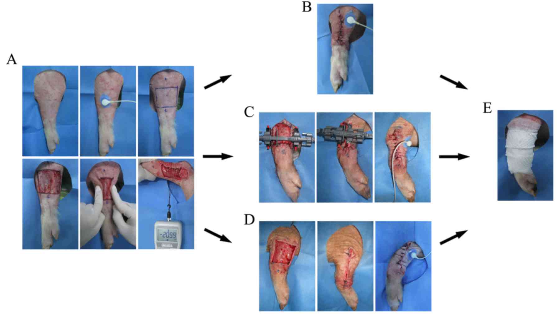
On the day of the operation, one skin defect (5×5
cm) was created by removing the intact skin and subcutaneous tissue
using a No. 10 blade on the lateral surface of each mid-forearm.
Furthermore, cutaneous microcirculation was measured based on the
transcutaneous partial pressure of oxygen (TcpO2) using
a PeriFlux System 5000 instrument (Perimed AB, Stockholm, Sweden)
(Fig. 2A). Pinch tests were used to
indicate high tension, and a push/pull gauge (Imada Co., Ltd.,
Toyohashi, Japan) measured the tension of each wound margin
(19.52–21.05 N) to standardize the wounds. Defects without any
undermining of the wound edges had lengths and widths of 5 cm.
For group A (suture), a direct simple interrupted
procedure was performed to cover the wound using 2/0 Mersilk
sutures (Ethicon Inc., Somerville, NJ, USA). Side holes were 0.5 cm
from the wound edges (Fig. 2B). For
group B (Fig. 2C), the
EASApprox® skin-stretching device was applied by
installing hook needles ~0.5 cm from the wound edge. In total,
three cycles of intermittent skin stretching (cycle loading) were
subsequently loaded. Forces of 1.0–3.0 kg were loaded for 4 min,
followed by 1-min periods of relaxation (13). After a second pinch test indicated
low tension, simple interruption by 2/0 Mersilk sutures was
performed between the two wound edges in group B. For group B
(Fig. 2D), two 2.0-mm Kirschner
wires were inserted along both wound sides ~0.5 cm from the wound
edges (13,14). Subsequently, intermittent skin
stretching was conducted similar to that applied in group B through
the traction of Kirschner wires. A further pinch test was applied
to estimate the actual tension of the wound. Subsequently, complete
closure was achieved by simple interrupted suturing with 2/0
Mersilk sutures. Finally, all wounds in groups A-C were bandaged
with sterile dressings and marked carefully on each pig (Fig. 2E).
Postoperative procedures
Following surgery, wound dressings were changed
every 2 days, and wound care and healing recording were conducted
at the same time. On postoperative day 10, all wound sutures were
removed.
Microcirculation measurements
Cutaneous microcirculation was assessed based on
TcpO2. Under general anesthesia, measurements of the
lateral surface of the mid-forearm were performed to detect the
baseline value for normal skin. Before suturing the wound in groups
A-C, TcpO2 was measured using the same protocol as
during wound closure. Variations of TcpO2 were recorded
and compared.
Clinical scoring of wound healing
Healing was observed following primary wound closure
in groups A, B and C; and the clinical score was recorded every two
days until suture removal on POD 10. A modified clinical scale was
used as follows: 1, no visible reaction; 2, minimal swelling or
erythema; 3, suture line inflammatory reaction at least 1-cm thick
with pain or redness; 4, seroma or abscess formation; and 5,
dehiscence, skin necrosis or impossible primary closure (15). Clinical scores were assessed by a
clinician who was blinded to the groupings of the present
study.
Microscopic and ultramicroscopic
structure evaluation
Samples for histological evaluation were taken at
suture removal (POD 10). Specimens were obtained using a No. 10
blade while the pigs were under general anesthesia, as previously
described. The area of resection included the healing area and the
adjacent skin at a distance of 0.5 cm from the wound margin or
suture line. Specimens were cut into two halves and pinned flat to
a piece of wood. One half was fixed in 10% neutral-buffered
formalin (Beijing Solarbio Science & Technology Co., Ltd.).
Following routine processing, which included dehydration in an
alcohol gradient series, clearing in xylene and paraffin embedding,
the samples were cut into 4-µm sections, mounted on glass slides
and stained with Verhoeff Van-Gieson (VVG) for elastin hematoxylin
and eosin (HE) for microvessels, and Masson and Sirius Red (all
Leagene Biotech Co., Ltd., Beijing, China) for collagen bundles.
All histological results were observed and analyzed using Image Pro
Plus 6.0 (Media Cybernetics, Inc., Silver Spring, MD, USA). The
remaining half of the specimens was fixed in 2.5% glutaraldehyde
(Beijing Solarbio Science & Technology Co., Ltd.) and treated
by desiccation and spraying with gold. Scanning electron microscopy
(SEM; SUPRA 55VP; Carl Zeiss AG, Oberkochen, Germany) was used to
study the collagen microstructure.
Statistical analysis
Statistical analysis was performed using SPSS 17.0
software (SPSS, Inc., Chicago, IL, USA). Data were statistically
analyzed based on the analysis of variance for repeated measures.
Data were expressed as the mean ± standard deviation (as
indicated), and the normality of the data distribution was assessed
using the Shapiro-Wilk test. Cross-group differences were analyzed
using the paired t-test. P≤0.05 was considered to indicate a
statistically significantly difference.
Results
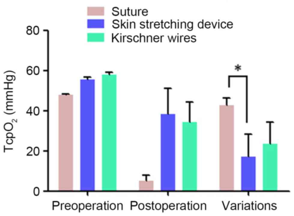
Microcirculation measurements
In different groups, cutaneous microcirculation was
assessed twice (Fig. 3). Before
creating the wound, basic TcpO2 was measured as
47.76±0.58, 55.33±1.53 and 57.67±1.53 mmHg, respectively, in groups
A-C. When wounds were closed by different methods in groups A-C,
TcpO2 decreased to 5.00±3.00, 38.33±12.74 and
34.33±10.02 mmHg, respectively. Finally, the differences in
TcpO2 between the groups were calculated and compared
using SPSS software. No statistically significant differences in
TcpO2 were observed between groups A and C or between
groups B and C. Significant differences were observed only between
groups A and B (P=0.036), and the difference between preoperative
and postoperative TcpO2 (skin microcirculation) was
smaller for the EASApprox® skin-stretching device.
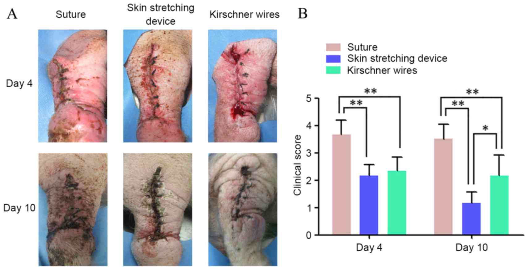
Clinical scoring of wound healing
Clinical scores for wounds during the healing period
(from wound closure to suture removal on POD 10) were recorded for
all groups. Table I and Fig. 4 show the condition of wound healing
on POD4 and 10. Wound healing in groups B and C was significantly
better than that in group A on POD4 and POD10 (P<0.01). Although
there were no significant differences between groups B and C in the
clinical scores on POD4, at the final observation period, the
outward appearances of wounds in group B significantly better than
those in group C (P<0.05).
 | Table I.Clinical scores of different
wounds. |
Table I.
Clinical scores of different
wounds.
|
| Suture | Skin stretching
device | Kirschner
wires |
|---|
|
|
|
|
|
|---|
| Serial number | Day 4 | Day 10 | Day 4 | Day 10 | Day 4 | Day 10 |
|---|
| 1 | 4 | 4 | 2 | 1 | 3 | 3 |
| 2 | 3 | 3 | 2 | 1 | 2 | 1 |
| 3 | 3 | 4 | 2 | 1 | 2 | 2 |
| 4 | 4 | 4 | 2 | 1 | 2 | 2 |
| 5 | 4 | 3 | 3 | 2 | 3 | 3 |
| 6 | 4 | 3 | 2 | 1 | 2 | 2 |
| Mean ± SD | 3.67±0.52 | 3.50±0.55 | 2.17±0.41 | 1.17±0.41 | 2.33±0.52 | 2.17±0.75 |
Microscopic structure evaluation
Histological staining of the three groups is shown
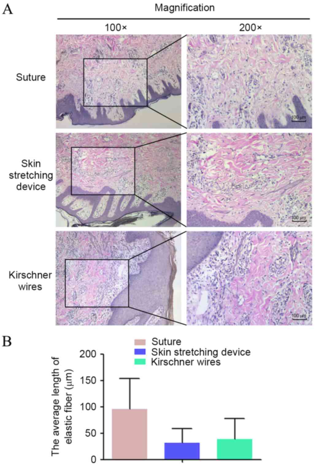
in Fig. 5. VVG staining for elastin
was performed (Fig. 5A). Overall,
elastin stained as a deep color that ran perpendicularly to the
epidermis. In group A, the overall length of elastin fiber was
longer than that of groups B and C. Pathological sections of group
C exhibited minimal long, straight elastin fibers; however, elastin
fragments were apparent in groups B and C. The mean lengths of
elastin in groups A-C are shown in Fig.
5B, with elastin length demonstrated to be the shortest in
group B.
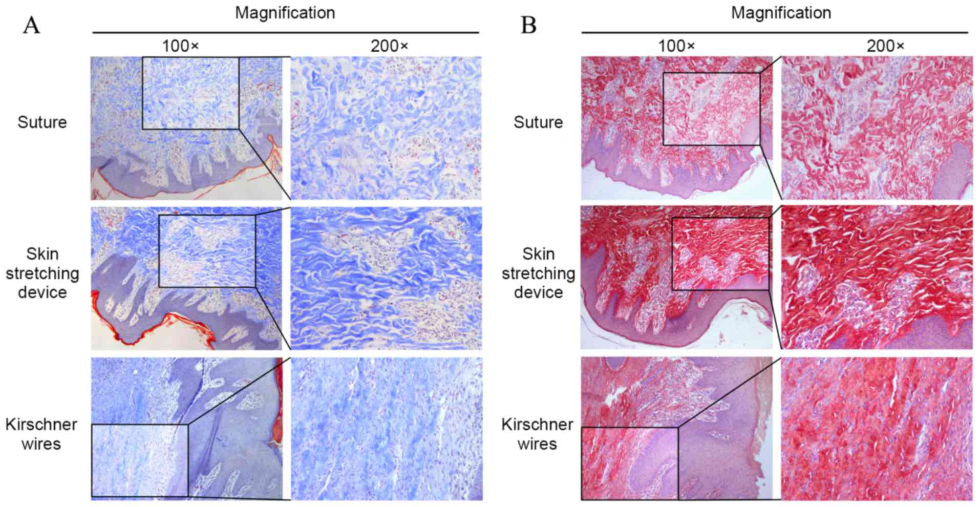
Masson and Sirius Red staining were used to assess
collagen bundles (Fig. 6A and B,
respectively). Under the microscope, collagen bundles appeared blue
after Masson staining and red after Sirius Red. In group A,
collagen was predominantly oriented perpendicular to the skin
surface. Most collagen bundles were disordered and exhibited
crimping. Collagen bundles in groups B and C were stretched and
oriented parallel to the epidermis, and the collagen bundles in
group B were straighter and longer than those in groups A and
C.
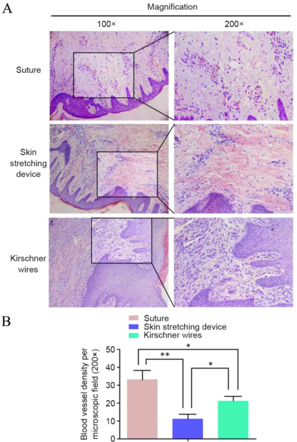
Microvessel density was calculated after HE
staining. Microscopically, microvessels comprised round vascular
walls and intravascular red blood cells. Significant differences
were observed among the three groups (P<0.05; Fig. 7). More microvessels were observed in
group A than in the other groups. HE staining indicated that the
pathological sections in group B had the lowest microvessel
density.
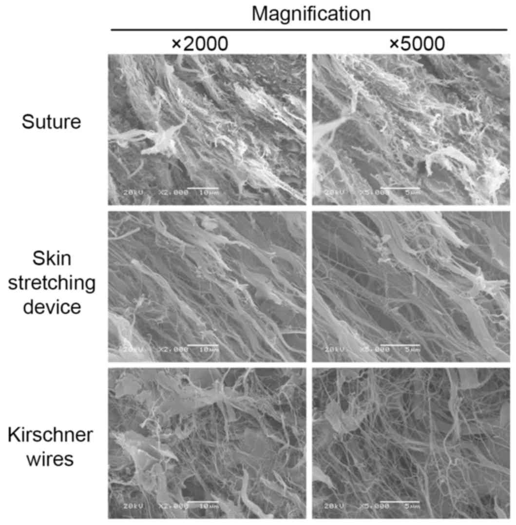
The specimen surfaces of samples from different
groups were scanned and observed on the SEM screen at
magnifications of ×2,000 and ×5,000. Images of representative areas
were captured (Fig. 8). The entire
shape of the collagen bundle was easily observed under SEM. In
group A, the collagen bundles presented with particularly small
fibers and were seriously fractured. However, the collagen bundles
in group B were bolder, straighter and more intact than those in
the groups A and C. Randomly arranged, very small fibers were
observed in specimens from group C.
Discussion
The closure of large skin defects is challenging for
surgeons worldwide. Large local excisions or acute trauma can
create defects that are usually not amenable to primary closure. In
the past, the treatment of large skin defects required the
application of a graft or flap. Due to advances in the research of
biomechanical characteristics of skin, presuturing techniques and
other mechanical means have been presented in the literature; these
methods facilitate wound closure by manipulating skin that is in
close proximity or adjacent to the margins of a wound or proposed
operative area (16–20). Among several impressive wound closure
techniques, the first skin-stretching device, which was designed by
Hirshowitz et al (7), has
been applied in many medical institutions with satisfactory
efficiency. This device skillfully exploits the viscoelastic and
stress relaxation properties of skin to stretch the tissue and
close the defect. However, notable complications can occur after
skin stretching, including wound dehiscence, hypertrophic scarring,
infection, marginal necrosis and delayed healing; these
complications limit the applications of this technique.
In order to facilitate the operating process and
avoid complications that result from skin stretching, a new
generation of skin-stretching devices has been designed. These
devices are simple to insert at the wound edge, and an indicator
measures wound tension. Compared with other devices, the new
EASApprox® skin-stretching device investigated in the
present study is able to mobilize large areas of skin that lie
adjacent to and more distant from the operative area through
cyclical stretching and relaxation. Consequently, skin can be
stretched from more than one region and direction in relation to
the surgical area.
Skin consists of the epidermis and dermis. The
thickness of the horny layer in Bama miniature pigs is similar to
that in humans and does not change with age. Liu et al
(21) considered Bama miniature pigs
a suitable animal model for studies of human skin; thus, these
animals were used in the present study. Before the surgery
commenced, the TcpO2 value of the forearms in the
different pig groups ranged between 47.76±0.58 and 57.67±1.53 mmHg.
Under the same resection of the entire skin, a pig model of a
5-cmx5-cm wound was achieved. The pinch test suggested that the
wound tension was too high to achieve primary closure. Furthermore,
the tension was ~20 N, representing unsafe conditions for acutely
closing the wound.
The efficiency of the new EASApprox® skin
stretching device was compared with two presuturing techniques.
Direct suture is the method originally used to achieve wound
closure, particularly for conditions without any tension. Skin
stretching using Kirschner wires, a prototype of the
skin-stretching device, is considered the most basic form of
primary closure. Wounds in groups B and C were manipulated by cycle
stretching. According to a safe threshold, the loaded force ranged
between 0.5 and 4 kg (7). During the
period of skin stretching, the tension adopted in group B ranged
from 1 to 3 kg to ensure skin integrity. Greater forces stretch
collagen bundles and small blood vessels, thereby damaging blood
perfusion, resulting in subsequent necrosis of the skin margins
(4,14). When used in combination with cycles
of stretching and relaxation loading, this method contributed to
skin extension, as previously described (6,22).
Furthermore, when a pinch test suggested that the wound was under
low tension following three sets of cycle stretching, primary
closure was performed. The TcpO2 value of a second
measurement indicated that the EASApprox®
skin-stretching device protected skin microcirculation to a greater
extent than direct suture. This protective function benefited from
the ingenious design, rational structure and use of the cycle
stretching method. Additionally, the sharply hooked needles were
convenient to pin the wound margins and reduced side injuries
caused by the surgery.
At the stage of wound healing (POD4 and POD10), the
clinical scores of the wounds that were treated using the
EASApprox® skin-stretching device were significantly
lower than those achieved using the other methods. In general, the
wounds showed light, visible reactions or minimal swelling without
any inflammatory reaction. The treatment using the Kirschner wires
group was the second most effective method. By contrast, direct
suture had a long-term impact on wound healing, which was
accompanied by evident inflammation or seroma. Therefore, treatment
using the EASApprox® skin-stretching device promoted
wound healing and prevented the occurrence of adverse
reactions.
Collagen, elastin and ground substance are the most
important structural components of the dermis (23). Among them, collagen constitutes the
main structural component of the skin, accounting for >50% of
its fat-free dry weight (3). Without
any stress loading, collagen fibers are arranged haphazardly;
however, when the skin is stretched, the fibers are aligned
parallel to each other. Mechanically, collagen bundles have high
tensile strength, are stiff and lack extensibility. These bundles
are the main source of structural support for the skin but are not
significant in its recoiling abilities (24,25).
According to observations made on skin that was
stained with Masson and Sirius Red stain, the collagen bundles were
longer and straighter in the skin-stretching device group than in
the other groups. In addition, stretching by both the new
EASApprox® device and Kirschner wires oriented the
collagen bundles parallel to the skin surface. Observations made
using microscopy were consistent with those obtained using SEM.
Under SEM, when the skin was stretched using the new
EASApprox® device the collagen bundles were bolder,
straighter and more parallel to each other than those in the other
groups. Furthermore, at the micro- and ultramicro-scales, the new
EASApprox® skin-stretching device made full use of the
skin viscoelasticity and achieved optimal stretching.
Different from collagen bundles, elastin comprises
<5% of the fat-free dry weight of skin and is characterized by
long-range elastic extensibility (3). Even after a maximal strain, elastin has
the capacity to return to its original shape. Furthermore, it has a
close association with collagen and promotes the return of collagen
to its wavy posture when it is in a state of rest. Thus, elastin
provides the ability of skin to recoil after the application of
deforming stresses; however, if sufficient stretching load is
applied, elastic fibers can fragment, resulting in a loss of recoil
(24,25). Therefore, the shape of elastin could
be treated as another stretch level.
Histological staining of sections by VVG
demonstrated that elastic fibers were fragmented after a stretching
load was applied. In the direct suture group, the mean length of
the elastic fibers was greater than those in the other groups. Due
to stretching by the new EASApprox® device, elastic
fibers were thoroughly fragmented. By analyzing the observations of
collagen bundles and elastic fibers, it was once more confirmed
that there is a correlation between collagen and elastin;
indicating that the EASApprox® skin-stretching device
supported greater stretching efficiency.
Microvessel density among the three different
methods was demonstrated to be in the order of: Direct suture>
EASApprox® skin-stretching >Kirschner wires.
Researchers consider high microvessel density beneficial for
healing (26,27). The present study proposed that local
inflammation following tissue damage can easily lead to
vascularization. According to this hypothesis, the high microvessel
density observed in the direct suture group indicated serious
tissue damage. Conversely, use of the EASApprox®
skin-stretching device resulted in the least tissue damage.
In conclusion, the EASApprox®
skin-stretching system is a safe and effective method to treat
large skin defects and has several potential advantages over other
devices currently used for wound closure; thus, this device can
eliminate the requirement for more costly flap, grafting or
tissue-expansion techniques. We suggest that cycle stretching
should be used in concert with the EASApprox® device
during surgery. Tension applied to the skin during stretching
(ranging between 1.0 and 3.0 kg) was found to be safe. Compared
with direct suture and stretching using Kirschner wires, skin
stretching by the device resulted in only minor side effects on
skin histology and microcirculation. Additionally, the
EASApprox® device may promote wound healing. In an era
of increasing medical investment, this device is particularly
attractive because it only requires a simple operation. However,
more clinical and preclinical studies of the EASApprox®
skin-stretching system are required.
Acknowledgements
The authors would like to thank Dr Wilhelm
Fleischmann [BIOWIM (China), Ltd.] for his theoretical guidance
during the execution of the present study, and BIOWIM (China),
Ltd., for providing the EASApprox® skin-stretching
system.
Glossary
Abbreviations
Abbreviations:
|
TcpO2
|
transcutaneous partial pressure of
oxygen
|
|
POD
|
postoperative day
|
|
VVG
|
Verhoeff Van-Gieson
|
|
SEM
|
scanning electron microscopy
|
References
|
1
|
Armstrong DG, Sorensen JC and Bushman TR:
Exploiting the viscoelastic properties of pedal skin with the sure
closure skin stretching device. J Foot Ankle Surg. 34:247–253.
1995. View Article : Google Scholar : PubMed/NCBI
|
|
2
|
Gibson T and Kenedi RM: Biochemical
properties of skin. Surg Clin North Am. 47:279–294. 1967.
View Article : Google Scholar : PubMed/NCBI
|
|
3
|
Hussain SH, Limthongkul B and Humphreys
TR: The biomechanical properties of the skin. Dermatol Surg.
39:193–203. 2013. View Article : Google Scholar : PubMed/NCBI
|
|
4
|
Wilhelmi BJ, Blackwell SJ, Mancoll JS and
Phillips LG: Creep vs. stretch: A review of the viscoelastic
properties of skin. Ann Plast Surg. 41:215–219. 1998. View Article : Google Scholar : PubMed/NCBI
|
|
5
|
Liang MD, Briggs P, Heckler FR and Futrell
JW: Presuturing-a new technique for closing large skin defects:
Clinical and experimental studies. Plast Reconstr Surg. 81:694–702.
1988. View Article : Google Scholar : PubMed/NCBI
|
|
6
|
Bashir AH: Wound closure by skin traction:
An application of tissue expansion. Br J Plast Surg. 40:582–587.
1987. View Article : Google Scholar : PubMed/NCBI
|
|
7
|
Hirshowitz B, Lindenbaum E and Har-Shai Y:
A skin-stretching device for the harnessing of the viscoelastic
properties of skin. Plast Reconstr Surg. 92:260–270. 1993.
View Article : Google Scholar : PubMed/NCBI
|
|
8
|
Marrero GM and Dufresne RG Jr: An
intraoperative skin-stretching device to close wounds in mohs
defects. Dermatol Surg. 22:546–550. 1996. View Article : Google Scholar : PubMed/NCBI
|
|
9
|
Signorini M, Blandini D, Rafanelli G,
Colonna M and Candiani P: Experience with the skin stretching
device. European J Plastic Surg. 19:63–68. 1996. View Article : Google Scholar
|
|
10
|
Verhaegen PD, Bloemen MC, van der Wal MB,
Vloemans AF, Tempelman FR, Beerthuizen GI and van Zuijlen PP: Skin
stretching for primary closure of acute burn wounds. Burns.
40:1727–1737. 2014. View Article : Google Scholar : PubMed/NCBI
|
|
11
|
Samis AJ and Davidson JS: Skin-stretching
device for intraoperative primary closure of radial forearm flap
donor site. Plast Reconstr Surg. 105:698–702. 2000. View Article : Google Scholar : PubMed/NCBI
|
|
12
|
Har-Shai Y and Hirshowitz B: The use of a
modified skin stretching device (SSD) together with a lower cheek
and neck flap for coverage of a large cheek skin defect. European J
Plastic Surg. 18:136–138. 1995.
|
|
13
|
Petro JA and Niazi ZBM: Immediate skin
expansion: An old concept by a novel and inexpensive technique. Ann
Plast Surg. 36:479–484. 1996. View Article : Google Scholar : PubMed/NCBI
|
|
14
|
Abramson DL, Gibstein LA and Pribaz JJ: An
inexpensive method of intraoperative skin stretching for closure of
large cutaneous wounds. Ann Plast Surg. 38:540–542. 1997.
View Article : Google Scholar : PubMed/NCBI
|
|
15
|
Freeman LJ, Pettit GD, Robinette JD,
Lincoln JD and Person MW: Tissue reaction to suture material in the
feline linea alba. A retrospective, prospective, and histologic
study. Vet Surg. 16:440–445. 1987. View Article : Google Scholar : PubMed/NCBI
|
|
16
|
Topaz M, Carmel NN, Silberman A, Li MS and
Li YZ: The TopClosure® 3S System, for skin stretching
and a secure wound closure. Eur J Plast Surg. 35:533–543. 2012.
View Article : Google Scholar : PubMed/NCBI
|
|
17
|
Pavletic MM: Use of an external
skin-stretching device for wound closure in dogs and cats. J Am Vet
Med Assoc. 217:350–354. 2000. View Article : Google Scholar : PubMed/NCBI
|
|
18
|
Nordström RE and Devin JW: Scalp
stretching with a tissue expander for closure of scalp defects.
Plast Reconstr Surg. 75:578–581. 1985. View Article : Google Scholar : PubMed/NCBI
|
|
19
|
Barnea Y, Gur E, Amir A, Leshem D,
Zaretski A, Shafir R and Weiss J: Our experience with Wisebands: A
new skin and soft-tissue stretch device. Plast Reconstr Surg.
113:862–871. 2004. View Article : Google Scholar : PubMed/NCBI
|
|
20
|
Macionis V: Noninvasive wound closure by
stretching skin with hook needles enveloped in double-sided
adhesive tape and stuck to the skin. Ann Plast Surg. 47:345–346.
2001. View Article : Google Scholar : PubMed/NCBI
|
|
21
|
Liu Y, Chen JY, Shang HT, Liu CE, Wang Y,
Niu R, Wu J and Wei H: Light microscopic, electron microscopic, and
immunohistochemical comparison of Bama minipig (Sus scrofa
domestica) and human skin. Comp Med. 60:142–148. 2010.PubMed/NCBI
|
|
22
|
Cohen BH and Cosmetto AJ: The suture
tension adjustment reel. A new device for the management of skin
closure. J Dermatol Surg Oncol. 18:112–123. 1992. View Article : Google Scholar : PubMed/NCBI
|
|
23
|
Uitto J: Biochemistry of the elastic
fibers in normal connective tissues and its alterations in
diseases. J Invest Dermatol. 72:1–10. 1979. View Article : Google Scholar : PubMed/NCBI
|
|
24
|
Chu DH: Chapter 7. Development and
Structure of SkinWolff K, Goldsmith LA, Katz SI, Gilchrest BA,
Paller AS and Leffell DJ: Fitzpatrick's dermatology in general
medicine. 7th. New York: McGraw-Hill; pp. 57–72. 2008
|
|
25
|
Wilkes GL, Brown IA and Wildnauer RH: The
biomechanical properties of skin. CRC Crit Rev Bioeng. 1:453–495.
1973.PubMed/NCBI
|
|
26
|
Guimarães GF, De Araújo VC, Nery JC,
Peruzzo DC and Soares AB: Microvessel density evaluation of the
effect of enamel matrix derivative on soft tissue after implant
placement: A preliminary study. Int J Periodontics Restorative
Dent. 35:733–738. 2014.
|
|
27
|
Kant V, Gopal A and Kumar D, Pathak NN,
Ram M, Jangir BL, Tandan SK and Kumar D: Curcumin-induced
angiogenesis hastens wound healing in diabetic rats. J Surg Res.
193:978–988. 2015. View Article : Google Scholar : PubMed/NCBI
|