Introduction
Chronic obstructive pulmonary disease (COPD) is
characterized by chronic inflammation affecting the airway, lung
parenchyma and pulmonary vascular system (1). Infiltration of inflammatory cells such
as alveolar macrophages, T lymphocytes, particularly
CD8+ T cells, and neutrophils leads to the release of
inflammatory mediators, including leukotriene B4 (LTB4),
interleukin-8 (IL-8) and tumor necrosis factor (TNF)-α (2,3). These
inflammatory mediators are considered to contribute to the
pathogenesis of COPD (4). In
addition, inhalation of toxic substances such as cigarette smoke
may further exacerbate airway inflammation-induced injury and
tissue remodeling (5,6).
Since COPD is characterized by chronic airway
inflammation and consequent tissue remodeling, anti-inflammatory
therapy is considered a priority for COPD treatment. In this
regard, glucocorticoids are among the most rapid and effective
anti-inflammatory drugs used for the treatment of chronic
inflammatory airway disease (6).
However, unlike asthma, COPD is often unresponsive to
glucocorticoid therapy (7,8). The mechanisms of corticosteroid
resistance may involve the imbalance of histone
acetylation/deacetylation, oxidative stress and genetic or
epigenetic alterations (9,10). In this study, histone deacetylase 2
(HDAC2) dysfunction is thought to play an important role in the
development of corticosteroid resistance in COPD (8,9,11,12).
Thus, increasing HDAC2 activity may be a promising strategy to
overcome corticosteroid resistance in COPD. Existing treatments
such as theophylline, nortriptyline, macrolides and selective
phosphatidylinositol-3-kinase-δ inhibitors have been reported to
increase HDAC2 activity effectively (13). In support of this concept, Hsieh
et al accessed two public traditional Chinese medicine (TCM)
databases and retrieved the chemical constituents and TCM
characteristics of 3,294 TCM medicinal agents (14). They identified that 1,170/3,294 (36%)
of the TCM medicinal agents interact with human histone-modifying
enzymes and that 56% of the histone-modifying materials promote
chromatin condensation. Furthermore, analysis of TCM formulas
revealed that 99% of 200 government-approved TCM formulas are
histone-modifying and the synergy of the TCM medicinal agents in a
formula involved mostly concurrent DNA methyltransferase (DNMT) and
HDAC inhibition, co-inhibition of histone acetylation and H3S10
phosphorylation, or co-inhibition of H3K4 demethylation and H3K36
demethylation (14).
In addition to Western medicine, traditional Chinese
herbal medicine (TCHM) has long been used for the clinical
treatment of COPD (15,16) and the mechanisms underlying the
effects of TCHM on COPD therapy have been widely studied in
pre-clinical animal models of COPD (17,18). In
this study, TCHM such as Bufei Jianpi, Bufei Yishen and Yiqi Zishen
granules, and Liuweibuqi capsule have been demonstrated to
effectively improve lung function, increase skeletal muscle
strength, bone mass density and bone mineral content, and reduce
airway inflammation through different mechanisms, including the
inhibition of inflammatory cytokines, regulation of matrix
metalloproteinases (MMPs) and their biological inhibitors (tissue
inhibitors of MMPs), and modulation of the transforming growth
factor-β1/Smad signal pathway (17–22).
Moreover, it has been shown that glucocorticoids used in
combination with certain TCHMs have a marked effect in the
prevention and treatment of COPD (23–27) or
asthma (28). In this regard, the
combination of a Xiang Sha Liu Jun Zi Tang, salmeterol xinafoate
and fluticasone propionate inhalation powder has been demonstrated
to improve the clinical symptoms and lung function in patients with
stable COPD (25). Moreover, the
TCHM Liujunzi decoction used in combination with salmeterol
xinafoate and fluticasone propionate inhalation powder
significantly improved the symptoms and lung function of patients
with COPD by upregulating airway HDAC (in the sputum) and
downregulating IL-8 and TNF-α (23).
Recently, it has been reported that a TCHM, You-Gui-Wan, reduced
inflammation and eosinophil infiltration into the lung tissues
through increasing HDAC and decreasing histone acetyltransferase
activities in memory T lymphocytes (28).
Therefore, in the present study, the effects of on
pulmonary function Jinwei Tang, that in TCM is considered to
‘support qi, nourish yin, activate blood circulation and dispel
phlegm’ were investigated. In addition, HDAC2 expression in lung
tissue, and protein levels of IL-8, TNF-α and HDAC2 in lung tissue
homogenates of rat models of COPD were analyzed.
Materials and methods
Animals
Fifty male Wistar rats (3 months old; body weight,
220±10 g) of specific pathogen-free grade were purchased from Si
Bei Fu Experimental Animal Technology (Beijing, China). All
experiments followed a protocol approved by the Animal Care Ethics
Committee of Beijing University of Traditional Chinese Medicine
(Beijing, China).
Materials and equipment
Lipopolysaccharide (LPS) was purchased from
Sigma-Aldrich (Merck KGaA, Darmstadt, Germany). Aminophylline was
purchased from Zizhu Pharmaceutical Co., Ltd. (Beijing, China).
Budesonide suspension for inhalation was purchased from AstraZeneca
(Shanghai, China). The TCHM Jinwei Tang was made by a combination
of Jinshui Liujun Jian and Weijing Tang, and consisted of Danggui
(Angelica sinensis root) 15 g, Shudi (Rehmannia root)
15 g, Chenpi (dried orange peel) 12 g, Qingbanxia (tuber of
pinellia) 12 g, Fulin (sclerotium of poria coco) 30 g, Lugen (reed
stems) 15 g, Taoren (peach seed) 12 g, Chaoyiyiren (coix seed) 15
g, Beishashen (littoralis root) 15 g, Maidong (Ophiopogon
japonicas root) 15 g, Shenghuangqi (Astragalus root) 30
g, Danshen (Salvia root) 30 g, Zhimahuang (Chinese ephedra)
6 g and Zhigancao (licorice root) 6 g. All herbs were purchased
from the Pharmacy of Traditional Chinese Medicine at the Third
Hospital of Beijing University of Traditional Chinese Medicine
(Beijing, China). The Rat IL-8 ELISA kit (cat. no. E02I0056) was
purchased from Shanghai BlueGene Biotech Co., Ltd. (Shanghai,
China). The Rat TNF-α ELISA kit (cat. no. 70-EK382) and rat HDAC2
ELISA kit (cat. no. SEC210Ra) were boh purchased from Beijing
Bioway Biotech Group Co., Ltd. (Beijing, China). PARI BOY SX
(085G3005) nebulizer was purchased from PARI Medical Holding GmbH
(Starnberg, Germany). Daqianmen cigarettes were purchased from
Shanghai Tobacco Group Ltd. (Shanghai, China). Image Pro Plus 6.0
data analysis software was purchased from Media Cybernetics
(Rockville, MD, USA).
Grouping and treatment
Rat models of COPD were prepared by intratracheal
instillation of LPS and passive smoke exposure as described
previously (24,25). The rats were divided randomly into
five groups (each n=10): COPD group, which received intratracheal
instillation of LPS (200 µl; 1 mg/ml) on days 1 and 14, plus
passive smoke exposure with 12 Daqianmen cigarettes in a 72-l
closed box for 30 min (smoke concentration, 5%) in the morning on
days 2–13 and 15–28; control group, which did not receive any
treatment; budesonide group, which underwent the same modeling
procedure as the COPD model group, and also inhaled budesonide (2
mg) every afternoon in a 28-l closed box connected to a PARI BOY SX
(085G3005) nebulizer from day 8 of the COPD model; theophylline +
budesonide group, which underwent the same modeling procedure as
the COPD model group, and also received aminophylline (25 mg/kg; 1
ml/100 g) treatment by gavage prior to passive smoke exposure daily
from day 8 of the COPD model followed by inhalation of budesonide
(2 mg) every afternoon in a 28-l closed box; TCHM + budesonide
group, which underwent the same modeling procedure as the COPD
model group, and also underwent treatment with granules of the TCHM
(3.6 g/kg) by gavage daily prior to passive smoke exposure from day
8 of the COPD model followed by inhalation of budesonide (2 mg)
every afternoon in a 28-l closed box.
Rat lung function test
Eight rats were randomly selected from each group
after 28 days of cigarette smoke exposure. A tracheotomy was
conducted following intraperitoneal anesthesia with 10%
pentobarbital sodium. Lung function was tested as described
previously (29). Briefly, following
the tracheotomy, the animal was ventilated and placed into a body
plethysmography box (Beijing Bestlab High-Tech Co., Ltd., Beijing,
China). Normal breathing was recorded first, and the forced vital
capacity (FVC) limit was set to five-fold higher than the tidal
volume (1 ml, 5 ml/kg). The animals were allowed to breath
normally, the FVC of air was then passively delivered to the lung
by a ventilator, and expiration was performed using negative
pressure (−25 cmH2O), which was considered as a one-time
lung function test. Following 20 sec of normal breathing, the FVC
was delivered again to measure the lung function. The lung
functions tested included the ratio of forced expiratory volume in
0.2 sec (FEV0.2) to FVC (FEV0.2/FVC),
FEV0.2, expiratory resistance (Re) and lung dynamic
compliance (Cydn). In total, the lung function test was performed
five times for each animal. Data were analyzed using AniRes 2005
software version 1 (Beijing Beilangbo Technology Co. Ltd., Beijing,
China (30).
Tissue sample collection and
homogenate preparation
Following the lung function test, the rats were
sacrificed by abdominal aorta venesection. The right lower lung
tissue was taken for pathology and immunohistochemistry of HDAC2 as
described below. The left lung was homogenized with protein
extraction buffer containing a cocktail of proteinase inhibitors
(cat. no. 78430; Thermo Fisher Scientific, Inc., Waltham, MA, USA)
at a final concentration of 10% homogenate (w/v) and used for
quantification of IL-8, TNF-α and HDAC2 by ELISA following the
manufacturer's protocol.
Histopathology and
immunohistochemistry
The right lung of each animal was fixed with 10%
formalin for 24 h at 4°C and embedded in paraffin. Lung tissue
slices (5 µm) were either stained with hematoxylin (5 min at room
temperature) and eosin (2 min at room temperature) for
histopathological examination or immunostained for HDAC2, using
anti-HDAC2 antibody (Wuhan Boster Biological Technology, Ltd.,
Wuhan, China). Briefly, tissues were deparaffinized and rehydrated
followed by washing with PBS. Endogenous oxygenase was blocked with
3% H2O2 and goat serum (Invitrogen; Thermo
Fisher Scientific, Inc.) for 20 min at room temperature and
incubated with anti-HDAC2 antibodies (cat. no. Q92769; 1:100
dilution) at 4°C overnight. Following washing, biotinylated
secondary antibodies (cat. no. 85-6643; 1:100 dilution; Invitrogen;
Thermo Fisher Scientifc, Inc.) were allowed to react at room
temperature for 15 min followed by binding to horseradish
peroxidase-avidin conjugation for 15 min. HDAC2 staining was
visualized by DAB development (Wuhan Boster Biological Technology,
Ltd.). Images were obtained under a Bx-41 fluorescence microscope
and Nikon Digital Sight Color CCD camera (both from (Olympus
Corporation, Tokyo, Japan). Intensity of the positive staining was
analyzed with Image Pro Plus software version 6.0 (Media
Cybernetics, Inc.).
Statistical analysis
Data are expressed as mean ± standard deviation and
analyzed with Statistics Product and Service Solutions 13.0
software (SPSS v.13.0 for Windows; SPSS, Inc., Chicago, IL, USA).
One-way analysis of variance followed by Tukey correction was used
to evaluate the statistical significance of differences among the
groups. Linear regression was performed to test for correlations
between HDAC2 and TNF-α or IL-8. P<0.05 was considered to
indicate a statistically significant difference.
Results
Lung function tests
Rat lung function was assessed and analyzed. As
shown in Table I,
FEV0.2/FVC, FEV0.2 and Cydn were
significantly decreased, while Re was significantly increased in
the COPD group compared with the control group (P<0.001). The
TCHM + budesonide and theophylline + budesonide treatments were
able to significantly block the changes of FEV0.2/FVC,
FEV0.2, Cydn and Re (P<0.01), while budesonide alone
slightly but not significantly improved the lung function
parameters.
 | Table I.Comparison of rat lung functions
(mean ± standard deviation, n=8). |
Table I.
Comparison of rat lung functions
(mean ± standard deviation, n=8).
| Group |
FEV0.2/FVC (%) | FEV0.2
(ml) | Re (sec·cm
H2O/ml) | Cydn (ml/cm
H2O) |
|---|
| Control |
93.25±4.86a |
2.99±0.31a |
0.52±0.06a |
0.209±0.017a |
| COPD |
84.36±2.74 |
2.27±0.27 |
0.69±0.13 |
0.144±0.032 |
| Budesonide |
85.47±2.99 |
2.37±0.30 |
0.66±0.04 |
0.153±0.031 |
| TCHM +
budesonide |
89.37±4.89b |
2.71±0.30c |
0.55±0.08c |
0.185±0.021c |
| Theophylline +
budesonide |
89.39±4.56b |
2.68±0.29c |
0.58±0.07c |
0.182±0.026c |
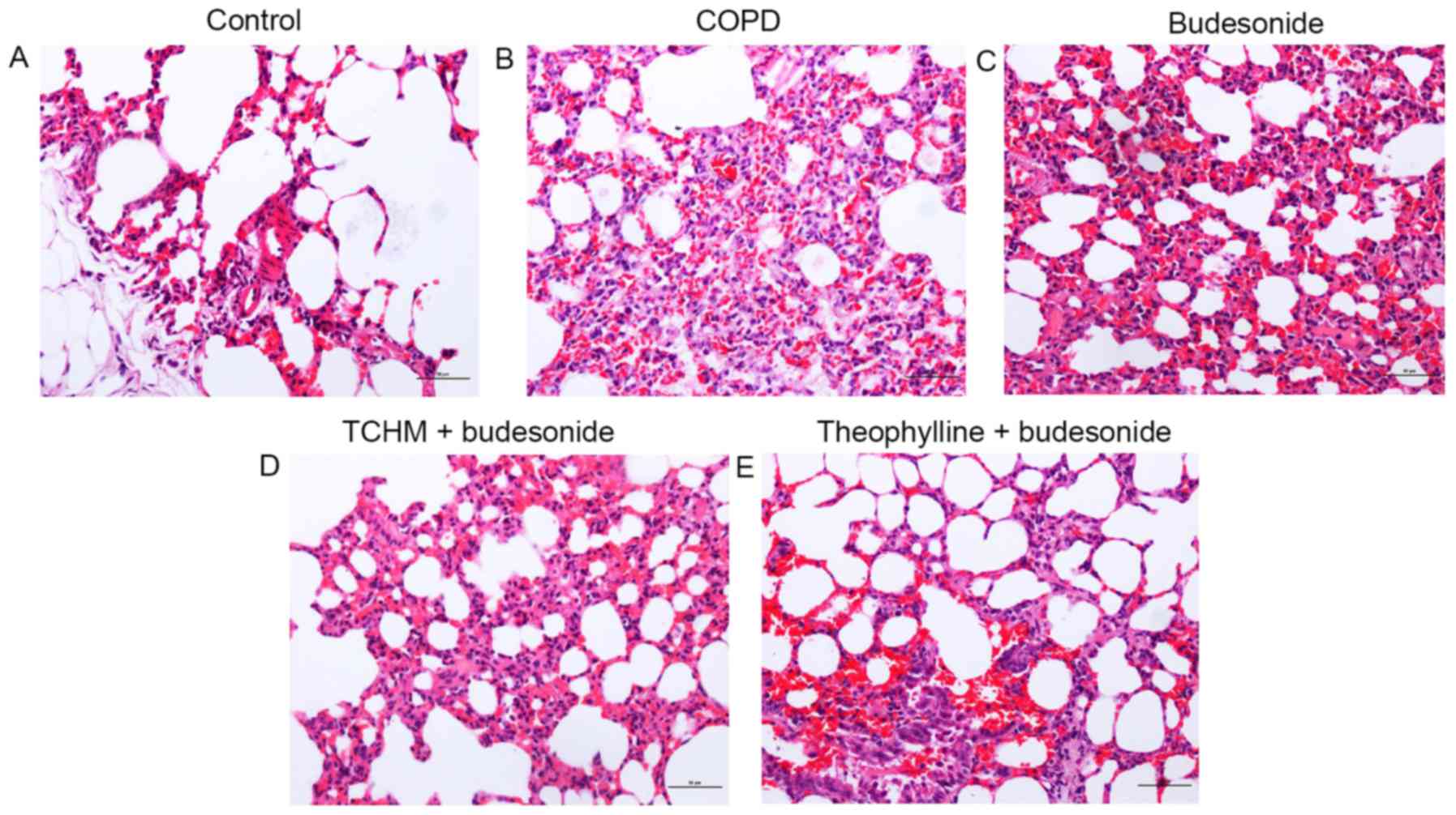
Lung tissue histopathology
Control group
The bronchial lumen and alveolar structure appeared
normal in this group (Fig. 1A).
Neither inflammatory cells in the submucosa nor inflammatory
exudate in the bronchial and alveolar cavity was observed (Fig. 1A).
COPD group
The bronchial lumen was deformed and damaged as
characterized by goblet cell hyperplasia and inflammatory cell
infiltration. There were exfoliated epithelial cells and
inflammatory exudate in the bronchial cavity. The alveolar septum
was thickened as a result of compensatory emphysema. Infiltration
of lymphocytes, plasma cells, neutrophils and eosinophils in the
alveolar wall and alveolar interstitial tissue was noted in this
group (Fig. 1B).
Budesonide group
The bronchial lumen was deformed and damaged. There
were exfoliated epithelial cells and inflammatory exudate in the
bronchial cavity with inflammatory cell infiltration. The alveolar
septum was significantly expanded, with the appearance of
bronchiectasis. Inflammation was severe in the alveolar
interstitial tissue (Fig. 1C).
TCHM + budesonide group
The small bronchial wall was intact. Exfoliated
epithelial cells and inflammatory exudate in the bronchial cavity
were significantly reduced. Inflammation of the alveolar walls was
also significantly reduced (Fig.
1D).
Theophylline + budesonide group
The bronchial lumen was deformed. However, there
were fewer exfoliated epithelial cells, less inflammatory exudate
in the bronchial cavity, and reduced infiltration of inflammatory
cells than were observed in the COPD group. The alveolar wall
collapsed due to emphysema, but pulmonary alveolar inflammation was
mitigated (Fig. 1E).
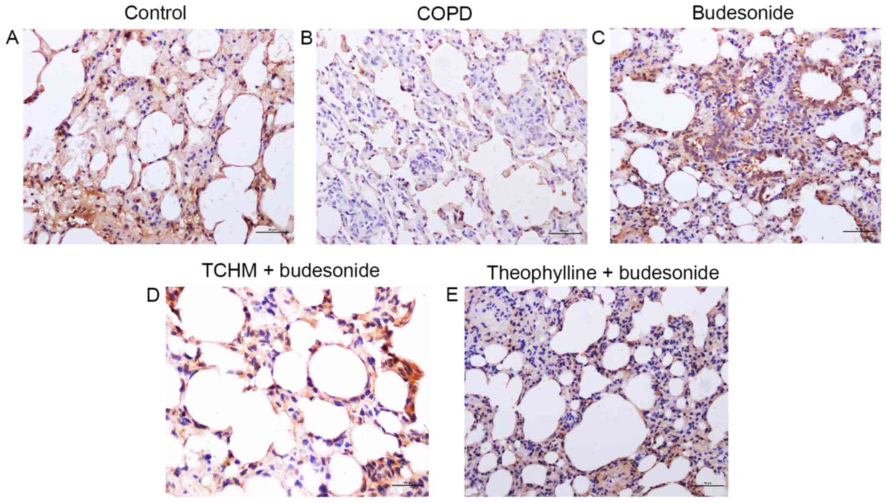
HDAC2 immunohistochemistry
The expression of HDAC2 in the lungs of each group
was determined by immunohistochemistry and is presented in Fig. 2. HDAC2 was strongly expressed in the
control rat lung (mean density, 0.077; Fig. 2A), while it was markedly suppressed
in the lungs of the COPD group (mean density, 0.012; Fig. 2B). While treatment with budesonide
alone partially blocked the suppression of HDAC2 expression in
response to LPS and cigarette smoke exposure (mean density, 0.032;
Fig. 2C), TCHM + budesonide not only
reduced changes of the alveolar structure but also further blocked
the reduction of HDAC2 expression in response to LPS and cigarette
smoke exposure (mean density, 0.039; Fig. 2D). Similarly, the regulatory effect
of theophylline + budesonide on HDAC2 expression (mean density,
0.037; Fig. 2E) in response to LPS
and cigarette smoke exposure was also evident.
Quantification of HDAC2, IL-8 and
TNF-α in rat lung tissue homogenate
As shown in Table
II, in the lung tissue homogenates of the COPD group compared
with the control group, HDAC2 expression was significantly
decreased while IL-8 and TNF-α levels were significantly increased
(P<0.001). Treatment with TCHM + budesonide or theophylline +
budesonide significantly blocked the suppression of HDAC2
expression by LPS plus cigarette smoke exposure (P<0.01), while
budesonide alone had no effect on HDAC2. Similarly, in the TCHM +
budesonide and theophylline + budesonide groups, the LPS plus
cigarette smoke-stimulated TNF-α expression was significantly
reduced (P<0.05 vs. the COPD group), while budesonide alone did
not significantly affect TNF-α levels. TCHM + budesonide
significantly inhibited IL-8 protein release in response to LPS and
cigarette smoke exposure (P<0.05), while neither budesonide
alone nor theophylline + budesonide had a significant effect on
IL-8.
| Table II.Levels of HDAC2, IL-8 and TNF-α in
rat lung tissue (mean ± standard deviation, n=8). |
Table II.
Levels of HDAC2, IL-8 and TNF-α in
rat lung tissue (mean ± standard deviation, n=8).
| Group | HDAC2 (ng/ml) | IL-8 (pg/ml) | TNF-α (pg/ml) |
|---|
| Control |
0.507±0.063a |
241.30±17.18a |
64.72±11.13a |
| COPD |
0.348±0.031 |
306.94±36.99 |
96.49±10.61 |
| Budesonide |
0.369±0.027 |
298.64±42.09 |
94.64±5.56 |
| TCHM +
budesonide |
0.444±0.066b |
274.87±21.96c |
77.57±8.54b |
| Theophylline +
budesonide |
0.442±0.068b |
278.74±20.79 |
78.86±9.02c |
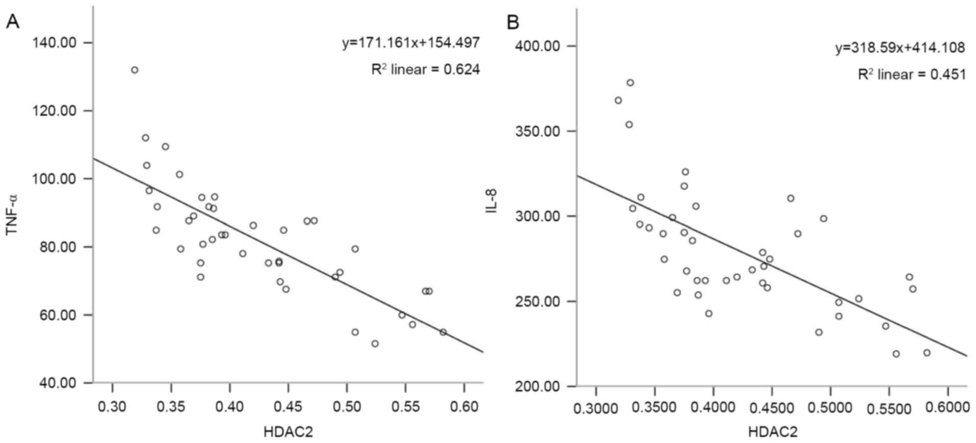
Correlation between HDAC2 and IL-8 or
TNF-α
The correlation of HDAC2 with IL-8 or TNF-α levels
in the rat lungs was analyzed. As shown in Fig. 3, the level of HDAC2 was negatively
correlated with the level of IL-8 (r=−0.672, P<0.001; Fig. 3A) and the level of TNF-α (r=−0.790,
P<0.001; Fig. 3B).
Discussion
In the present study, in order to investigate the
role of Jinwei Tang in regulating the effect of budesonide on
airway inflammation, a COPD model characterized by airway
inflammation was prepared in rats by a combination of intratracheal
LPS instillation and cigarette smoke exposure. As demonstrated by
lung function tests as well as by histopathological examination,
the rat COPD model was successfully created in the current study.
In addition, the expression of inflammatory cytokines (IL-8 and
TNF-α) and HDAC2 was significantly altered in the lungs of COPD
models compared with those in control lungs. Furthermore, the
expression of IL-8 and TNF-α in the lung tissue was negatively
correlated with HDAC2 expression, suggesting that HDAC2 is involved
in the modulation of inflammatory cytokine release in the context
of airway inflammation.
Since HDAC dysfunction may be associated with
corticosteroid resistance in COPD (8,9,11,12), in
the present study, the synergetic effect of the TCHM and
glucocorticoid on the expression of HDAC2 as well as inflammatory
mediators (IL-8 and TNF-α) in the lungs of the COPD rat model was
further explored. The TCHM plus budesonide significantly blocked
the suppression of HDAC2 expression in the lungs in response to the
intratracheal instillation of LPS plus cigarette smoke exposure,
while budesonide treatment alone did not. Theophylline plus
budesonide had a similar effect as the TCHM plus budesonide. In
addition, the TCHM plus budesonide significantly inhibited the
synthesis of the inflammatory mediators IL-8 and TNF-α in the lungs
of rats exposed to LPS and cigarette smoke. These findings suggest
that the TCHM augmented the anti-inflammatory effect of the
glucocorticoid through modulating HDAC2 expression in lungs
affected by COPD.
The dynamic balance between histone acetylation by
histone acetyltransferase and histone deacetylation by HDAC is
crucial in maintaining homeostasis, particularly in chronic
inflammation. It is known that the anti-inflammatory effect of
glucocorticoid largely depends on the HDAC2 level and its activity
and that the reduced expression and activity of HDAC2 contribute to
the development of glucocorticoid resistance in COPD (9). In this study, the expression and
activity of HDAC2 protein as well as HDAC mRNA have been found to
be reduced in bronchial biopsies, bronchoalveolar lavage
macrophages and peripheral lung tissue obtained from patients with
COPD; moreover, the reduction in HDAC2 correlates with disease
severity (31). Consistent with
this, the current study demonstrated that HDAC2 was significantly
suppressed in response to intratracheal LPS instillation and
cigarette smoke exposure.
It has been reported that when HDAC2 activity is
decreased, corticosteroid function is also reduced; and when the
activity of HDAC2 is restored or protected, the sensitivity to
corticosteroid is recovered (28).
Selective activation of HDAC2 can be achieved with low therapeutic
concentrations of theophylline, which restores HDAC2 activity in
macrophages from patients with COPD to normal and reverses
corticosteroid resistance (8,10,13).
Consistent with these studies, the ability of theophylline plus
budesonide to restore HDAC2 expression in the lung tissue of rat
COPD models was also observed in the current study. The TCHM plus
budesonide was also able to rescue HDAC2 expression in the lung
tissue of rats in response to LPS and cigarette smoke exposure,
suggesting that the TCHM targets HDAC2 as well.
TCHMs have been demonstrated to serve an important
role in the prevention and treatment of COPD (21,32).
Previous experiments with animal models of COPD have shown that
TCHM, including Bufei Yishen granules and Liujunzi Tang, are able
to improve pulmonary function in the COPD rat model as well as
reduce pathological changes resulting from airway remodeling
(32,33). The current study demonstrated that
budesonide plus the TCHM, Jinwei Tang significantly decreased the
secretion of IL-8 and TNF-α and inhibited the aggregation of
neutrophils, suggesting that the TCHM relieves inflammation in the
COPD model. Another TCHM, Jiawei Yuepi plus Banxia decoction, has
been shown to reduce lung inflammation in a rat COPD model through
inhibiting IL-8 and TNF-α in a concentration-dependent manner
(34). In addition, the TCHM
Baibunongjian decoction inhibited the release of TNF-α, IL-8 and
LTB4 and regulated airway remodeling in a rat COPD model (35). Moreover, the rectal delivery of
another TCHM (Pingchuan decoction) significantly inhibited the
inflammatory cytokines TNF-α and IL-8 but stimulated vascular
endothelial growth factor in a rat model of COPD exacerbation
(36). Furthermore, the TCHMs
Ailuokechuanning and Shenhayifei capsule have been reported to
inhibit airway inflammation through regulating phospho-p38
mitogen-activated protein kinase signaling and myeloperoxidase
activity as well as reducing the release of IL-17 and TNF-α in rat
models of COPD (37,38). The present study further demonstrated
that the TCHM, Jinwei Tang modulates HDAC2 expression in response
to LPS and cigarette smoke exposure by attenuating glucocorticoid
resistance in the treatment of chronic airway inflammation.
In conclusion, the current study demonstrated that
the TCHM, Jinwei Tang was able to rescue HDAC2 expression from the
inhibitory effect of LPS plus cigarette smoke exposure, and augment
the inhibitory effect of glucocorticoid on airway inflammation as
evidenced by significant suppression of IL-8 and TNF-α in the lungs
of a rat model of COPD. The findings of the current study indicate
that this TCHM can effectively modulate the anti-inflammatory
effect of glucocorticoid in the context of chronic airway
inflammation, including COPD.
Acknowledgements
The present study was supported by Beijing
University of Traditional Chinese Medicine Foundation (grant no.
2014-JYBZZ-JS-066).
References
|
1
|
Han MK, Quibrera PM, Carretta EE, Barr RG,
Bleecker ER, Bowler RP, Cooper CB, Comellas A, Couper DJ, Curtis
JL, et al: Frequency of exacerbations in patients with chronic
obstructive pulmonary disease: An analysis of the SPIROMICS cohort.
Lancet Respir Med. 5:619–626. 2017. View Article : Google Scholar : PubMed/NCBI
|
|
2
|
O'Donnell R, Breen D, Wilson S and
Djukanovic R: Inflammatory cells in the airways in COPD. Thorax.
61:448–454. 2006. View Article : Google Scholar : PubMed/NCBI
|
|
3
|
Papi A, Luppi F, Franco F and Fabbri LM:
Pathophysiology of exacerbations of chronic obstructive pulmonary
disease. Proc Am Thorac Soc. 3:pp. 245–251. 2006; View Article : Google Scholar : PubMed/NCBI
|
|
4
|
MacNee W: Pathogenesis of chronic
obstructive pulmonary disease. Proc Am Thorac Soc. 2:pp. 258–266.
2005; View Article : Google Scholar : PubMed/NCBI
|
|
5
|
Crotty Alexander LE, Shin S and Hwang JH:
Inflammatory diseases of the lung induced by conventional cigarette
smoke: A review. Chest. 148:1307–1322. 2015. View Article : Google Scholar : PubMed/NCBI
|
|
6
|
Newton R: Anti-inflammatory
glucocorticoids: Changing concepts. Eur J Pharmacol. 724:231–236.
2014. View Article : Google Scholar : PubMed/NCBI
|
|
7
|
Ammit AJ: Glucocorticoid insensitivity as
a source of drug targets for respiratory disease. Curr Opin
Pharmacol. 13:370–376. 2013. View Article : Google Scholar : PubMed/NCBI
|
|
8
|
Malhotra D, Thimmulappa RK, Mercado N, Ito
K, Kombairaju P, Kumar S, Ma J, Feller-Kopman D, Wise R, Barnes P
and Biswal S: Denitrosylation of HDAC2 by targeting Nrf2 restores
glucocorticosteroid sensitivity in macrophages from COPD patients.
J Clin Invest. 121:4289–4302. 2011. View
Article : Google Scholar : PubMed/NCBI
|
|
9
|
Barnes PJ: Corticosteroid resistance in
patients with asthma and chronic obstructive pulmonary disease. J
Allergy Clin Immunol. 131:636–645. 2013. View Article : Google Scholar : PubMed/NCBI
|
|
10
|
Hakim A, Adcock IM and Usmani OS:
Corticosteroid resistance and novel anti-inflammatory therapies in
chronic obstructive pulmonary disease: Current evidence and future
direction. Drugs. 72:1299–1312. 2012. View Article : Google Scholar : PubMed/NCBI
|
|
11
|
Koenderman L and Chilvers ER: Future
treatment in patients with chronic obstructive pulmonary disease:
To reverse or not to reverse steroid resistance - that is the
question. J Allergy Clin Immunol. 134:323–324. 2014. View Article : Google Scholar : PubMed/NCBI
|
|
12
|
Sun X, Li Q, Gong Y, Ren L, Wan H and Deng
W: Low-dose theophylline restores corticosteroid responsiveness in
rats with smoke-induced airway inflammation. Can J Physiol
Pharmacol. 90:895–902. 2012. View Article : Google Scholar : PubMed/NCBI
|
|
13
|
Barnes PJ: Development of new drugs for
COPD. Curr Med Chem. 20:1531–1540. 2013. View Article : Google Scholar : PubMed/NCBI
|
|
14
|
Hsieh HY, Chiu PH and Wang SC: Histone
modifications and traditional Chinese medicinals. BMC Complement
Altern Med. 13:1152013. View Article : Google Scholar : PubMed/NCBI
|
|
15
|
Haifeng W, Hailong Z, Jiansheng L, Xueqing
Y, Suyun L, Bin L, Yang X and Yunping B: Effectiveness and safety
of traditional Chinese medicine on stable chronic obstructive
pulmonary disease: A systematic review and meta-analysis.
Complement Ther Med. 23:603–611. 2015. View Article : Google Scholar : PubMed/NCBI
|
|
16
|
Coyle M, Shergis JL, Liu S, Wu L, Zhang
AL, Guo X, Lu C and Xue CC: Safety of chinese herbal medicine for
chronic obstructive pulmonary disease. Evid Based Complement
Alternat Med. 2015:3806782015. View Article : Google Scholar : PubMed/NCBI
|
|
17
|
Dong Y, Li Y, Sun Y, Mao J, Yao F, Tian Y,
Wang L, Li L, Li S and Li J: Bufei Jianpi granules improve skeletal
muscle and mitochondrial dysfunction in rats with chronic
obstructive pulmonary disease. BMC Complement Altern Med.
15:512015. View Article : Google Scholar : PubMed/NCBI
|
|
18
|
Tian Y, Li Y, Li J, Xie Y, Wang M, Dong Y,
Li L, Mao J, Wang L and Luo S: Bufei Yishen granule combined with
acupoint sticking improves pulmonary function and morphormetry in
chronic obstructive pulmonary disease rats. BMC Complement Altern
Med. 15:2662015. View Article : Google Scholar : PubMed/NCBI
|
|
19
|
Li J, Yang L, Yao Q, Li Y, Tian Y, Li S,
Jiang S, Wang Y, Li X and Guo Z: Effects and mechanism of bufei
yishen formula in a rat chronic obstructive pulmonary disease
model. Evid Based Complement Alternat Med. 2014:3819762014.
View Article : Google Scholar : PubMed/NCBI
|
|
20
|
Li Y, Li JS, Li WW, Li SY, Tian YG, Lu XF,
Jiang SL and Wang Y: Long-term effects of three Tiao-Bu Fei-Shen
therapies on NF-κB/TGF-β1/smad2 signaling in rats with chronic
obstructive pulmonary disease. BMC Complement Altern Med.
14:1402014. View Article : Google Scholar : PubMed/NCBI
|
|
21
|
Wang C, Li Z, Liu X, Peng Q, Li F, Li D
and Wang C: Effect of Liuweibuqi capsule, a Chinese patent
medicine, on the JAK1/STAT3 pathway and MMP9/TIMP1 in a chronic
obstructive pulmonary disease rat model. J Tradit Chin Med.
35:54–62. 2015. View Article : Google Scholar : PubMed/NCBI
|
|
22
|
Yange T, Ya L, Jiansheng L, Suyun L, Suli
J, Ying W, Xiaofan L and Weiwei L: Effects of therapies for
regulating and reinforcing lung and kidney on osteoporosis in rats
with chronic obstructive pulmonary disease. J Tradit Chin Med.
35:175–183. 2015. View Article : Google Scholar : PubMed/NCBI
|
|
23
|
Feng Y, Dai Y and Zhou LH: Clincal
Observation on TCM Combined with western medicine in treatment of
COPD with qi deficiency and blood stasis syndrome. J Emerg Traditi
Chin Med. 22:301–302. 2013.
|
|
24
|
Chen L, Zhang GL, Chen M, Ji JX and Wang
SG: The curative effect of Liujunzi decoction in the treatment of
chronic obstructive pulmonary disease patients in peroid with lung
sleep deficiency type. Zhong Yi Yao Dao Bao. 21:79–81. 2015.(In
Chinese).
|
|
25
|
Pei XJ, Lu YX, Zhang LD, Tan MC and Shi W:
Clinical observation of modified Liujunzi decoction for respiratory
muscle fatigue of stable phase patients with chronic obstructive
pulmonary disease. Xin Zhong Yi. 46:59–61. 2014.(In Chinese).
|
|
26
|
Shan L: The intervention effect of
supplemeting qi, activating blood circulation, resolving phlegm and
its different combination regimens on COPD Rats. Guangzhou Univ
Chin Med. 70:12012.
|
|
27
|
Wang LD and Pan W: Treatment for 36 cases
of chronic obstructive pulmonary disease at mild and remission
stages with salmeterol fluticasone and mixture of replenising Qi
and nourishing yin. West J Tradit Chin Med. 25:76–77. 2012.
|
|
28
|
Zhang HP, Fu JJ, Fan T, Zhang WB, Wang ZL,
Wang L and Wang G: Histone deacetylation of memory T lymphocytes by
You-Gui-Wan alleviates allergen-induced eosinophilic airway
inflammation in asthma. Chin Med. 10:92015. View Article : Google Scholar : PubMed/NCBI
|
|
29
|
Tazaki G, Kondo T, Tajiri S, Tsuji C,
Shioya S and Tanigaki T: Functional residual capacity and airway
resistance in rats of COPD model induced by systemic hyaluronidase.
Tokai J Exp Clin Med. 31:125–127. 2006.PubMed/NCBI
|
|
30
|
An ZP: BS: Lung function test in wistar
rats. Chin J Lab Anim Sci. 12:102–104. 2013.
|
|
31
|
Mercado N, To Y, Ito K and Barnes PJ:
Nortriptyline reverses corticosteroid insensitivity by inhibition
of phosphoinositide-3-kinase-δ. J Pharmacol Exp Ther. 337:465–470.
2011. View Article : Google Scholar : PubMed/NCBI
|
|
32
|
Zhou R, Luo F, Lei H, Zhang K, Liu J, He
H, Gao J, Chang X, He L, Ji H, et al: Liujunzi Tang, a famous
traditional Chinese medicine, ameliorates cigarette smoke-induced
mouse model of COPD. J Ethnopharmacol. 193:643–651. 2016.
View Article : Google Scholar : PubMed/NCBI
|
|
33
|
Li Y, Tian YG, Li JS, Dong YQ, Wang MH,
Feng SX, Li LL, Mao J, Wang LL and Luo S: Bufei Yishen granules
combined with acupoint sticking therapy suppress oxidative stress
in chronic obstructive pulmonary disease rats: Via regulating
peroxisome proliferator-activated receptor-gamma signaling. J
Ethnopharmacol. 193:354–361. 2016. View Article : Google Scholar : PubMed/NCBI
|
|
34
|
Wu J, Li X and Qin Y: Exploration of
regularity of treating AECOPD based on cluster analysis. J Emerg
Tradit Chin Med. 23:1436–1437. 2014.
|
|
35
|
Wang ZY and Gu C: Effect of BaiBuNong
decoction on rat COPD model histopathology and inflammatory
mediators. J Shandong Tradit Chin Med. 33:1010–1013. 2014.
|
|
36
|
Wang YX, XM and Zhao QP: Effect of rectal
delivery Pingchuan decoction on TNF-α, IL-8 and VEGF in rat COPD
model. J Guangming Tradit Chin Med. 29:705–707. 2014.
|
|
37
|
Lei ZL, GY and Zhong HW: Effect of
ShenHaYiFei capsule on TNF-α expression in rat COPD model. Henan
Tradit Chin Med. 34:822–825. 2014.
|
|
38
|
Shang LX, WY and Zhang LZ: Effect of
AiLuoKeChuanNing on inflammatory mediators and oxidative stress in
rat COPD model. Chin J Exp Tradit Med Formulae. 24:168–171.
2014.
|