Introduction
Benign prostatic hyperplasia (BPH) is a common
disease in men >50 years of age as the incidence of the disease
increases with age (1). BPH can
seriously affect the quality of life of patients with common
clinical manifestations, including dysuria, urinary frequency,
urgency, urinary incontinence, and other lower urinary tract
symptoms (2). Severe cases may cause
urinary tract infections, urinary tract obstruction, bladder
stones, renal failure and other adverse effects (2). Epidemiological studies have suggested
that risk factors for BPH include heredity, nutrition, and immunity
(3,4). Recent studies suggest that BPH is also
closely associated with metabolic syndromes, such as obesity,
hyperglycemia, dyslipidemia and hypertension; it is also associated
with secondary urinary tract syndrome secondary to BPH (5).
At present, treatment of BPH consists primarily of
drug therapy and surgical treatment. Given the critical role of
androgens, derived largely from the testis with a small amount
(~1%) from the adrenal gland (6), in
regulating prostate tissue growth, 5-alpha reductase inhibitors
(5-ARI) constitute the only drug that reduces the size of the
prostate gland by lowering the level of dihydrotestosterone.
However, 5-ARI therapy is slow, requiring at least three months
before it works, and only reduces prostate volume by 20% (7,8).
The development of new therapeutic approaches
requires a detailed examination of the underlying pathogenic
processes. Although the pathogenesis of BPH has not been fully
elucidated at present, pathological changes associated with BPH
include prominent glandular hyperplasia with stromal hyperplasia
(9). The most commonly used animal
model is a rat model of androgen-induced prostate hyperplasia. A
study by Scolnik et al (10)
demonstrated that Sprague Dawley (SD) or Wistar rats injected with
exogenous androgen after castration resulted in the proliferation
of rat prostate tissue, an effect that was stable with good
reproducibility.
The present study was undertaken to investigate the
role of androgens in androgen-induced BPH in castrated rats and to
evaluate the role of the phosphoinositide 3-kinase/protein kinase
B/mechanistic target of rapamycin (PI3K/Akt/mTOR) pathway in this
process. The role of autophagy in androgen-induced BPH was also
determined. In the present study, androgens induced glandular
hyperplasia, which may be mediated by inhibiting prostate cell
apoptosis and promoting the proliferation of prostate cells. A role
for the PI3K/Akt/mTOR signaling pathway in androgen-induced BPH was
also demonstrated. These results may form the basis of further
clinical studies analyzing these pathways as potential therapeutic
targets for BPH treatment.
Materials and methods
Animals
A total of 40 healthy male SD rats (age, 8 weeks;
weight, 250±10 g) were provided by the Experimental Animal Center
of Xiamen University (Xiamen, China). Rats were housed in an
air-conditioned atmosphere at 22°C and 50% relative humidity in a
specific pathogen-free controlled room with a 12 h light/dark cycle
and provided with unrestricted amount of rodent chow and water. All
experimental procedures were conducted in conformity with
institutional guidelines for the care and use of laboratory
animals, and protocols were approved by the Institutional Animal
Care and Use guidelines in The First Affiliated Hospital of Xiamen
University (Xiamen, China). The study was approved by the Ethics
Committee of The First Affiliated Hospital of Xiamen University
(Xiamen, China).
Male SD rats were randomly divided into four groups
(n=10 per group): The testosterone group (received bilateral
testicular resection and subcutaneous injection of testosterone),
rapamycin group (received bilateral testicular resection,
subcutaneous injection of testosterone and intraperitoneal
injection of rapamycin), 3-MA group (received bilateral testicular
resection, subcutaneous injection of testosterone and
intraperitoneal injection of 3-MA) and control group [received
bilateral testicular resection, subcutaneous injection with solvent
(90% olive oil and 10% ethanol) and intraperitoneal injection of
normal saline]. To establish a BPH model, rats in all of the groups
underwent bilateral testicular resection following administration
of anaesthetic, as previously described (6,11). At
day 25 following surgery, rats in the testosterone, rapamycin and
3-MA groups were injected with 0.5 mg/day testosterone propionate
(Sigma-Aldrich; Merck KGaA Darmstadt, Germany) in the hind leg.
Following establishment of the BPH model, the testosterone group
was injected intraperitoneally with normal saline (1 mg/kg/day),
the rapamycin group was injected intraperitoneally with rapamycin
(1 mg/kg/day; Sigma-Aldrich Merck KGaA) and the 3-MA group was
injected intraperitoneally with 3-MA (1 mg/kg/day; Sigma-Aldrich;
Merck KGaA). In the control group, the scrotum skin was sutured
after the testes of rats were detached, and the rats received an
intraperitoneal injection of 1 mg/day normal saline as well as a
subcutaneous injection in the hind leg with 1 ml solvent (90% olive
oil and 10% ethanol) immediately following the surgery. Treatments
were administered for 28 days.
Investigation of prostate index
Rats in each group were weighed 28 days after
feeding. Rats were sacrificed by intraperitoneal injection using
100 mg/kg Nembutal (Beijing Genia Biotechnology, Co., Ltd.,
Beijing, China) and the prostate tissues were removed. The weight
of the prostate tissue after cleaning the blood with filter paper
was measured and the volume of the prostate was measured using the
displacement method after submerging the tissue in a water-filled
graduated cylinder. The left and right ventral lobes of the
prostate were stored in liquid nitrogen or 10% formaldehyde
solution, respectively.
Hematoxylin and eosin (HE)
staining
Prostate tissues from rats in each group were fixed
in 10% formalin at 4°C overnight, and were then washed with PBS.
The blocks were then dehydrated in graded alcohols, cleared in
xylene and embedded in paraffin wax. The paraffin blocks were cut
it into sections of 6 µm thickness and stained with HE. Briefly,
the sections were dewaxed with xylene and dehydrated using graded
ethanol, followed by washing with distilled water. The sections
were sequentially transferred and washed after each step as
follows: hematoxylin solution for 1 min, 1% hydrochloric acid
solution for 10 sec, 1% ammonia complex blue for 30 sec, and 0.5%
eosin solution for 2 min in thermostat with temperature 40°C. After
ethanol dehydration, the extent of hyperplasia, as a percentage of
hyperplastic tissue in the whole section was observed using an
optical microscope (Nikon Eclipse 50i, Nikon Corporation, Tokyo,
Japan).
Western blotting to determine protein
expression levels of B-cell lymphoma 2 (Bcl-2) and
microtubule-associated protein 1 light chain 3 (LC3)-II in rat
prostate tissues
Proteins were isolated from rat prostate tissues
following snap freezing by immersion in liquid nitrogen. For 5 mg
of tissue, 300 µl of ice-cold RIPA lysis buffer (cat no. 89900;
Thermo Fisher Scientific, Inc.) was rapidly added. Following
homogenization with a blade that was rinsed twice (200 µl each)
with lysis buffer, the homogenate was maintained under constant
agitation for 2 h at 4°C. Protein concentrations were determined
using a Pierce BCA Protein assay kit, according to the
manufacturer's protocol. After the addition of protein loading
buffer and boiling for 10 min, 50 µg of lysate was loaded and
separated using 10–15% SDS-PAGE and transferred to nitrocellulose
membranes. All further incubations were performed at room
temperature on a platform shaker. Membranes were blocked with TBS
supplemented with 0.1% Tween-20 (v/v) and 5% (w/v) non-fat dry milk
for 30 min at room temperature prior to incubation with the
following primary antibodies for 12 h: Anti-LC3 (cat no. 3868; Cell
Signaling Technology, Danvers, MA, USA; 1:1,000); anti-Beclin-1
(cat no. ab62557; Abcam; Cambridge, MA, USA, 1:1,000) and
anti-caspase-3 (cat no. ab32351; Abcam; 1:1,000). The blots were
washed three times with wash buffer (5–10 min each). Subsequently,
the membranes were incubated with anti-rabbit immunoglobulin
horseradish peroxidase-conjugated secondary antibodies (cat no.
sc-2030 and cat no. sc-2005; Multisciences Biotech Co., Ltd.,
Hangzhou, China, 1:10,000) for 2 h. GAPDH (cat no. ab9485; Abcam,
1:1,000) was used as an internal reference. After washing the blot
four times with wash buffer (5–10 min each), immunoreactive
proteins were visualized by enhanced chemiluminescence (Perkin
Elmer, Shelton, CT, USA).
Detection of Bcl-2 and Beclin1 mRNA
expression levels in rat prostate tissues by reverse
transcription-quantitative polymerase chain reaction (RT-qPCR)
RNAs were isolated and purified from rat prostate
tissues using a RNeasy Mini kit (cat no. 74104; Qiagen AB,
Sollentuna, Sweden). For Complementary DNA synthesis, reverse
transcription was performed using the Prime Script RT Reagent kit
(Takara Bio Inc., Otsu, Japan) according to the manufacturer's
protocols. For reverse transcription, the reaction mixtures was
incubated under the following conditions: 1 cycle of 37°C for 15
min; 1 cycle of 85°C for 5 sec and 1 cycle of 4°C for 10 min.
Primers were designed by the GenScript PCR Primer Design Tool
(http://www.genscript.com/) and were
synthesized by Shanghai GenePharma Co., Ltd. (Shanghai, China). The
primer sequences were as follows: β-actin, forward,
5′-GAAGATCAAGATCATTGCTCCT-'3 and reverse,
5′-TACTCCTGCTTGCTGATCCA-3′; Bcl-2, forward,
5′-TAAGCTGTCACAGAGGGGCT-'3 and reverse, 5′-GCGACGAGAGAAGTCATCCC-3′;
and Beclin1, forward, 5′-GGCTGAGAGACTGGATCAGG-3′ and reverse,
5′-CTGCGTCTGGGCATAACG-3′. qPCR was performed using SYBR Premix EX
TaqII (Takara, Shiga, Japan) following the manufacturer's
instructions. Each 30-µl reaction contained 0.4 µM primer pairs,
100 ng cDNA, 15 µl SYBR Green, and 0.6 µl ROX as a fluorescence
internal control. The amplification program comprised of two
stages. The first Tag activation stage began with an initial 95°C
for 10 min followed by 40 cycles of denaturation at 95°C for 5 sec
and annealing at 60°C for 40 sec. Subsequently, a melting curve
analysis was performed by collecting fluorescence data. The
relative T/S values were calculated according to the
2−∆∆Cq method (12).
Detection of apoptosis in rat prostate
tissue by terminal deoxynucleotidyl transferase dUTP nick-end
labeling (TUNEL) assay
TUNEL was performed according to the instructions of
the TUNEL cell apoptosis detection kit (cat no. 11684795910; Roche
Molecular Diagnostics, Branchburg, NJ, USA). Exposed 3′-OH of
genomic DNA were broken and terminal deoxynucleotidyl transferase
enzyme catalysis and DUTP (fluorescein-dUTP) coupled with
fluorescein labeling (fluorescein isothiocyanate) was used.
Detection was performed using fluorescence microscopy (Leica DMI
4000B; Leica Microsystems, Inc., Buffalo Grove, IL, USA). Green
fluorescence was observed at 520±20 nm using a standard
fluorescence filter.
Observation of autophagosomes in
tissue sections by scanning transmission electron microscopy
Prostate tissues were cut into tissue blocks (1
mm3) and then fixed in 2.5% glutaraldehyde in 0.01 mol/l
phosphate buffer at 4°C for 2 h. Following incubation in 2% osmium
tetroxide at 4°C for additional 2 h, tissue blocks were dehydrated
in graded ethanol solutions. Subsequently, ethanol was substituted
with propylene oxide for 30 min at room temperature and embedded in
epoxy resin. Ultrathin sections (0.1 µm) were double-stained with
1% uranyl acetate and 0.2% lead citrate for 15 min at room
temperature and analyzed by transmission electron microscope
(JEM-1220; JEOL, LTD., Tokyo, Japan) at 80 kV.
Statistical analysis
All experiments were repeated three times and
similar results were obtained. All data of normal distribution were
presented as the mean ± standard deviation as indicated and data
were analyzed by one-way analysis of variance with Bonferroni post
hoc analysis. Data of non-normal distribution were analyzed by
Kruskal-Wallis H-tests. Differences between groups were further
analyzed by the Nemenyi test. P<0.05 was considered to indicate
a statistically significant difference. SPSS v. 11.5 statistical
software (SPSS, Inc., Chicago, IL, USA) was used for all
statistical analyses.
Results
Measurement of prostate index
The prostate wet weight, volume and prostatic gland
exponent (%) in the testosterone group were significantly higher
than those in the control group (P<0.05; Table I). The prostate wet weight, volume
and prostatic gland exponent (%) in the rapamycin group was
significantly reduced compared with those in the testosterone group
(P<0.05; Table I).
 | Table I.Rat weight and wet weight, volume and
prostate index of prostate tissue of the rats in each group. |
Table I.
Rat weight and wet weight, volume and
prostate index of prostate tissue of the rats in each group.
| Group | n | Rat weight, g | Prostatic gland
weight, g | Prostatic gland
volume, ml | Prostatic gland
exponent, % |
|---|
| Testosterone | 10 | 413±29a |
1.09±0.21a |
0.75±0.32a |
0.26±0.04a |
| Rapamycin | 10 | 329±27 |
0.48±0.10b |
0.40±0.08b |
0.14±0.03b |
| 3-MA | 10 | 428±30 | 0.84±0.18 | 0.70±0.15 | 0.19±0.05 |
| Control | 10 | 357±30 | 0.54±0.09 | 0.45±0.08 | 0.15±0.02 |
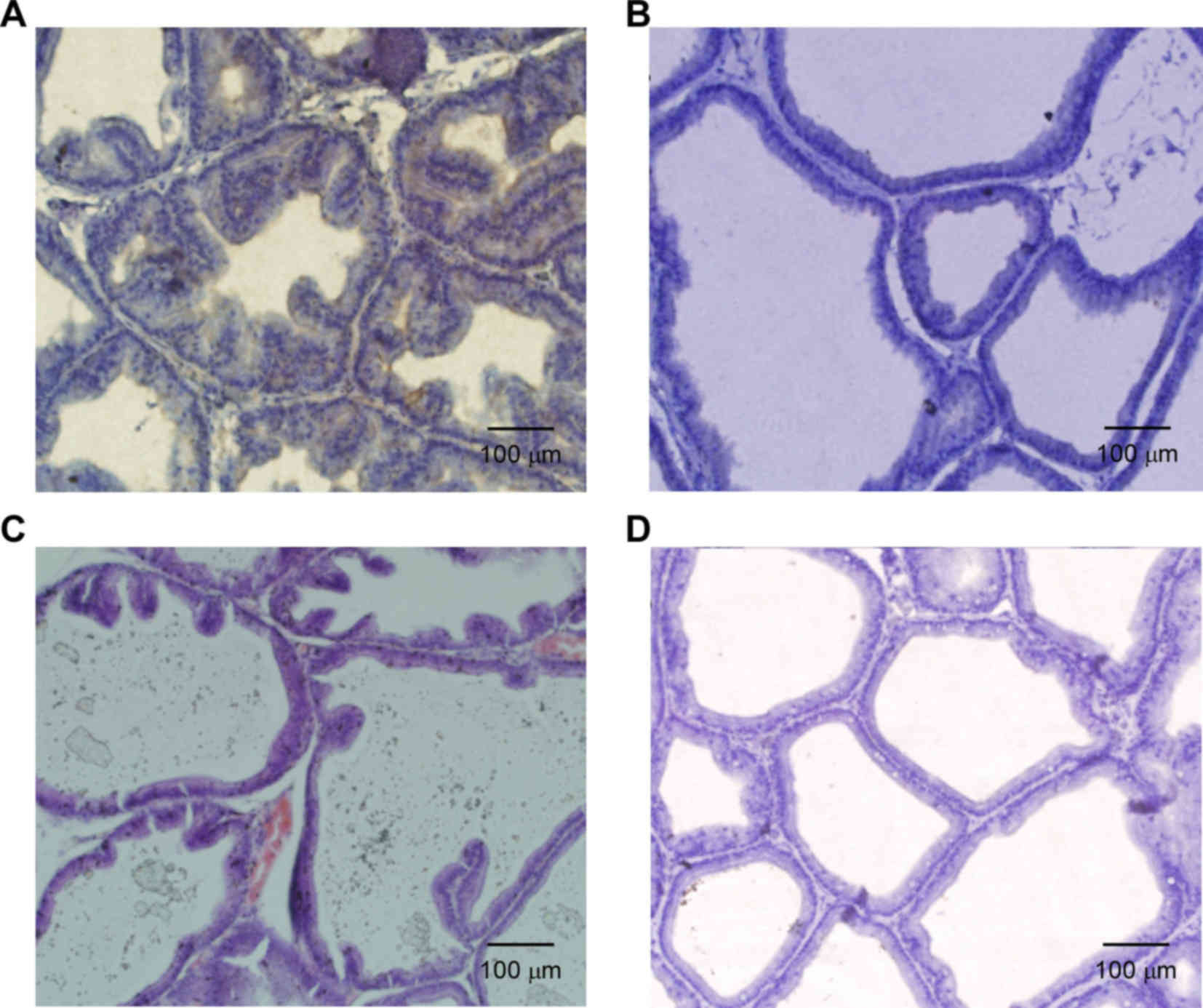
HE detection of the morphology of rat
prostate tissue
Under the microscope, the prostate glands of rats in
the testosterone group (Fig. 1A)
were compact. Additionally, part of the cavity was expanded, part
of the epithelium was pseudostratified, the glandular epithelium
was thickened, part of the glandular epithelium was papillary and
prominent in the glandular cavity, more stromal cells were present
and the small blood vessels were notably expanded compared with the
control group. The morphology of the prostate tissue of the
rapamycin group was similar to that of the control group with
normal arrangement and no obvious thickening of the glandular
epithelium; however, the glandular cavity was dilated, deformed and
exhibited a small amount of interstitial proliferation (Fig. 1B). The prostate gland in the 3-MA
group was enlarged and exhibited an enlarged gland cavity, the
gland epithelium was thicker and part of the glandular epithelium
was papillary and prominent within the glandular cavity; however,
the extent of this was less than that of the testosterone group.
Furthermore, interstitial composition of the prostate tissue in the
3-MA group was increased and congestion and edema were observed
(Fig. 1C). In the control group, the
structure of the prostate gland was clear, the glandular epithelium
was a single layer in columnar arrangement and few epithelia were
protruding into the lumen of gland epithelium. Interstitial
composition of the prostate tissue was relatively small and no
congestion or edema was indicated in the control group (Fig. 1D).
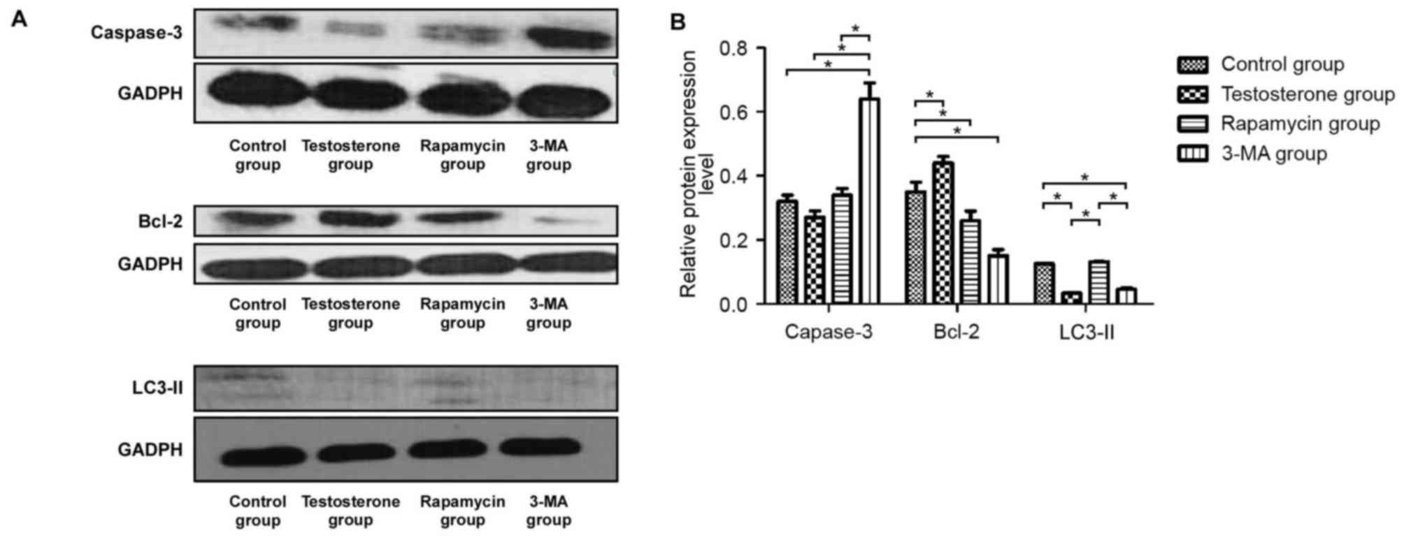
Protein expression levels of Bcl-2,
LC3-II and caspase-3 in rat prostate tissue according to western
blotting
As demonstrated in Fig.
2A and B, western blotting results indicated that the protein
expression levels of Bcl-2 in the testosterone group were
significantly higher compared with that in the control group
(P<0.05). Bcl-2 protein expression levels in the prostate tissue
of the 3-MA group were the lowest of all groups and the protein
expression levels of caspase-3 in the prostate tissue of the 3-MA
group were significantly higher than those in the other three
groups (P<0.05). Furthermore, the protein expression levels of
LC3-II in the rapamycin group were similar to the control group but
significantly higher than that in the 3-MA and testosterone groups
(P<0.05).
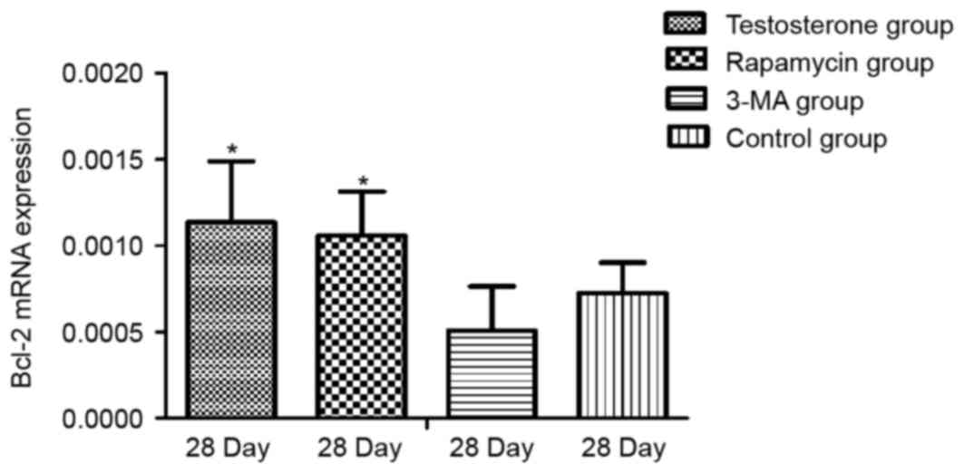
mRNA expression levels of Bcl-2 and
Beclin-1 in prostate tissue according to RT-qPCR
As demonstrated in Fig.
3, RT-qPCR results revealed that the mRNA expression levels of
Bcl-2 at 28 days were significantly higher in the testosterone and
rapamycin groups than that in the control group (P<0.05). The
mRNA expression levels of Bcl-2 after 28 days in the 3-MA group
were lower than that in the control group, however the result was
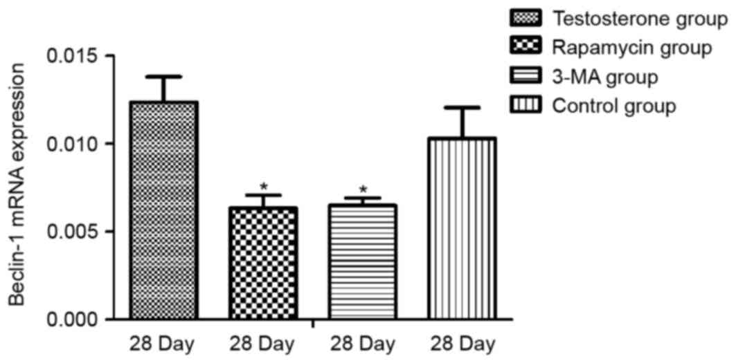
not significant. As demonstrated in Fig.
4, there was no significant difference in the expression levels
of Beclin-1 mRNA between the control and the testosterone groups.
The mRNA expression levels of Beclin-1 were significantly lower in
the rapamycin and the 3-MA groups than that in the control group
(P<0.05).
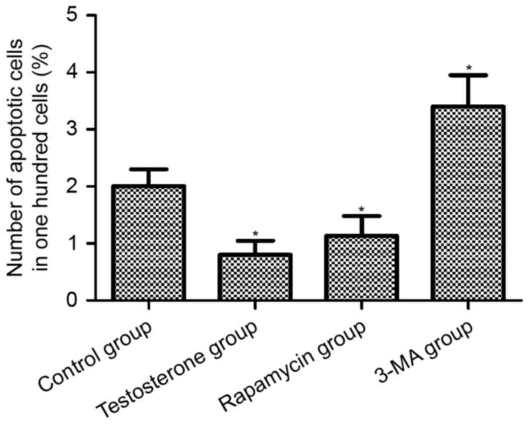
Level of apoptosis in rat prostate
tissues
TUNEL analysis was performed to examine the level of
apoptosis in the prostate tissues (Fig.
5). Quantitative analysis of the images revealed that the
apoptotic rate of the 3-MA group was significantly increased
compared with the control group (P<0.05), whereas the apoptotic
rate of prostate tissues in the testosterone and rapamycin groups
was significantly decreased compared with the control group
(P<0.05; Fig. 6).
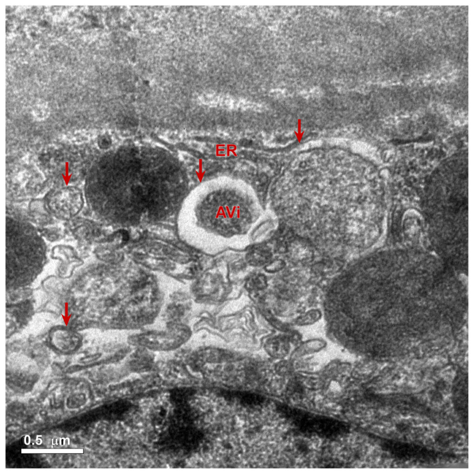
Scanning transmission electron
microscope observation
At present, it has been suggested that autophagy may
be observed using an electron microscope, which is the gold
standard to judge the extent of autophagy (13–15). The
formation of a lysosome, a vesicle structure with a double
membrane, is a vital process of autophagy (14). Some damaged organelles, proteins or
glycogen are present inside vesicles (16). When the vesicles are fused with the
lysosomal membrane, the inner membrane of the vesicle is hydrolyzed
(17). Fusion of the outer membrane
of the vesicle and the lysosomal membrane form the autophagy
autophagolysosome (17). In the
present study, electron microscopy of the rapamycin group revealed
that the autophagolysosome contained a single layer of membrane.
The ultrathin sections of rat prostate tissue in the rapamycin
group were observed under a magnification of ×5,000 and revealed
that the rat prostate epithelial cells contained
autophagolysosomes. In addition, some of the organelles within the
autophagosomes in the rapamycin group were not fully digested
(Fig. 7). Following injection with
3-MA, the formation of autophagy bodies was not observed in the
prostate epithelial cells. Collectively, these results revealed
that the level of autophagy was highly activated in rat prostate
tissues after rapamycin treatment compared with the control
group.
Discussion
BPH is a common disease in men >50 years of age
and the incidence of the disease increases with age (1). The pathogenesis of BPH is not yet
clear; however, it is closely associated with androgens (18). The growth and development of prostate
tissue is associated with the action of androgens and the
maintenance of normal prostate tissue morphology must therefore
also depend on the role of androgens (19,20).
Previous studies have indicated that increased prostate volume is
not due to excessive proliferation of prostate tissue, but is
instead related to the decrease of apoptosis in prostate tissue
(21–23). In adult castrated rats, androgen
treatment induces prostatic regrowth, proliferation and increases
prostate size (24,25). This method has become a common
applied method for the construction of the animal model of BPH
(25). In the present study, the wet
weight and volume of the prostate of rats after 28 days were
measured and the prostate indices of rats in each group were
calculated. Results demonstrated that the prostate wet weights,
volumes and prostate index in the testosterone group were
significantly higher than those in the control group. The results
indicated that testosterone successfully induced rat prostate
hyperplasia. HE staining results further revealed that, compared
with the control group, the prostate glands of the rats in the
testosterone group were compact with partially expanded cavities
and partially pseudostratified epithelium, the glandular epithelium
was thickened, part of the glandular epithelium was papillary and
prominent in the glandular cavity with increased stromal cells and
expansion of small blood vessels compared with the control group.
Prostate tissues in the 3-MA group also exhibited similar changes
compared to the testosterone group; however, to a lesser extent.
Results demonstrated that the establishment of the present model
was successful. In the rapamycin group, the structure of the
prostate was normal, the glands were closely arranged, the size of
the cavity was normal and the glandular epithelium was not raised.
Compared with the control group, the interstitial composition in
the rapamycin group was slightly increased; however, no vascular
congestion was detected and the epithelial cells were in a single
arrangement with no pseudostratified epithelial formation. Compared
with the testosterone and control groups, no notable prostate
tissue proliferation was observed in the prostate tissue of the
rapamycin group. Therefore, we propose that rapamycin may inhibit
the proliferation of prostate tissue in rats induced by
testosterone.
Currently, it is not known whether androgen-induced
prostate hyperplasia in rats is promoted by the excessive
proliferation of prostate tissue or by the reduction of prostate
tissue apoptosis; therefore, this was a primary investigation in
the present study. Western blotting results demonstrated that,
compared with the control group, androgen was able to decrease the
expression of caspase-3, increase the expression of Bcl-2 protein
and reduce the expression of LC3-II protein in prostate tissue.
TUNEL assay results demonstrated that, compared with the control
group, the rate of apoptosis in the prostate tissues of rats in the
testosterone and the rapamycin groups were significantly decreased.
These results indicated that androgen was able to decrease the rate
of apoptosis and autophagy in the prostate tissue of castrated
rats.
Androgen regulates the growth, proliferation and
death of prostate cancer cells, which may be associated with
androgen receptor (AR) function and the phosphoinositide
3-kinase/protein kinase B/mechanistic target of rapamycin
(PI3K/Akt/mTOR) signaling pathway (26). Both AR and PI3K/Akt/mTOR signaling
influence the proliferation and death of prostate cancer cells and
feedback to each other (27).
Kinkade et al (28) have
identified that targeting Akt/mTOR and ERK/MAPK signaling pathways
inhibits androgen-independent prostate tumors in the mouse model.
This research proposed that inhibition of the Akt/mTOR and ERK/MAPK
signaling pathways produces a wide range of therapeutic effects on
prostate cancer, particularly concerning androgen-resistant
prostate cancer, in the advanced stages (28). In order to verify the assumption that
androgen is related to the PI3K-1/Akt/mTOR pathway, testosterone
propionate was excessively injected into rats of the testosterone
group, and rapamycin was used to inhibit the PI3K/Akt pathway in
the rapamycin group. The present results revealed that rapamycin
was able to inhibit the effect of androgen. Compared with the
control group, the weight of rats was significantly increased 28
days after injections with testosterone propionate, which may be
related to the role of androgen in promoting the growth of rat
skeletal muscle (29,30). Rapamycin was used to block the
PI3K-1/Akt/mTOR pathway and the same dose of testosterone was
injected in rats of the rapamycin group. Compared with the control
group, there was no significant change in the weight of the rats in
the rapamycin group. Western blotting results demonstrated that the
expression levels of caspase-3 and Bcl-2 protein in the prostate
cells of the rapamycin group were higher and lower, respectively,
than those in the testosterone group. The role of testosterone may
be related to the PI3K-1/Akt/mTOR pathway (31).
The present study demonstrated that rapamycin
blocked the PI3K/Akt/mTOR pathway in vivo, which may affect
the role of testosterone in promoting skeletal muscle growth. These
results were similar to findings from a corresponding in
vitro study (28) and further
supported that the signaling pathway for androgen is related to the
PI3K-1/Akt/mTOR pathway. Androgen may improve the level of protein
synthesis and promote cell growth and proliferation in prostate
tissue by activating the PI3K-1/Akt/mTOR pathway. This may
therefore result in a decrease in the rate of apoptosis.
The relationship between autophagy and apoptosis is
complex. Increasing the level of autophagy in tumor tissue may
decrease the rate of apoptosis in tumor cells (32). The reason is that the survival
ability of tumor cells in an unfavorable environment, such as that
created by radiotherapy and chemotherapy, is enhanced due to the
enhanced autophagy (33). The rate
of prostate cancer cell apoptosis appears to bottleneck in response
to endocrine therapy characterized by a decrease that is not
sustained (34). Therefore, various
researchers have proposed autophagy inhibition as a possible
clinical strategy to counteract therapeutic resistance in prostate
cancer (34).
In the process of androgen-induced prostatic
hyperplasia in rats, the extent to which the level of autophagy
affects the role of androgen has not been reported. Rapamycin is a
common inducer of autophagy and also has the ability to block the
PI3K-1/Akt/mTOR pathway (35).
Rapamycin is able to inhibit mTOR protein and therefore promote
autophagy. At present, the formation of autophagy bodies is the
gold standard to judge the occurrence of autophagy (36,37). Rat
prostate tissue was observed using an electron microscope after
injection of testosterone and rapamycin in rats for 28 days. In the
rapamycin group, autophagolysosomes were observed. Some of the
organelles had not been fully digested and were located inside the
autophagolysosome. In the control, testosterone and 3-MA groups,
autophagolysosomes were not observed. Western blotting results
demonstrated that the expression level of LC3-II protein in the rat
prostate tissue of the rapamycin group was similar to that in the
control group, which suggested that rapamycin induces autophagy
activation. In contrast, autophagy was inhibited in rat prostate
tissue in the 3-MA group by 3-MA. Results indicated that rapamycin
inhibited the role of androgen in promoting the proliferation of
prostate tissue in rats by stimulating autophagy. Compared with the
control group, there was no obvious prostate hyperplasia in the
rapamycin group. However, autophagy inhibition by 3-MA promoted
hyperplasia in the 3-MA-treated rat prostate tissues. In addition,
the level of apoptosis in the rat prostate tissues was increased
compared with that in the control group.
In conclusion, the results of the present study
suggested that androgen-induced prostate hyperplasia in rats is not
only related to testosterone inhibition of prostate cell apoptosis,
it is also possible that testosterone promotes excessive
proliferation of prostate tissue in rats. Furthermore, rapamycin
may promote the level of autophagy and inhibit the proliferation of
prostate tissue in rats, which is not induced by the promotion of
apoptosis.
Acknowledgements
The present study was supported by the Medical Elite
Cultivation Program of Fujian (Fujian, China; grant no.
2014-ZQN-ZD-33).
References
|
1
|
Dhingra N and Bhagwat D: Benign prostatic
hyperplasia: An overview of existing treatment. Indian J Pharmacol.
43:6–12. 2011. View Article : Google Scholar : PubMed/NCBI
|
|
2
|
Lepor H: Pathophysiology, epidemiology and
natural history of benign prostatic hyperplasia. Rev Urol.
6:S3–S10. 2004.
|
|
3
|
Suzuki S, Platz EA, Kawachi I, Willett WC
and Giovannucci E: Intake of energy and macronutrients and the risk
of benign prostatic hyperplasia. Am J Clin Nutr. 75:689–697. 2002.
View Article : Google Scholar : PubMed/NCBI
|
|
4
|
Golda R, Wolski Z, Wyszomirska-Golda M,
Madaliński K and Michałkiewicz J: The presence and structure of
circulating immune complexes in patients with prostate tumors. Med
Sci Monit. 10:CR123–CR127. 2004.PubMed/NCBI
|
|
5
|
Vignozzi L, Rastrelli G, Corona G, Gacci
M, Forti G and Maggi M: Benign prostatic hyperplasia: A new
metabolic disease? J Endocrinol Invest. 27:1380–1384. 2014.
|
|
6
|
Turcu A, Smith JM, Auchus R and Rainey WE:
Adrenal androgens and androgen precursors: Definition, synthesis,
regulation and physiologic actions. Compr Physiol. 4:1369–1381.
2014. View Article : Google Scholar : PubMed/NCBI
|
|
7
|
Bhutia SK, Das SK, Azab B, Dash R, Su ZZ,
Lee SG, Dent P, Curiel DT, Sarkar D and Fisher PB: Autophagy
switches to apoptosis in prostate cancer cells infected with
melanoma differentiation associated gene-7/interleukin-24
(mda-7/il-24). Autophagy. 7:1076–1077. 2011. View Article : Google Scholar : PubMed/NCBI
|
|
8
|
Lian J, Wu X, He F, Karnak D, Tang W, Meng
Y, Xiang D, Ji M, Lawrence TS and Xu L: A natural bh3 mimetic
induces autophagy in apoptosis-resistant prostate cancervia
modulating bcl-2-beclin1 interaction at endoplasmic reticulum. Cell
Death Differ. 18:60–71. 2011. View Article : Google Scholar : PubMed/NCBI
|
|
9
|
Wang Y, Shao JC and Zhang SW:
Histomorphological studies on hyperplastic prostate of castrated
rat caused by androgen. Zhonghua Nan Ke Xue. 8:190–193. 2001.(In
Chinese).
|
|
10
|
Scolnik MD, Servadio C and Abramovici A:
Comparative study of experimentally induced benign and atypical
hyperplasia in the ventral prostate of different sat strains. J
Androl. 15:287–297. 1994.PubMed/NCBI
|
|
11
|
Wei XY, Zhang JK, Li J and Chen SB: Effect
of bilateral testicular resection on thymocyte and its
microenvironment in aged mice. Asian J Androl. 3:271–275.
2001.PubMed/NCBI
|
|
12
|
Livak KJ and Schmittgen TD: Analysis of
relative gene expression data using real-time quantitative PCR and
the 2(-Delta Delta C(T)) method. Methods. 25:402–408. 2001.
View Article : Google Scholar : PubMed/NCBI
|
|
13
|
Bizargity P and Schröppel B: Autophagy:
Basic principles and relevance to transplant immunity. Am J
Transplant. 14:1731–1739. 2014. View Article : Google Scholar : PubMed/NCBI
|
|
14
|
Todde V, Veenhuis M and van der Klei IJ:
Autophagy: Principles and significance in health and disease.
Biochim Biophys Acta. 1792:3–13. 2009. View Article : Google Scholar : PubMed/NCBI
|
|
15
|
Mizushima N: Autophagy: Process and
function. Genes Dev. 21:2861–2873. 2007. View Article : Google Scholar : PubMed/NCBI
|
|
16
|
Wrighton KH: Eating up damaged lysosomes.
Nat Rev Mol Cell Biol. 14:4652013. View
Article : Google Scholar
|
|
17
|
Münz C: The Autophagic machinery in viral
exocytosis. Front Microbiol. 8:2692017. View Article : Google Scholar : PubMed/NCBI
|
|
18
|
Moore A, Butcher MJ and Köhler TS:
Testosterone replacement therapy on the natural history of prostate
disease. Curr Urol Rep. 16:512015. View Article : Google Scholar : PubMed/NCBI
|
|
19
|
Singh M, Jha R, Melamed J, Shapiro E,
Hayward SW and Lee P: Stromal androgen receptor in prostate
development and cancer. Am J Pathol. 184:2598–2607. 2014.
View Article : Google Scholar : PubMed/NCBI
|
|
20
|
Zhou Y, Bolton EC and Jones JO: Androgens
and androgen receptor signaling in prostate tumorigenesis. J Mol
Endocrinol. 54:R15–R29. 2015. View Article : Google Scholar : PubMed/NCBI
|
|
21
|
Quiles MT, Arbós MA, Fraga A, de Torres
IM, Reventós J and Morote J: Antiproliferative and apoptotic
effects of the herbal agent pygeumafricanumon cultured prostate
stromal cells from patients with benign prostatic hyperplasia
(BPH). Prostate. 70:1044–1053. 2010. View Article : Google Scholar : PubMed/NCBI
|
|
22
|
Boya P and Kroemer G: Beclin 1: A BH3-only
protein that fails to induce apoptosis. Oncogene. 28:2125–2127.
2009. View Article : Google Scholar : PubMed/NCBI
|
|
23
|
Ciechomska IA, Goemans GC, Skepper JN and
Tolkovsky AM: Bcl-2 complexed with Beclin-1 maintains full
anti-apoptotic function. Oncogene. 28:2128–2141. 2009. View Article : Google Scholar : PubMed/NCBI
|
|
24
|
Nicholson TM and Ricke WA: Androgens and
estrogens in benign prostatic hyperplasia: Past, present and
future. Differentiation. 82:184–199. 2011. View Article : Google Scholar : PubMed/NCBI
|
|
25
|
Oudot A, Oger S, Behr-Roussel D, Caisey S,
Bernabé J, Alexandre L and Giuliano F: A new experimental rat model
of erectile dysfunction and lower urinary tract symptoms associated
with benign prostatic hyperplasia: The testosterone-supplemented
spontaneously hypertensive rat. BJU Int. 110:1352–1358. 2012.
View Article : Google Scholar : PubMed/NCBI
|
|
26
|
Liao RS, Ma S, Miao L, Li R, Yin Y and Raj
GV: Androgen receptor-mediated non-genomic regulation of prostate
cancer cell proliferation. Transl Androl Urol. 2:187–196.
2013.PubMed/NCBI
|
|
27
|
Floc'h N and Abate-Shen C: The promise of
dual targeting Akt/mTOR signaling in lethal prostate cancer.
Oncotarget. 3:1483–1484. 2012. View Article : Google Scholar : PubMed/NCBI
|
|
28
|
Kinkade CW, Castillo-Martin M, Puzio-Kuter
A, Yan J, Foster TH, Gao H, Sun Y, Ouyang X, Gerald WL,
Cordon-Cardo C and Abate-Shen C: Targeting AKT/mTOR and ERK MAPK
signaling inhibits hormone-refractory prostate cancer in a
preclinical mouse model. J Clin Investig. 118:3051–3064.
2008.PubMed/NCBI
|
|
29
|
Michel G and Baulieu EE: Androgen receptor
in rat skeletal muscle: Characterization and physiological
variations. Endocrinology. 107:2088–2098. 1980. View Article : Google Scholar : PubMed/NCBI
|
|
30
|
Fu R, Liu J, Fan J, Li R, Li D, Yin J and
Cui S: Novel evidence that testosterone promotes cell proliferation
and differentiation via G protein-coupled receptors in the rat L6
skeletal muscle myoblast cell line. J Cell Physiol. 227:98–107.
2012. View Article : Google Scholar : PubMed/NCBI
|
|
31
|
Basualto-Alarcón C, Jorquera G, Altamirano
F, Jaimovich E and Estrada M: Testosterone signals through mTOR and
androgen receptor to induce muscle hypertrophy. Med Sci Sports
Exerc. 45:1712–1720. 2013. View Article : Google Scholar : PubMed/NCBI
|
|
32
|
Lorenzo PI and Saatcioglu F: Inhibition of
apoptosis in prostate cancer cells by androgens is mediated through
downregulation of c-Jun N-terminal kinase activation. Neoplasia.
10:418–428. 2008. View Article : Google Scholar : PubMed/NCBI
|
|
33
|
Wang Z, Du T, Dong X, Li Z, Wu G and Zhang
R: Autophagy inhibition facilitates erlotinib cytotoxicity in lung
cancer cells through modulation of endoplasmic reticulum stress.
Int J Oncol. 48:2558–2566. 2016. View Article : Google Scholar : PubMed/NCBI
|
|
34
|
Ziparo E, Petrungaro S, Marini ES, Starace
D, Conti S, Facchiano A, Filippini A and Giampietri C: Autophagy in
prostate cancer and androgen suppression therapy. Internat J Mol
Sci. 14:12090–12106. 2013. View Article : Google Scholar
|
|
35
|
Heras-Sandoval D, Pérez-Rojas JM,
Hernández-Damián J and Pedraza-Chaverri J: The role of
PI3K/AKT/mTOR pathway in the modulation of autophagy and the
clearance of protein aggregates in neurodegeneration. Cell Signal.
26:2694–2701. 2014. View Article : Google Scholar : PubMed/NCBI
|
|
36
|
Klionsky DJ, Abdelmohsen K, Abe A, Abedin
MJ, Abeliovich H, Acevedo Arozena A, Adachi H, Adams CM, Adams PD,
Adeli K, et al: Guidelines for the use and interpretation of assays
for monitoring autophagy (3rd edition). Autophagy. 12:1–222. 2016.
View Article : Google Scholar : PubMed/NCBI
|
|
37
|
Huang CC, Lee CC, Lin HH, Chen MC, Lin CC
and Chang JY: Autophagy-regulated ROS from xanthine oxidase acts as
an early effector for triggering late mitochondria-dependent
apoptosis in cathepsin S-targeted tumor cells. PLoS One.
10:e01280452015. View Article : Google Scholar : PubMed/NCBI
|