Introduction
Cervical cancer is one of the most common types of
malignant tumor within gynecology. Behind Chile, China is the
country with the second highest incidence of cervical cancer
worldwide (1). In 2017, statistics
released by the National Cancer Center (2) revealed that cervical cancer is the
seventh most common malignant tumor in women living in urban areas
and patients with cervical cancer are getting younger. Although the
morbidity and mortality of cervical cancer have declined in recent
years with advances in medical science and technology, there are
150,000 new cases of cervical cancer each year in China and
>80,000 patients succumb to the disease (3). Similarly to other malignant tumors,
invasion and metastasis of tumor cells are the primary cause of
disease onset and mortality in patients with cervical cancer
(4,5).
Tissue factor (TF), also called platelet coagulation
factor III, is an important member of the human coagulation factor
family. TF initiates the blood coagulation cascade in vivo
by binding with coagulation factor VII/VIIa (6,7). It has
been confirmed that the expression of TF is a biomarker for
preoperative diagnosis and the prognosis of malignant tumors
(6,7). However, TF also affects the clinical
progression of malignant tumors by affecting the proliferation,
infiltration and metastasis of cancer cells (8–10); for
this reason, TF is a potential target for the treatment of
malignant tumors (11). However, few
previous studies have reported the expression of TF in cervical
cancer. In the present study, the expression of TF in cervical
cancer was detected using immunohistochemistry and western blot
analysis and its impact on the progression of cervical cancer was
investigated.
Patients and methods
Study patients
A total of 258 pairs of fresh cervical cancer
tissues and adjacent normal tissues (>3 cm) were collected at
the West China Second Hospital (Chengdu, China) between September
2014 and September 2016. All cases of suspected cervical cancer
were confirmed by postoperative pathology. Two pieces of fresh
tissue, the size of soybean granules (diameter range, 4–6 mm), were
removed from cervix during a pathological biopsy and preserved in
liquid nitrogen (−196°C). This surgery/biopsy was part of the
patients' general treatment. The 258 patients with cervical cancer
were aged 28–67 years with a median age of 45 years and a mean age
of (46.2±7.9) years. There were 203, 45 and 10 cases of squamous
cell carcinoma, adenocarcinoma and adenosquamous carcinoma,
respectively. Further clinical data, including clinical stage,
tumor differentiation and HPV infection are listed in Table I. All patients included in the
present study provided written informed consent prior to their
inclusion and the study was approved by the Ethics Committee of
West China Second Hospital.
 | Table I.Baseline data of the 258 patients with
cervical cancer. |
Table I.
Baseline data of the 258 patients with
cervical cancer.
| Characteristic | Total | Percentage |
|---|
| Age (years) |
|
|
| ≥45 | 106 | 41.09 |
|
<45 | 152 | 58.91 |
| Clinical
classification |
|
|
| Squamous
cell carcinoma | 203 | 78.69 |
|
Adenocarcinoma | 45 | 17.44 |
|
Adenosquamous carcinoma | 10 | 3.87 |
| Tumor growth
type |
|
|
|
Cauliflower | 160 | 62.02 |
|
Nodular | 52 | 20.16 |
|
Ulcer | 28 | 10.85 |
|
Erosive | 18 | 6.97 |
| Tumor size (cm) |
|
|
| ≤4 | 77 | 29.84 |
|
>4 | 181 | 70.16 |
| Differentiation |
|
|
| Poor | 47 | 18.22 |
|
Moderate | 77 | 29.84 |
| High | 134 | 51.94 |
| Lymph node
metastasis |
|
|
| Yes | 153 | 59.30 |
| No | 105 | 40.70 |
| Distant
metastasis |
|
|
| Yes | 142 | 55.04 |
| No | 116 | 44.96 |
| FIGO stage |
|
|
|
Ia1-Ib2 | 115 | 44.57 |
|
IIa1-IIb | 125 | 48.45 |
|
IIIa | 18 | 6.98 |
| Type of HPV
infection |
|
|
|
HPV16+ | 135 | 52.33 |
|
HPV18+ | 100 | 38.76 |
|
Other | 23 | 8.91 |
Instruments and reagents
Anti-TF (ab104513) and goat anti-rabbit
immunoglobulin G H&L antibodies (ab150084) were purchased from
Abcam (Cambridge, UK). The VECTASTAIN® Elite®
ABC kit (PK-6010; Vector Laboratories, Inc., Burlingame, CA, USA),
radioimmunoprecipitation (RIPA) lysis liquid (strong), RNAiso Plus
(RS0754), real-time fluorescence quantitative polymerase chain
reaction (PCR) kit (RR066A; both Takara Bio, Inc., Otsu, Japan),
the M-MLV Reverse Transcriptase RNase H Minus kit (M5301; Promega
Corporation, Madison, WI, USA) and the BCA protein concentration
determination kit (P0009; Beyotime Institute of Biotechnology,
Haimen, China) were used in the present study.
The manual precision microtome (CUT4062; SLEE
medical GmbH, Mainz, Germany), the light microscope (BX51; Olympus
Corporation, Tokyo, Japan), polyvinylidene difluoride (PVDF)
transfer membranes (GE Healthcare Life Sciences, Little Chalfont,
UK) and real-time fluorescence quantitative PCR (Agilent
Technologies, Inc., Santa Clara, CA, USA) were also used in the
present study.
Immunohistochemistry
The fresh tissue sections were dehydrated using the
following ascending alcohol series: Dehydration in 75% ethanol
overnight, 85% ethanol for 3 h, 95% ethanol I for 1.5 h, 95%
ethanol II for 1.5 h, 100% ethanol I for 1 h and 100% ethanol II
for 1 h. Xylene was then added to the ethanol mixture for 25 min at
room temperature and the samples were embedded in paraffin. A
thermostat microtome was used to slice the tissues into sections
(1–2 µm) and they were rinsed three times with 0.01 M preheated PBS
(10 min each time). TF protein expression was detected by
VECTASTAIN® Elite® ABC kit according to the
manufacturer's protocol. The tissue sections were incubated
overnight at 4°C with anti-TF antibodies (dilution 1:200) and then
washed three times with PBS (10 min each time). PBS was used as the
negative control instead of the primary antibodies. The sections
were subsequently incubated with the goat anti-rabbit secondary
antibodies (dilution 1:1,000) for 4 h at room temperature.
The TF immunohistochemistry results were assessed
according to a previous study by Zhang et al (12). Initially, the percentage of positive
cells ≤25%, 25–75% or ≥75%, were scored 0, 1 and 2 respectively.
Next, the cells were stained colorless, light yellow, brown and
tan, and scored 0, 1, 2 and 3 respectively. The TF
immunohistochemistry score was determined by multiplying the
positive cell rate score with the staining intensity score. In the
present study, a TF immunohistochemistry score of ≥4 represented
high expression and <4 represented low expression.
Western blot analysis
The tissue samples frozen in liquid nitrogen were
removed and placed in a mortar. Using a liquid nitrogen-assisted
freeze-thawing procedure, the tissue samples were ground rapidly.
When the tissue was sufficiently ground, a RIPA lysate buffer with
1 mM added phenylmethylsulfonyl fluoride, was used to extract the
total proteins from the tissue. The RIPA lysate was transferred
into an EP tube, resuspended, placed on ice for 10 min and
centrifuged at 6,000 × g for 10 min in 4°C. The supernatant was
collected and added to a 20% SDS buffer solution to a final SDS
concentration of 1%. This solution was then boiled at 100°C for 5
min. A BCA protein concentration determination kit was used to
determine the protein concentration. A total of 75 mg total protein
was loaded into each lane, separated by 15% SDS-PAGE and
transferred to PVDF membranes. Membranes were blocked with 5%
bovine serum albumin (P007; Beyotime Institute of Biotechnology)
for 1 h in room temperature. Then membranes were incubated with
anti-TF or anti-β-actin antibodies overnight at 4°C (both dilution
1:1,000) and then washed three times with PBS (each time 10 min).
Following this the membranes were incubated with goat anti-rabbit
secondary antibodies (dilution 1:3,000) for 1 h at room
temperature. The ECL luminescent liquid was used for visualization.
The expression of the target protein was analyzed by Image J v1.8.0
software (National Institutes of Health, Bethesda, MD, USA) and the
relative expression level of the target protein was characterized
by the gray value of the target protein gray level compared with
the β-actin protein band.
Statistical analysis
SPSS 20.0 software (IBM Corp., Armonk, NY, USA) was
used for the statistical analysis of the data. The data were
presented as the mean ± standard deviation. A paired t-test was
performed to compare the TF expression differences between cervical
cancer tissues and the adjacent normal tissues. An independent
sample t-test was performed two compare two samples and one-way
analysis of variance, followed by Ducan's test, was used to compare
differences between multiple groups. A χ2 test was also
used to compare differences between groups. P<0.05 was
considered to indicate a statistically significant difference.
Results
Detection of TF expression by
immunohistochemistry
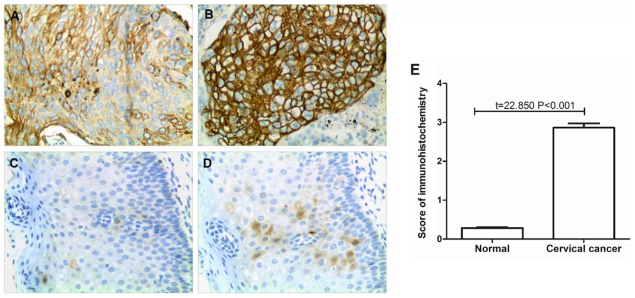
TF proteins were observed as being localized to the
cell membrane. The expression of TF in 258 paired cervical cancer
and adjacent normal tissues was detected by immunohistochemistry.
The results revealed that TF expression was significantly reduced
in the adjacent normal tissues compared with the cervical cancer
tissues (P<0.001; Fig. 1). In 73
normal tissue samples the immunohistochemical score was 1 and in
the remaining 185 samples the score was 0; the normal tissue mean
immunohistochemical score was 0.28±0.45. TF had low expression in
171 of the cervical cancer samples, while it was highly expressed
in 87 cases. There were 15, 50, 57, 49, 47 and 40 cases with
immunohistochemical scores of 0, 1, 2, 3, 4 and 6, respectively.
The average score of immunohistochemistry was 2.86±1.76 in the
cervical cancer tissues.
Association between TF expression and
the characteristics of patients with cervical cancer
The 258 cervical cancer tissue samples were divided
into two groups according to their expression of TF as determined
by immunohistochemistry. There were 87 cases in the TF high
expression group (immunohistochemical score ≥4) and 171 cases in
the TF low expression group (immunohistochemical score <4). The
results revealed that the expression of TF in cervical cancer
tissue was not significantly associated with age, HPV infection
type, tumor classification, tumor growth type, tumor size or the
degree of differentiation (Table
II). However, TF expression was significantly associated with
the metastasis of cervical cancer and the FIGO clinical stages
(P<0.05; Table II).
| Table II.Association between TF expression and
clinical characteristics in patients with cervical cancer. |
Table II.
Association between TF expression and
clinical characteristics in patients with cervical cancer.
|
| TF expression |
|
|
|---|
|
|
|
|
|
|---|
| Characteristic | Low (n=171) | High (n=87) | χ2 | P-value |
|---|
| Age (years) |
|
|
|
|
|
≥45 | 69 | 37 | 0.113 | 0.737 |
|
<45 | 102 | 50 |
|
|
| Clinical
classification |
|
|
|
|
|
Squamous cell carcinoma | 139 | 64 | 2.417 | 0.299 |
|
Adenocarcinoma | 27 | 18 |
|
|
|
Adenosquamous carcinoma | 5 | 5 |
|
|
| Tumor growth
type |
|
|
|
|
|
Cauliflower | 107 | 53 | 1.716 | 0.633 |
|
Nodular | 32 | 20 |
|
|
|
Ulcer | 21 | 7 |
|
|
|
Erosive | 11 | 7 |
|
|
| Tumor size
(cm) |
|
|
|
|
| ≤4 | 49 | 28 | 5.343 | 0.058 |
|
>4 | 122 | 59 |
|
|
| Differentiation
degree |
|
|
|
|
|
Poor | 38 | 9 | 5.656 | 0.059 |
|
Moderate | 50 | 27 |
|
|
|
High | 83 | 51 |
|
|
| Lymph node
metastasis |
|
|
|
|
|
Yes | 90 | 63 | 9.350 | 0.002 |
| No | 81 | 24 |
|
|
| Distal
metastasis |
|
|
|
|
|
Yes | 84 | 58 | 7.127 | 0.007 |
| No | 87 | 29 |
|
|
| FIGO stage |
|
|
|
|
|
Ia1-Ib2 | 83 | 32 | 7.952 | 0.019 |
|
IIa1-IIb | 81 | 44 |
|
|
|
IIIa | 7 | 11 |
|
|
| Type of HPV
infection |
|
|
|
|
|
HPV16+ | 89 | 46 | 2.693 | 0.260 |
|
HPV18+ | 70 | 30 |
|
|
|
Other | 12 | 11 |
|
|
Association between TF expression and
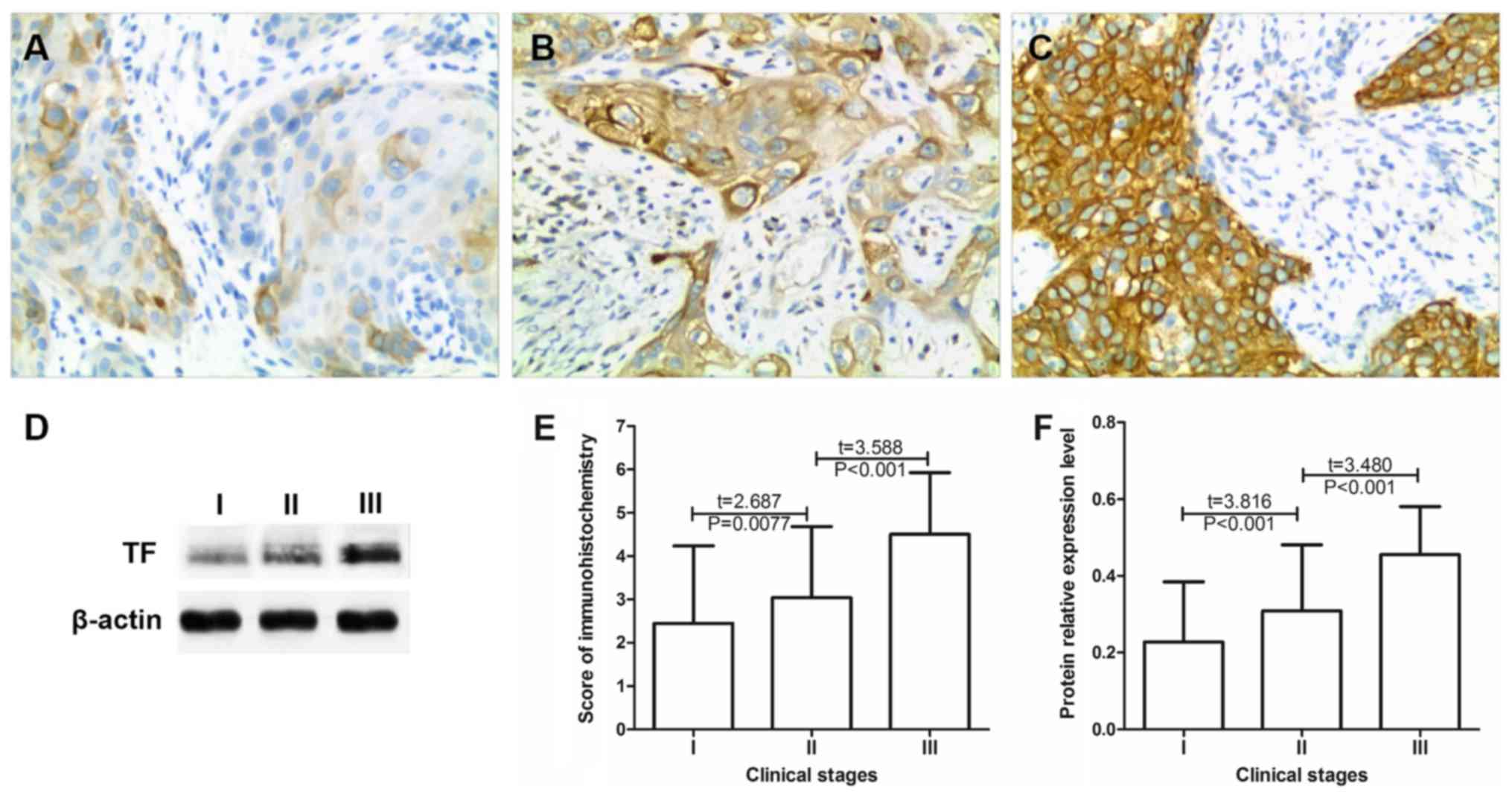
the clinical stage of cervical cancer
The expression of TF in 115 cases of phase I, 125
cases of phase II and 18 cases of phase III cervical cancer were
compared by immunohistochemistry (Table III). The results revealed that the
mean immunohistochemical scores in stage I, II and III cervical
cancer tissues, were 2.35±1.84, 3.08±1.65 and 4.50±1.42,
respectively. The immunohistochemical score significantly increased
as the cancer stage increased (P<0.01; Fig. 2E) Western blot analysis revealed that
the relative expression of TF in the stage I, II and III cervical
cancer tissues were 0.23±0.17, 0.31±0.21 and 0.46±0.32,
respectively (Fig. 2F). As the
cancer stage increased the protein expression of TF significantly
increased (P<0.01; Fig. 2). These
results indicated that increased TF expression may promote cervical
cancer progression.
| Table III.Tissue factor expression in cervical
cancer tissue at different clinical stages. |
Table III.
Tissue factor expression in cervical
cancer tissue at different clinical stages.
|
| Immunohistochemical
score |
|---|
|
|
|
|---|
| FIGO stage | 0 | 1 | 2 | 3 | 4 | 6 |
|---|
|
Ia1-Ib2 | 15 | 25 | 28 | 15 | 20 | 12 |
|
IIa1-IIb | 0 | 25 | 29 | 27 | 24 | 20 |
|
IIIa | 0 | 0 | 0 | 7 | 3 | 8 |
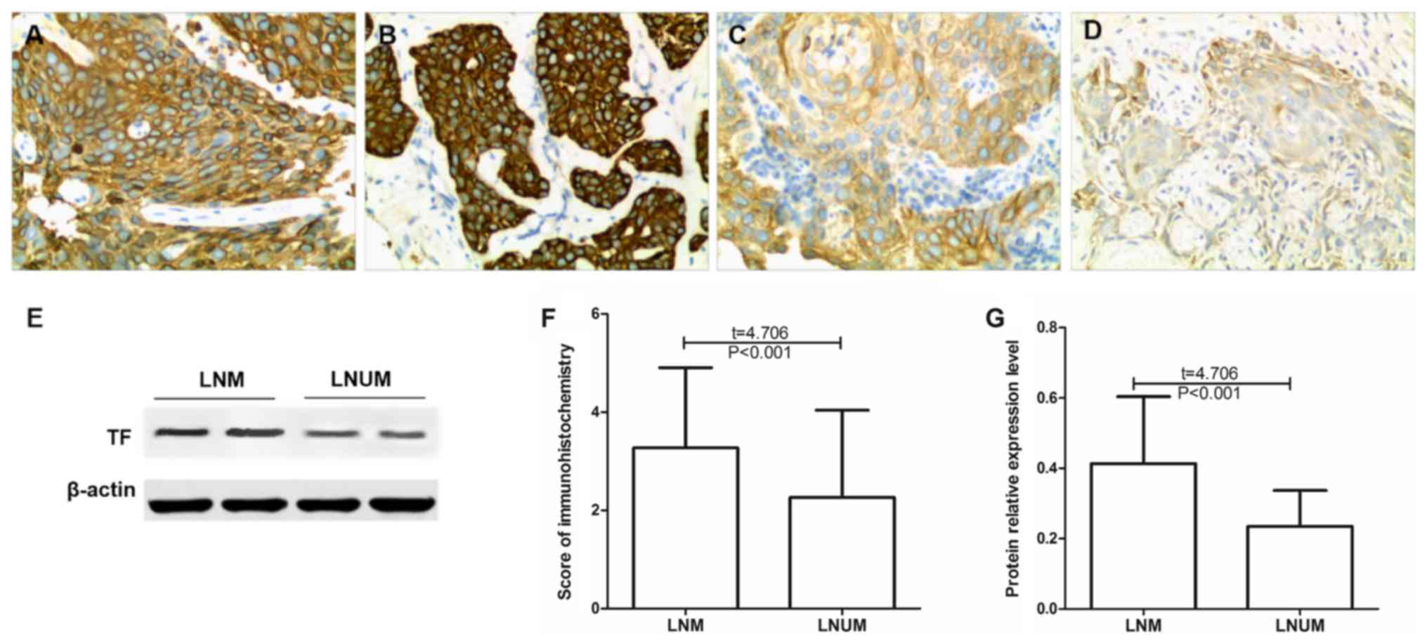
Association between TF expression and
cervical cancer cell lymph node metastasis
The expression of TF was compared between cervical
cancer tissues from patients with or without lymph node metastasis
(Table IV). The results of the
immunohistochemistry analysis revealed that the mean expression of
TF in cervical cancer with lymph node metastasis was 3.55±1.48,
which was significantly increased compared with individuals without
lymph node metastasis, whose mean TF expression was 2.27±1.77
(P<0.05; Fig. 3). The results of
the western blot analysis demonstrated that the relative protein
expression of TF in tumor tissues from patients with lymph node
metastasis was 0.41±0.19, which was significantly increased
compared with patients without lymph node metastasis (0.23±0.18;
P<0.05; Fig. 3).
| Table IV.Tissue factor expression in patients
with and without lymph node metastasis of cervical cancer. |
Table IV.
Tissue factor expression in patients
with and without lymph node metastasis of cervical cancer.
|
| Immunohistochemical
score |
|---|
|
|
|
|---|
| Lymph node
metastasis | 0 | 1 | 2 | 3 | 4 | 6 |
|---|
| Yes | 2 | 20 | 31 | 37 | 35 | 28 |
| No | 13 | 30 | 26 | 12 | 12 | 12 |
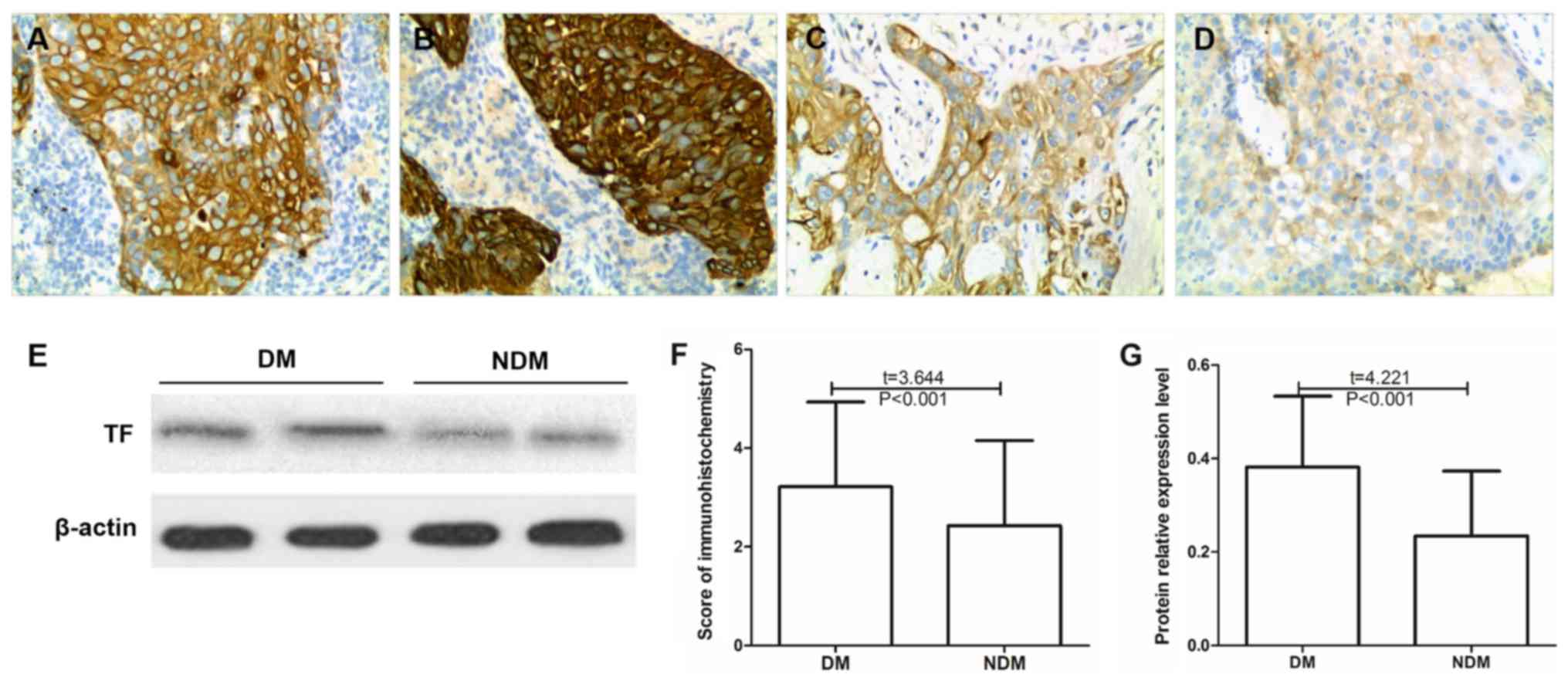
Association between TF expression and
distant metastasis in cervical cancer cells
The expression of TF was compared between cervical
cancer tissues from patients with and without distant metastasis
(Table V). The results of the
immunohistochemistry analysis revealed that the mean TF expression
scores in the tumor tissues of patients with distant metastasis of
cervical cancer was 3.22±1.71, which was significantly increased
compared with patients without metastasis, who had a mean score of
2.43±1.72 (P<0.05; Fig. 4). The
results of the western blot analysis demonstrated that the relative
protein expression of TF in tumor tissues with distant metastasis
of cervical cancer was 0.38±0.15, which was significantly increased
compared with samples from patients without distal metastases
(0.23±0.16; P<0.05; Fig. 4).
| Table V.Tissue factor expression in patients
with and without distant metastasis of cervical cancer. |
Table V.
Tissue factor expression in patients
with and without distant metastasis of cervical cancer.
|
| Immunohistochemical
score |
|---|
|
|
|
|---|
| Distant
metastasis | 0 | 1 | 2 | 3 | 4 | 6 |
|---|
| Yes | 4 | 21 | 27 | 32 | 31 | 27 |
| No | 11 | 29 | 30 | 17 | 16 | 13 |
Discussion
Within the human body TF has the strongest influence
on the promotion of blood coagulation. Under normal physiological
conditions the blood mononuclear and vascular endothelial cells do
not express TF (13). However, when
tissue injury occurs, cytokine stimulation or inflammatory
reactions cause the abundant expression of TF (13). TF is the only transmembrane protein
expressed on the surface of the cell, which serves a role in
coagulation and also serves a non-coagulation function as a
receptor protein (13). TF in the
blood of patients with malignant tumors may come from cancer cells
(13). Although the specific
molecular mechanisms associated with the development of malignant
tumors are not clear, it has been determined that the high
expression of TFs in the peripheral blood is closely associated
with the invasion, metastasis, clinical stage and prognosis of
malignant tumors (14).
In the present study it was revealed that the
expression of TF was significantly increased in cervical cancer
tissue compared with adjacent normal tissue. TF is a type of trace
protein, which is widely expressed in various normal tissues; it is
a transmembrane protein that consists of three parts,
extracellular, transmembrane and cytoplasm (6,7). Under
normal physiological conditions, TF on the cell surface is in a
silent state (6,7). However, it has been suggested that TF
is induced by tumor necrosis factor, tumor cells or the
tumor-specific microenvironment and activated on the surface of
tumor cells in ovarian cancer (15),
gastric cancer (16) and breast
cancer (17) and therefore exhibits
abnormally high expression on the surface of tumor cells. Lo et
al (16) observed that TF was
highly expressed in gastric cancer tissues. Lo et al also
demonstrated that the TF expression level increased with the
clinical stage in gastric cancer, which suggests that the
expression of TF in malignant tumor tissues may be associated with
the progression of malignant tumors. Therefore, the present study
further analyzed the potential association between TF expression
and patient characteristics in cervical cancer tissues.
The present study demonstrated that TF expression
was significantly associated with metastasis of cervical carcinoma
cells (lymph node and distal metastasis) and FIGO stage. The
protein expression of TF was significantly increased in patients
with metastatic cervical carcinoma compared with patients with
non-metastatic carcinoma. The occurrence and development of
malignant tumors is a multi-step process, which is caused by
multiple internal factors, including gene mutation, and external
factors, including drug effects and environmental pollution.
In recent years, it has been reported that TF
primarily affects the invasion and migration of tumor cells via the
coagulation and non-coagulation pathways (18). Tumor cells may activate the
coagulation system in patients, resulting in the development of a
hypercoagulable state in patients with malignant tumors,
particularly those at an advanced stage (19). In these patients, increased blood
viscosity and slowed blood flow reduce the mechanical damage of
tumor cells due to microvascular compression, and the
microenvironment is more favorable to the formation of tumor
thrombus and invasion and metastasis of cancer cells (20,21).
This may enhance the development of malignant tumors, which
suggests that the activation of the coagulation system in certain
patients with malignant tumors is closely associated with clinical
stage and prognosis (13,22). TFs facilitate the signal transduction
of transmembrane proteins in and out of tumor cells and therefore
have a greater impact compared with non-transmembrane proteins on
the invasion and metastasis of tumor cells (23,24).
Previous studies have demonstrated that TF may be activated by
forming complexes with coagulation factor VII or VIIa; together
with protease activated receptor (PAR)-1 or PAR-2 these complexes
may mediate signal transduction within and outside tumor cells
(23,24). Chanakira et al (23) revealed that the TF/VIIa complex
activates PAR-2 to increase the chemotactic migration of epithelial
ovarian tumor cells, and in turn promotes disease progression. In
addition, in different tumor tissues TF is activated by different
signals and activates different PAR proteins, which mediates the
conduction of different signaling pathways (25,26).
Previous studies have demonstrated that TF may mediate signaling
pathways, including nuclear factor-kb, mitogen-activated protein
kinase and alanine aminotransferase to promote tumor cell
metastasis (25,26).
In conclusion, TF is highly expressed in cervical
cancer tissues and the high expression of TF may promote cervical
cancer progression by enhancing the invasive and migratory
abilities of cervical cancer cells.
Acknowledgements
Not applicable.
Funding
No funding was received.
Availability of data and materials
The datasets used and/or analyzed during the current
study are available from the corresponding author on reasonable
request.
Authors' contributions
XiaZ contributed to the conception and design of the
study. XitZ analyzed and interpreted the patient data regarding the
hematological disease and the transplant. CC, JG, TY, YQ and XD
performed the histological examination of cervical cancer tissues.
XiaZ and XitZ were major contributors in writing the manuscript.
All authors read and approved the final manuscript.
Ethics approval and consent to
participate
All patients included in the present study provided
written informed consent prior to their inclusion. The study was
approved by the Ethics Committee of West China Second Hospital.
Patient consent for publication
Not applicable.
Competing interests
The authors declare that they have no competing
interests.
References
|
1
|
Chen W, Zheng R, Baade PD, Zhang S, Zeng
H, Bray F, Jemal A, Yu XQ and He J: Cancer statistics in china,
2015. CA Cancer J Clin. 66:115–132. 2016. View Article : Google Scholar : PubMed/NCBI
|
|
2
|
Chen W, Zheng R, Zhang S, Zeng H, Zuo T,
Xia C, Yang Z and He J: Cancer incidence and mortality in china in
2013: An analysis based on urbanization level. Chin J Cancer Res.
29:1–10. 2017. View Article : Google Scholar : PubMed/NCBI
|
|
3
|
Di J, Rutherford S and Chu C: Review of
the cervical cancer burden and population-based cervical cancer
screening in China. Asian Pac J Cancer Prev. 16:7401–7407. 2015.
View Article : Google Scholar : PubMed/NCBI
|
|
4
|
Wang W, Chu HJ, Liang YC, Huang JM, Shang
CL, Tan H, Liu D, Zhao YH, Liu TY and Yao SZ: FABP5 correlates with
poor prognosis and promotes tumor cell growth and metastasis in
cervical cancer. Tumour Biol. 37:14873–14883. 2016. View Article : Google Scholar : PubMed/NCBI
|
|
5
|
Li Z, Yang Y, Gao Y, Wu X, Yang X, Zhu Y,
Yang H, Wu L, Yang C and Song L: Elevated expression of flotillin-1
is associated with lymph node metastasis and poor prognosis in
early-stage cervical cancer. Am J Cancer Res. 6:38–50.
2015.PubMed/NCBI
|
|
6
|
Claussen C, Rausch AV, Lezius S,
Amirkhosravi A, Davila M, Francis JL, Hisada YM, Mackman N,
Bokemeyer C, Schmalfeldt B, et al: Microvesicle-associated tissue
factor procoagulant activity for the preoperative diagnosis of
ovarian cancer. Thromb Res. 141:39–48. 2016. View Article : Google Scholar : PubMed/NCBI
|
|
7
|
Otero LL, Alonso DF, Castro M, Cinat G,
Gabri MR and Gomez DE: Tissue factor as a novel marker for
detection of circulating cancer cells. Biomarkers. 16:58–64. 2011.
View Article : Google Scholar : PubMed/NCBI
|
|
8
|
Yu YJ, Hou XD and Li YM: Effect of tissue
factor knockdown on the growth, invasion, chemoresistance and
apoptosis of human gastric cancer cells. Exp Ther Med. 7:1376–1382.
2014. View Article : Google Scholar : PubMed/NCBI
|
|
9
|
Xu Y, Qin X, Zhou J, Tu Z, Bi X, Li W, Fan
X and Zhang Y: Tissue factor pathway inhibitor-2 inhibits the
growth and invasion of hepatocellular carcinoma cells and is
inactivated in human hepatocellular carcinoma. Oncol Lett.
2:779–783. 2011.PubMed/NCBI
|
|
10
|
Geddings JE, Hisada Y, Boulaftali Y, Getz
TM, Whelihan M, Fuentes R, Dee R, Cooley BC, Key NS, Wolberg AS, et
al: Tissue factor-positive tumor microvesicles activate platelets
and enhance thrombosis in mice. J Thromb Haemost. 14:153–166. 2016.
View Article : Google Scholar : PubMed/NCBI
|
|
11
|
Eisenreich A, Bolbrinker J and Leppert U:
Tissue factor: A conventional or alternative target in cancer
therapy. Clin Chem. 62:563–570. 2016. View Article : Google Scholar : PubMed/NCBI
|
|
12
|
Zhang Y, You L, Chen J and Mao C:
Expression of kinesin family member 3B is associated with poor
prognosis in epithelial ovarian cancer patients. Int J Clin Exp
Pathol. 10:2834–2842. 2017.
|
|
13
|
Kasthuri RS, Taubman MB and Mackman N:
Role of tissue factor in cancer. J Clin Oncol. 27:4834–4838. 2009.
View Article : Google Scholar : PubMed/NCBI
|
|
14
|
Unruh D, Sagin F, Adam M, Van Dreden P,
Woodhams BJ, Hart K, Lindsell CJ, Ahmad SA and Bogdanov VY: Levels
of alternatively spliced tissue factor in the plasma of patients
with pancreatic cancer may help predict aggressive tumor phenotype.
Ann Surg Oncol. 3 Suppl 22:S1206–S1211. 2015. View Article : Google Scholar
|
|
15
|
Cocco E, Varughese J, Buza N, Bellone S,
Lin KY, Bellone M, Todeschini P, Silasi DA, Azodi M, Schwartz PE,
et al: Tissue factor expression in ovarian cancer: Implications for
immunotherapy with hI-con1, a factor VII-IgGF(c) chimeric protein
targeting tissue factor. Clin Exp Metastasis. 28:689–700. 2011.
View Article : Google Scholar : PubMed/NCBI
|
|
16
|
Lo L, Valentine H, Harrison J, Hayes S,
Welch I, Pritchard S, West C and Ang Y: Tissue factor expression in
the metaplasia–adenoma-carcinoma sequence of gastric cancer in a
european population. Br J Cancer. 107:1125–1130. 2012. View Article : Google Scholar : PubMed/NCBI
|
|
17
|
Schaffner F, Versteeg HH, Schillert A,
Yokota N, Petersen LC, Mueller BM and Ruf W: Cooperation of tissue
factor cytoplasmic domain and PAR2 signaling in breast cancer
development. Blood. 116:6106–6113. 2010. View Article : Google Scholar : PubMed/NCBI
|
|
18
|
Golino P, Ragni M, Cimmino G and Forte L:
Role of tissue factor pathway inhibitor in the regulation of tissue
factor-dependent blood coagulation. Cardiovasc Drug Rev. 20:67–80.
2002. View Article : Google Scholar : PubMed/NCBI
|
|
19
|
Mego M, Karaba M, Minarik G, Benca J,
Sedlácková T, Tothova L, Vlkova B, Cierna Z, Janega P, Luha J, et
al: Relationship between circulating tumor cells, blood
coagulation, and urokinase-plasminogen-activator system in early
breast cancer patients. Breast J. 21:155–160. 2015. View Article : Google Scholar : PubMed/NCBI
|
|
20
|
Koizume S, Jin MS, Miyagi E, Hirahara F,
Nakamura Y, Piao JH, Asai A, Yoshida A, Tsuchiya E, Ruf W and
Miyagi Y: Activation of cancer cell migration and invasion by
ectopic synthesis of coagulation factor VII. Cancer Res.
66:9453–9460. 2006. View Article : Google Scholar : PubMed/NCBI
|
|
21
|
Ma YY, He XJ, Wang HJ, Xia YJ, Wang SL, Ye
ZY and Tao HQ: Interaction of coagulation factors and
tumor-associated macrophages mediates migration and invasion of
gastric cancer. Cancer Sci. 102:336–342. 2011. View Article : Google Scholar : PubMed/NCBI
|
|
22
|
Li W, Xu S, Lin S and Zhao W:
Overexpression of runt-related transcription factor-2 is associated
with advanced tumor progression and poor prognosis in epithelial
ovarian cancer. J Biomed Biotechnol. 2012:4565342012. View Article : Google Scholar : PubMed/NCBI
|
|
23
|
Chanakira A, Westmark PR, Ong IM and
Sheehan JP: Tissue factor-factor VIIa complex triggers protease
activated receptor 2-dependent growth factor release and migration
in ovarian cancer. Gynecol Oncol. 145:167–175. 2017. View Article : Google Scholar : PubMed/NCBI
|
|
24
|
Ribeiro FS, Simão TA, Amoêdo ND, Andreollo
NA, Lopes LR, Acatauassu R, Rumjanek FD, Albano RM, Pinto LF and
Monteiro RQ: Evidence for increased expression of tissue factor and
protease-activated receptor-1 in human esophageal cancer. Oncol
Rep. 21:1599–1604. 2009.PubMed/NCBI
|
|
25
|
Luan L, Yang Z and Tan J: Tissue factor
and circulating tumor cells. Tumor. 35(5)2015.
|
|
26
|
Yang HP, Yue L, Jiang WW, Liu Q, Kou JP
and Yu BY: Diosgenin inhibits tumor necrosis factor-induced tissue
factor activity and expression in THP-1 cells via down-regulation
of the NF-κB, Akt, and MAPK signaling pathways. Chin J Nat Med.
11:608–615. 2013. View Article : Google Scholar : PubMed/NCBI
|