Introduction
As the second most common malignancy of the
genitourinary tract, urinary bladder cancer affects more than 2
million people worldwide (1). The
incidence of urinary bladder cancer is increased in developed
countries compared with less developed regions. It has been
demonstrated that urinary bladder cancer accounts for >5% of
newly diagnosed tumors in European countries (2). With the growth of the aging population
and changes in exposure to risk factors, the incidence of urinary
bladder cancer demonstrates an increasing trend and the age of
onset is decreasing (1). With the
efforts made in the prevention and treatment of urinary bladder
cancer, the 5-year survival rate of patients with this disease is
60% (3). However, the prognosis of
patients with metastatic urinary bladder cancer is usually poor
(4). Therefore, early diagnosis and
treatment remains critical for the survival of patients with
urinary bladder cancer.
In addition to messenger RNAs (mRNAs) that encode
protein products, the human genome also transcribes a large set of
non-coding RNAs that serve pivotal roles in normal physiological
and pathological processes (5). The
comparison of non-coding RNA expression under physiological and
pathological conditions provides references for the diagnosis and
prognosis of human diseases (6).
MicroRNA (miRNA) is a subgroup of non-coding RNA molecules, each
measuring ~22 nucleotides (7).
Studies from previous decades have indicated that miRNAs are
involved in almost all aspects of critical biological processes in
the human body (8). miRNA-373 serves
different roles in different types of cancer (9,10).
miRNA-373 is likely an oncogene in testicular germ cell tumors, and
the overexpression of miRNA-373 in tumor tissues promotes tumor
progression (9). By contrast,
miRNA-373 serves a tumor suppression role in estrogen receptor
negative breast cancer by targeting nuclear factor
kappa-light-chain-enhancer of activated B cells and transforming
growth factor-β signaling pathways (10). The contradictory functions of
miRNA-373 reveal the complexity of its regulatory role in cancer
biology. In the present study, it was identified that miRNA-373
promoted the proliferation, migration and invasion of urinary
bladder cancer by upregulating epidermal growth factor receptor
(EGFR). The present study provided novel insights for the diagnosis
and treatment of urinary bladder cancer.
Patients and methods
Patients
From January 2015 to January 2018, a total of 55
patients who were pathologically diagnosed with urinary bladder
cancer and treated in Shengjing Hospital of China Medical
University (Shenyang, China) were included. Those patients included
40 males and 15 females, and the ages ranged from 28–69 years, with
a mean age of 45±10.1 years. Concurrently, a total of 45 healthy
people were also included to serve as control group. The control
group included 37 males and 8 females, and the ages ranged from
27–70 years, with a mean age of 44±10.9 years. No significant
differences in age and sex were identified between the two groups.
The present study was approved by the Ethics Committee of Shengjing
Hospital of China Medical University. All patients signed informed
consent.
Tissue collection and processing
Tumor tissues and adjacent healthy tissues within 2
cm around the tumors were collected from all patients during
surgical resection. Blood (10 ml) was extracted from the elbow vein
of the patients with urinary bladder cancer and healthy controls.
Blood was maintained at room temperature for 2 h, followed by
centrifugation of blood samples for 10 min at 1,875 × g at room
temperature to collect serum. All tissues were stored in liquid
nitrogen prior to use.
Cell lines and cell culture
Urinary bladder cancer HT-1376 (ATCC®
CRL-1472™) and HT-1197 (ATCC® CRL-1473™) cell lines were
purchased from the American Type Culture Collection (ATCC). Cells
were cultured with Eagle's Minimum Essential Medium (cat. no.
30-2003; ATCC) containing 10% fetal bovine serum (Sigma-Aldrich;
Merck KGaA, Darmstadt, Germany) at 37°C with 5% CO2.
Cell transfection
miR-373 mimic hsa-miR-373* (HMI0531) and negative
control 1 miRNA (GGUUCGUACGUACACUGUUCA; HMC0002) were purchased
from Sigma-Aldrich; Merck KGaA. Prior to transfection, cells were
cultured at 37°C with 5% CO2 overnight to reach 80–90%
confluence. Lipofectamine 2000® reagent (11668-019;
Invitrogen; Thermo Fisher Scientific, Inc., Waltham, MA, USA) was
used to transfected 50 nM miRNA into 5×10⁵ cells. Cells
without transfection were control cells (C). Cells transfected with
negative control miRNA were negative control cells (NC). Subsequent
experiments were performed at 24 h after transfection.
Cell proliferation assay
A Cell Counting kit-8 assay kit (Sigma-Aldrich;
Merck KGaA) was used to evaluate cell proliferation ability.
Briefly, cells were collected and centrifuged at 600 × g for 5 min
at room temperature and were used to prepare a cell suspension with
a density of 5×10⁴ cells per well, and 100 µl cell
suspension containing 5×10³ was added into each well of
96-well plates. Cells were cultured in an incubator at 37°C with 5%
CO2, and 10 µl CCK-8 solution was added at 12, 24, 48,
72 and 96 h. Subsequent to incubation at 37°C for an additional 4
h, optical density values at 450 nm were measured using a
microplate reader (Bio-Rad Laboratories, Inc., Hercules, CA,
USA).
Transwell cell migration and invasion
assay
A Transwell cell migration assay kit (BD
Biosciences, Franklin Lakes, NJ, USA) was used to evaluate cell
migration ability. Briefly, cells were collected and centrifuged at
600 × g for 5 min at room temperature and were used to prepare a
cell suspension with a density of 5×10⁴ cells per well,
and 100 µl cell serum-free suspension containing 5×10³
cells was added to the upper chamber. Then, RPMI-1640 medium
(Thermo Fisher Scientific, Inc.) containing 20% fetal calf serum
(Sigma-Aldrich; Merck KGaA) was added into the lower chamber.
Following incubation at 37°C with 5% CO2 for 24 h,
membranes were collected and stained with 0.5% crystal violet
(Sigma-Aldrich; Merck KGaA) at room temperature for 15 min. Stained
cells were counted under an optical microscope (Olympus
Corporation, Tokyo, Japan) at magnification, ×20. A cell invasion
assay was performed using the same protocol, but the upper chamber
was pre-coated with Matrigel (EMD Millipore, Billerica, MA, USA) at
4°C for 4 h.
Reverse transcription quantitative
polymerase chain reaction (RT-qPCR)
A miRNeasy kit (Qiagen GmbH, Hilden, Germany) was
used for all miRNA extraction from both cells and tissues. cDNA was
prepared using the miScript II RT kit (Qiagen GmbH) under the
following conditions: 25°C for 5 min, 50°C for 25 min and 75°C for
10 min. All protocols were performed according to the
manufacturer's instructions. An miScript SYBR-Green PCR kit (Qiagen
GmbH) was used to assay miRNA-373 expression using RNU6 (miRNA) as
an endogenous control. PCR reaction conditions were: 95°C for 1
min, then 95°C for 15 sec and 59°C for 40 sec for 40 cycles. All
primers were purchased directly from Beyotime Institute of
Biotechnology (Jiangsu, China). Data were analyzed using the
2−ΔΔCt method (11).
Western blot analysis
Total protein extraction from HT-1376 and HT-1197
cell lines was performed using a radioimmunoprecipitation assay
lysis solution (Thermo Fisher Scientific, Inc.), and BCA assay was
used for protein quantification. Subsequently, 20 µg protein from
each sample was subjected to 10% SDS-PAGE gel electrophoresis,
followed by gel transfer to polyvinylidene fluoride membranes.
Membranes were blocked with 5% skimmed milk at room temperature for
2 h, followed by washing with PBS and incubation with primary
antibodies including rabbit anti-human EGFR (1:2,000; cat. no.
ab131498; Abcam, Cambridge, UK) and rabbit anti-human GAPDH
(1:1,000; cat. no. ab8245; Abcam) overnight at 4°C. Following
washing with PBS, the membranes were incubated with an anti-rabbit
IgG-horseradish peroxidase secondary antibody (1:1,000; cat. no.
MBS435036; MyBioSource, Inc., San Diego, CA, USA) at room
temperature for 1 h. Then, enhanced chemiluminescent reagents
(Sigma-Aldrich; Merck KGaA) were added to detect the signals.
Membranes were scanned using MYECL™ Imager (Thermo Fisher
Scientific, Inc.), and ImageJ v1.46 software (National Institutes
of Health, Bethesda, MD, USA) was used to normalize the relative
expression level of EGFR to the endogenous control GAPDH.
Statistical analysis
All experiments were performed in triplicate. Data
were processed using SPSS 19.0 (IBM Corp., Armonk, NY, USA). Count
data are expressed as rate and were compared using a χ2
test. Measurement data were expressed as mean ± standard deviation,
and comparisons among multiple groups were performed using a
one-way analysis of variance followed by Tukey's test. Receiver
operating characteristic (ROC) curve analysis was performed to
evaluate the diagnostic value of serum miRNA-373 for urinary
bladder cancer. P<0.05 was considered to indicate a
statistically significant difference.
Results
Expression of miRNA-373 in tumor
tissues and adjacent healthy tissues of patients with urinary
bladder cancer
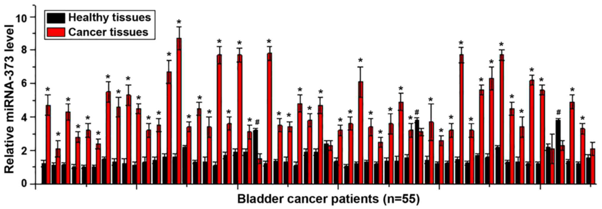
Analysis of the expression of miRNA-373 in tumor
tissues and adjacent healthy tissues of 55 patients with urinary
bladder cancer using RT-qPCR, which indicated that the expression
of miRNA-373 was significantly upregulated in tumor tissues
compared with adjacent healthy tissues in 49 out of 55 patients
(P<0.05; Fig. 1), accounting for
89.0% of this cohort. By contrast, the expression of miRNA-373 was
significantly downregulated in tumor tissues compared with adjacent
healthy tissues in 3 patients (P<0.05; Fig. 1), accounting for 5.5%. No significant
differences were identified in the remaining 3 cases, accounting
for 5.5%. Those data suggest that the upregulation of miRNA-373 is
likely to be involved in the pathogenesis of urinary bladder
cancer.
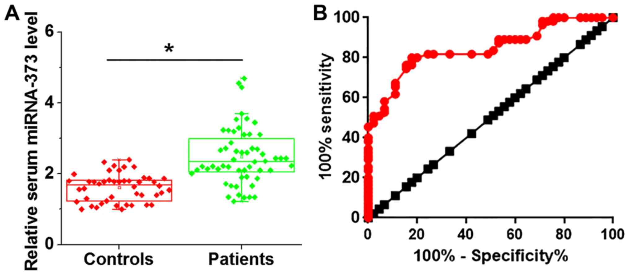
Comparison of serum levels of
miRNA-373 and the diagnostic values
As demonstrated in Fig.
2A, the serum levels of miRNA-373 were significantly increased
in patients with urinary bladder cancer compared with the healthy
controls. Receiver operating characteristic (ROC) curve analysis
was performed to evaluate the diagnostic value of serum miRNA-373
for urinary bladder cancer. As indicated in Fig. 2B, the area under the curve was
0.8473, with a 95% confidence interval of 0.7722–0.9223
(P<0.001). These data suggest that the upregulation of serum
miRNA-373 may serve as a potential diagnostic marker for urinary
bladder cancer.
Association between serum levels of
miRNA-373 and clinicopathological data of patients with urinary
bladder cancer
Patients were divided into high expression (n=28)
and low expression groups (n=27) according to the median serum
level of miRNA-373 (> or <2.56, respectively). The
associations between serum levels of miRNA-373 and
clinicopathological data of patients with urinary bladder cancer
were analyzed by χ2 test. As summarized in Table I, serum levels of miRNA-373 were not
significantly associated with the sex, age, or drinking and smoking
habits (P>0.05), but were significantly associated with the
diameter of primary tumors and distant metastasis (P<0.05).
 | Table I.Association between serum levels of
miRNA-373 and clinicopathological data of patients with urinary
bladder cancer. |
Table I.
Association between serum levels of
miRNA-373 and clinicopathological data of patients with urinary
bladder cancer.
|
|
|
| miRNA-373 expression
groups |
|
|
|---|
|
|
|
|
|
|
|
|---|
| Patient
characteristics | Groups | Cases, n | High | Low | χ2 | P-value |
|---|
| Sex | Male | 40 | 19 | 21 | 0.68 | 0.41 |
|
| Female | 15 | 9 | 6 |
|
|
| Age, years | ≥45 | 27 | 15 | 12 | 0.46 | 0.50 |
|
| <45 | 28 | 13 | 15 |
|
|
| Primary tumor
diameter, cm | ≥2 | 32 | 20 | 12 | 4.11 | 0.04 |
|
| <2 | 23 | 8 | 15 |
|
|
| Distant
metastasis | Yes | 26 | 18 | 8 | 6.62 | 0.01 |
|
| No | 29 | 10 | 19 |
|
|
| Smoking | Yes | 31 | 15 | 16 | 0.18 | 0.67 |
|
| No | 24 | 13 | 11 |
|
|
| Drinking | Yes | 38 | 20 | 18 | 0.15 | 0.70 |
|
| No | 17 | 8 | 9 |
|
|
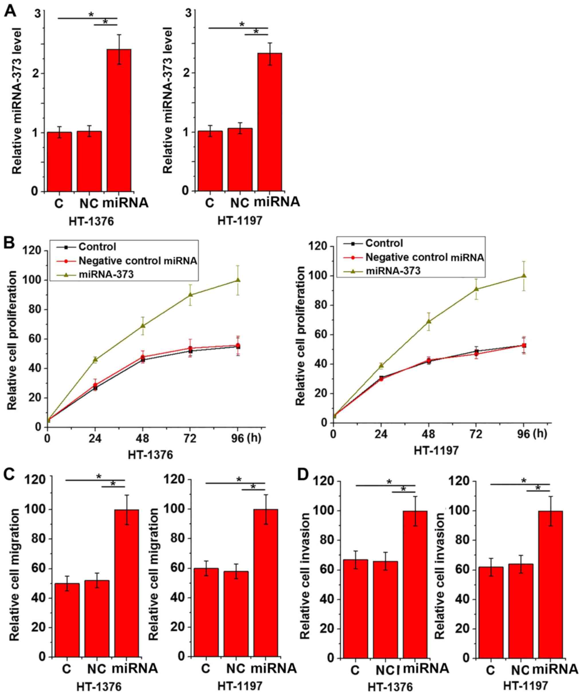
Effects of miRNA-373 overexpression of
urinary bladder cancer cell proliferation, migration and
invasion
The aforementioned data suggested that upregulation
of miRNA-373 may be associated with tumor growth and distant
metastasis of urinary bladder cancer. To additionally investigate
the role of miRNA-373 in urinary bladder cancer proliferation and
migration, miRNA-373 mimics were transfected into urinary bladder
cancer HT-1376 and HT-1197 cell lines. The overexpression of
miRNA-373 was confirmed by RT-qPCR (Fig.
3A). As indicated in Fig. 3,
miRNA-373 mimic transfection significantly promoted the
proliferation (Fig. 3B), migration
(Fig. 3C) and invasion (Fig. 3D) of the two cell lines.
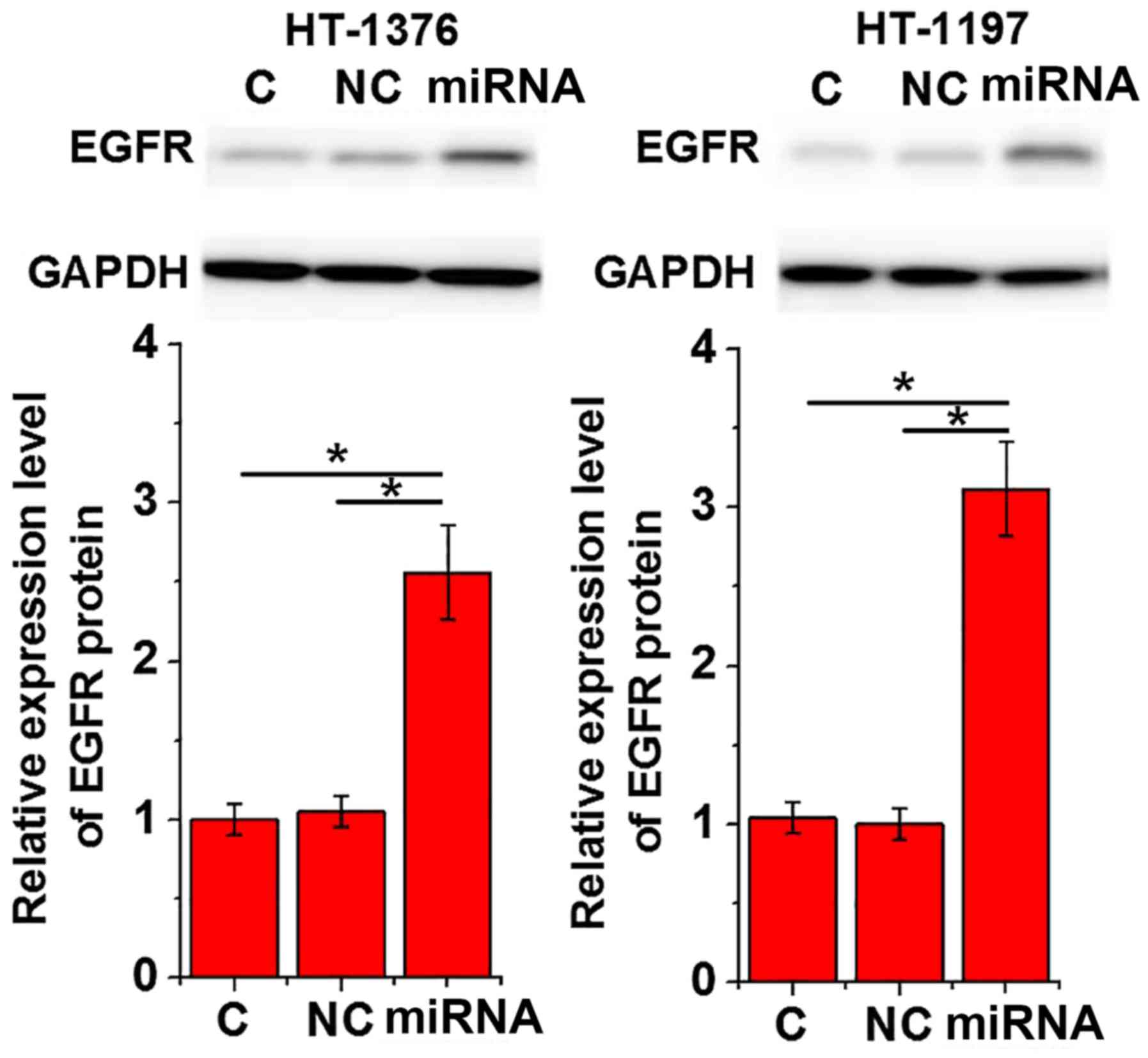
Effects of miRNA-373 overexpression on
EGFR expression
The overexpression of EGFR stimulates cell
proliferation, migration and invasion in certain types of cancer
cells (12). Therefore, the effects
of miRNA-373 overexpression on EGFR expression were investigated.
As indicated in Fig. 4, transfection
of miRNA-373 mimics significantly promoted the expression of EGFR
in cells of the two urinary bladder cancer cell lines
(P<0.05).
Discussion
miRNA-373, as a human embryonic stem cell-specific
miRNA, has been demonstrated to be involved the regulation of cell
proliferation, senescence, apoptosis, migration and invasion
(13). In addition, miRNA-373 also
serves a pivotal role in DNA damage repair following hypoxia stress
(14). The altered expression of
miR-373 has been observed in several types of human cancer,
indicating the role of miR-373 as an oncogene or tumor suppression
gene in those diseases. In the study of testicular germ cell
tumors, Voorhoeve et al (9)
suggested that miRNA-373 was upregulated in tumors and is likely to
serve an oncogenic role. However, a contrasting study identified
that miRNA-373 was downregulated in estrogen receptor negative
breast cancer, suggesting its role as a tumor suppression gene in
this disease (10). In the present
study, the upregulation of miRNA-373 expression in tumor tissues
compared with adjacent healthy tissues was observed in the majority
of patients with urinary bladder cancer. In addition, serum levels
of miRNA-373 were also increased in patients with urinary bladder
cancer compared with the healthy controls. These data suggest that
upregulation of miRNA-373 is likely involved in the pathogenesis of
urinary bladder cancer.
Due to the poor prognosis of patients with advanced
urinary bladder cancer (3), early
diagnosis and treatment of this disease remains critical.
Development of human diseases is usually accompanied by changes in
certain factors in the blood circulation system and comparison of
those factors under physiological and pathological conditions may
provide valuable references for the diagnosis of diseases (15). In the present study, ROC curve
analysis revealed that serum miRNA-373 may be an accurate biomarker
for urinary bladder cancer. It has been established previously that
the expression pattern of certain miRNAs may be altered by smoking
(16), alcohol consumption (17) and aging (18), which may affect the reliability of
miRNAs in the diagnosis of diseases. In the present study, no
significant associations between serum levels of miRNA-373 with
patient age, sex or behavioral habits including smoking and
drinking were observed. These data suggest that miRNA-373 is a
reliable and effective biomarker for urinary bladder cancer.
However, it is worth noting that an altered expression of miRNA-373
has been observed in different human diseases (9,10).
Therefore, multiple biomarkers should be combined to improve the
diagnostic accuracy.
The present study also indicated that serum levels
of miRNA-373 were closely associated with tumor size and distant
tumor metastasis. Previous studies have confirmed that miRNA-373 is
involved in the regulation of cell proliferation, migration and
invasion of several types of human cancer (19–21). In
the present study, transfection with miRNA-373 significantly
promoted cell proliferative, migratory and invasive abilities of
two urinary bladder cancer cell lines, indicating that the role of
miRNA-373 was a promotor of urinary bladder cancer growth and
metastasis. The overexpression of EGFR stimulates cell
proliferation, migration and invasion of certain types of cancer
cells, and EGFR is considered a target for the treatment of several
malignancies, including urinary bladder cancer (12,22). In
the present study, transfection with miRNA-373 significantly
promoted the expression of EGFR in two urinary bladder cancer cell
lines. These data suggest that miRNA-373 may promote the growth and
metastasis of urinary bladder cancer, and that this function may be
associated with the upregulation of EGFR.
Notably, Zhang et al (23) revealed an expression pattern and
functionality of miRNA-373 in bladder cancer contradictory to that
described in the present study. The discordance in these
observations may be due to the different cell lines used in the
present study and individual and/or regional differences across
patients. In addition, the present study only examined
miRNA-373-EGFR signaling in bladder cancer. The data obtained did
not provide any evidence on whether this interaction was direct or
indirect. Future studies should aim to reveal more information
regarding the signaling pathway. An additional limitation of the
present study was the small sample size. Future studies with bigger
sample sizes are required to additionally confirm the results. In
addition, more cells lines of different subtypes should be used to
investigate the role of miRNA-373 in bladder cancer.
In conclusion, miRNA-373 expression was upregulated
in urinary bladder cancer. Serum miRNA-373 may serve as an
effective and reliable diagnostic marker for urinary bladder
cancer. miRNA-373 overexpression promoted tumor cell proliferation,
migration and invasion, and EGFR expression. The present study
concluded that miRNA-373 overexpression may promote urinary bladder
cancer cell proliferation, migration and invasion by upregulating
EGFR. However, the mechanism of miRNA-373-mediated upregulation of
EGFR remains unknown. Additional studies are required.
Acknowledgements
Not applicable.
Funding
Not applicable.
Availability of data and materials
All data generated or analyzed during this study are
included in this published article.
Authors' contributions
YW and XW designed experiments. YW and ZX performed
experiments and collected data. YW and XW analyzed and interpreted
data. XW drafted the manuscript. All authors read and approved the
manuscript.
Ethics approval and consent to
participate
The present study was approved by the Ethics
Committee of Shengjing Hospital of China Medical University and all
participants provided written informed consent.
Patient consent for publication
Not applicable.
Competing interests
The authors declare that they have no competing
interests.
References
|
1
|
Ploeg M, Aben KK and Kiemeney LA: The
present and future burden of urinary bladder cancer in the world.
World J Urol. 27:289–293. 2009. View Article : Google Scholar : PubMed/NCBI
|
|
2
|
Volanis D, Kadiyska T, Galanis A, Delakas
D, Logotheti S and Zoumpourlis V: Environmental factors and genetic
susceptibility promote urinary bladder cancer. Toxicol Lett.
193:131–137. 2010. View Article : Google Scholar : PubMed/NCBI
|
|
3
|
Marcos-Gragera R, Mallone S, Kiemeney LA,
Vilardell L, Malats N, Allory Y and Sant M: EUROCARE-5 Working
Group: Urinary tract cancer survival in Europe 1999–2007: Results
of the population-based study EUROCARE-5. Eur J Cancer.
51:2217–2230. 2015. View Article : Google Scholar : PubMed/NCBI
|
|
4
|
Mahmoud-Ahmed AS, Suh JH, Kupelian PA,
Klein EA, Peereboom DM, Dreicer R and Barnett GH: Brain metastases
from bladder carcinoma: Presentation, treatment and survival. J
Urol. 167:2419–2422. 2002. View Article : Google Scholar : PubMed/NCBI
|
|
5
|
Mattick JS and Makunin IV: Non-coding RNA.
Human Mol Genet. 15:R17–R29. 2006. View Article : Google Scholar
|
|
6
|
Martens-Uzunova ES, Jalava SE, Dits NF,
van Leenders GJ, Møller S, Trapman J, Bangma CH, Litman T,
Visakorpi T and Jenster G: Diagnostic and prognostic signatures
from the small non-coding RNA transcriptome in prostate cancer.
Oncogene. 31:978–991. 2012. View Article : Google Scholar : PubMed/NCBI
|
|
7
|
Ha M and Kim VN: Regulation of microRNA
biogenesis. Nat Rev Mol Cell Biol. 15:509–524. 2014. View Article : Google Scholar : PubMed/NCBI
|
|
8
|
Bartel DP: MicroRNAs: Genomics,
biogenesis, mechanism, and function. Cell. 116:281–297. 2004.
View Article : Google Scholar : PubMed/NCBI
|
|
9
|
Voorhoeve PM, Le Sage C, Schrier M, Gillis
AJ, Stoop H, Nagel R, Liu YP, van Duijse J, Drost J, Griekspoor A,
et al: A genetic screen implicates miRNA-372 and miRNA-373 as
oncogenes in testicular germ cell tumors. Cell. 124:1169–1181.
2006. View Article : Google Scholar : PubMed/NCBI
|
|
10
|
Keklikoglou I, Koerner C, Schmidt C, Zhang
JD, Heckmann D, Shavinskaya A, Allgayer H, Gückel B, Fehm T,
Schneeweiss A, et al: MicroRNA-520/373 family functions as a tumor
suppressor in estrogen receptor negative breast cancer by targeting
NF-κB and TGF-β signaling pathways. Oncogene. 31:4150–4163. 2012.
View Article : Google Scholar : PubMed/NCBI
|
|
11
|
Livak KJ and Schmittgen TD: Analysis of
relative gene expression data using real-time quantitative PCR and
the 2(-Delta Delta C(T)) method. Methods. 25:402–408. 2001.
View Article : Google Scholar : PubMed/NCBI
|
|
12
|
Andl CD, Mizushima T, Nakagawa H, Oyama K,
Harada H, Chruma K, Herlyn M and Rustgi AK: Epidermal growth factor
receptor mediates increased cell proliferation, migration, and
aggregation in esophageal keratinocytes in vitro and in vivo. J
Biol Chem. 278:1824–1830. 2003. View Article : Google Scholar : PubMed/NCBI
|
|
13
|
Wei F, Cao C, Xu X and Wang J: Diverse
functions of miR-373 in cancer. J Transl Med. 13:1622015.
View Article : Google Scholar : PubMed/NCBI
|
|
14
|
Crosby ME, Kulshreshtha R, Ivan M and
Glazer PM: MicroRNA regulation of DNA repair gene expression in
hypoxic stress. Cancer Res. 69:1221–1229. 2009. View Article : Google Scholar : PubMed/NCBI
|
|
15
|
Borovecki F, Lovrecic L, Zhou J, Jeong H,
Then F, Rosas HD, Hersch SM, Hogarth P, Bouzou B, Jensen RV and
Krainc D: Genome-wide expression profiling of human blood reveals
biomarkers for Huntington's disease. Proc Natl Acad Sci USA.
102:11023–11028. 2005. View Article : Google Scholar : PubMed/NCBI
|
|
16
|
Marczylo EL, Amoako AA, Konje JC, Gant TW
and Marczylo TH: Smoking induces differential miRNA expression in
human spermatozoa: A potential transgenerational epigenetic
concern? Epigenet. 7:432–439. 2012. View Article : Google Scholar
|
|
17
|
Mamdani M, Williamson V, McMichael GO,
Blevins T, Aliev F, Adkins A, Hack L, Bigdeli T, van der Vaart AD,
Web BT, et al: Integrating mRNA and miRNA weighted gene
co-expression networks with eQTLs in the nucleus accumbens of
subjects with alcohol dependence. PLoS One. 10:e01376712015.
View Article : Google Scholar : PubMed/NCBI
|
|
18
|
Olivieri F, Capri M, Bonafè M, Morsiani C,
Jung HJ, Spazzafumo L, Viña J and Suh Y: Circulating miRNAs and
miRNA shuttles as biomarkers: Perspective trajectories of healthy
and unhealthy aging. Mech Ageing Dev. 165:162–170. 2017. View Article : Google Scholar : PubMed/NCBI
|
|
19
|
Huang Q, Gumireddy K, Schrier M, le Sage
C, Nagel R, Nair S, Egan DA, Li A, Huang G, Klein-Szanto AJ, et al:
The microRNAs miR-373 and miR-520c promote tumour invasion and
metastasis. Nat Cell Biol. 10:202–210. 2008. View Article : Google Scholar : PubMed/NCBI
|
|
20
|
Li X, Zhang Y, Zhang H, Liu X, Gong T, Li
M, Sun L, Ji G, Shi Y, Han Z, et al: miRNA-223 promotes gastric
cancer invasion and metastasis by targeting tumor suppressor
EPB41L3. Mol Cancer Res. 9:824–833. 2011. View Article : Google Scholar : PubMed/NCBI
|
|
21
|
Lee KH, Goan YG, Hsiao M, Lee CH, Jian SH,
Lin JT, Chen YL and Lu PJ: MicroRNA-373 (miR-373)
post-transcriptionally regulates large tumor suppressor, homolog 2
(LATS2) and stimulates proliferation in human esophageal cancer.
Exp Cell Res. 315:2529–2538. 2009. View Article : Google Scholar : PubMed/NCBI
|
|
22
|
Weintraub MD, Vourganti S, Li Q, Apolo AB,
Metwalli AR and Agarwal PK: Targeting the epidermal growth factor
receptor in bladder cancer. J Carcinog Mutag. 4:10–30. 2013.
|
|
23
|
Zhang Q, Wang C, Miao S, Li C, Chen Z and
Li F: Enhancing E-cadherin expression via promoter-targeted miR-373
suppresses bladder cancer cells growth and metastasis. Oncotarget.
8:93969–93983. 2017.PubMed/NCBI
|