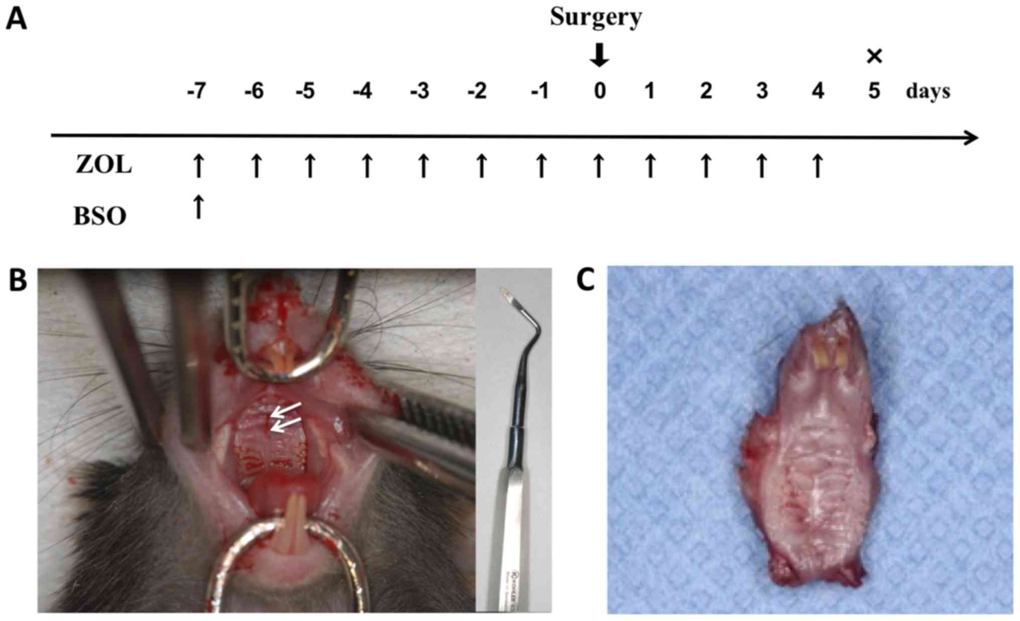
|
1
|
Migliorati CA, Casiglia J, Epstein J,
Jacobsen PL, Siegel MA and Woo SB: Managing the care of patients
with bisphosphonate-associated osteonecrosis: An American Academy
of Oral Medicine position paper. J Am Dent Assoc. 136:1658–1668.
2005. View Article : Google Scholar : PubMed/NCBI
|
|
2
|
Cheng A, Mavrokokki A, Carter G, Stein B,
Fazzalari NL, Willson DF and Goss AN: The dental implications of
bisphosphonates and bone disease. Aust Dent J. 50 (4 Suppl
2):S4–S13. 2005. View Article : Google Scholar : PubMed/NCBI
|
|
3
|
Bertoldo F, Santini D and Lo Cascio V:
Bisphosphonates and osteomylelitis of the jaw: A pathogenic puzzle.
Nat Clin Pract Oncol. 4:711–721. 2007. View Article : Google Scholar : PubMed/NCBI
|
|
4
|
Licata AA: Discovery, clinical
development, and therapeutic uses of bisphosphonates. Ann
Pharmacother. 39:668–677. 2005. View Article : Google Scholar : PubMed/NCBI
|
|
5
|
Michaelson MD and Smith MR:
Bisphosphonates for treatment and prevention of bone metastases. J
Clin Oncol. 23:8219–8224. 2005. View Article : Google Scholar : PubMed/NCBI
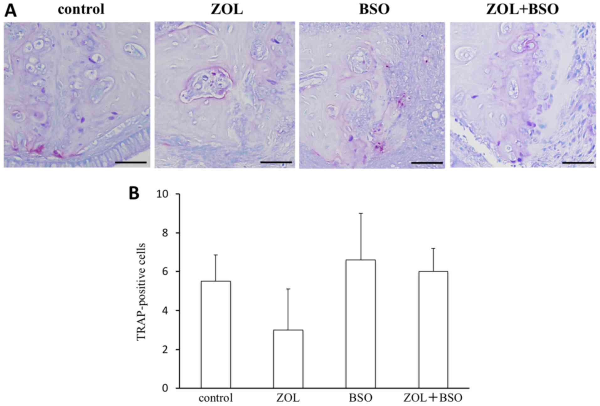
|
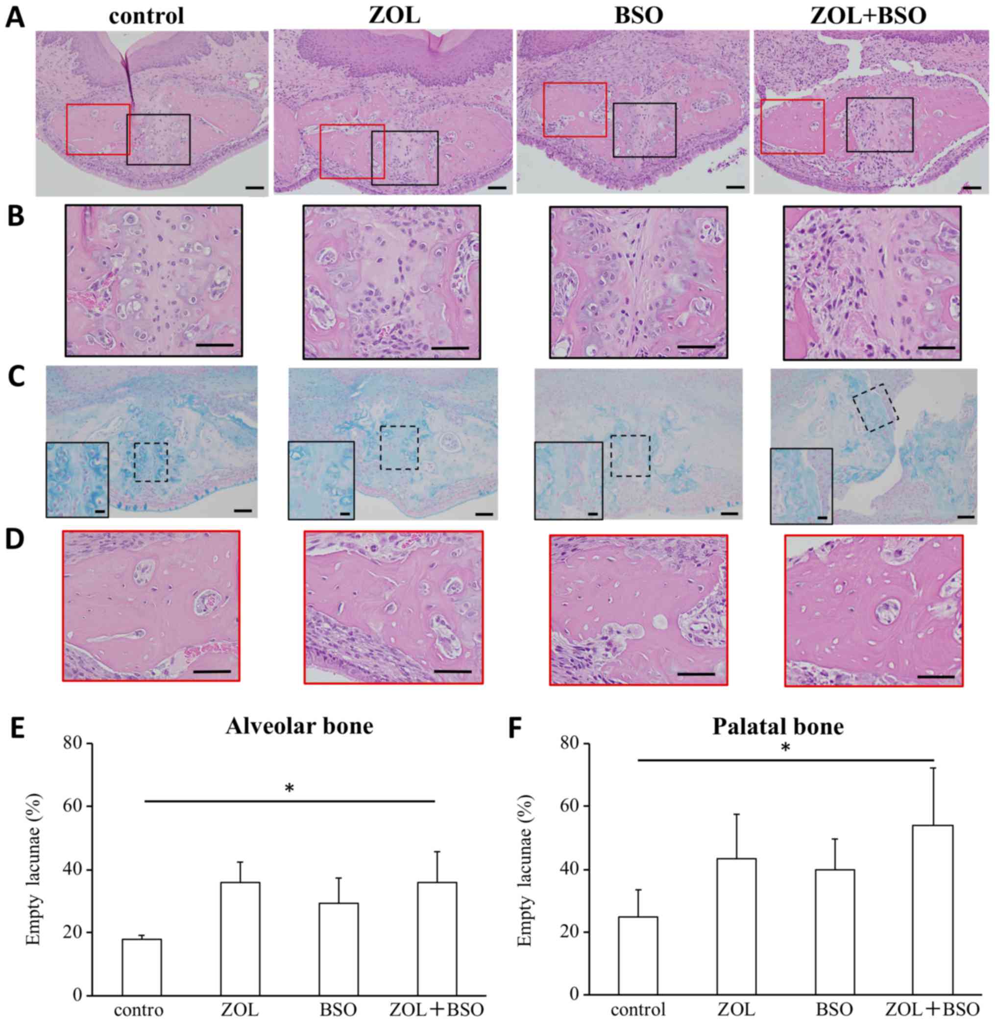
|
6
|
Ruggiero SL, Dodson TB, Assael LA,
Landesberg R, Marx RE and Mehrotra B: American Association of Oral
and Maxillofacial Surgeons: American Association of Oral and
Maxillofacial Surgeons position paper on bisphosphonate-related
osteonecrosis of the jaws-2009 update. J Oral Maxillofac Surg. 67
Suppl 5:2–12. 2009. View Article : Google Scholar : PubMed/NCBI
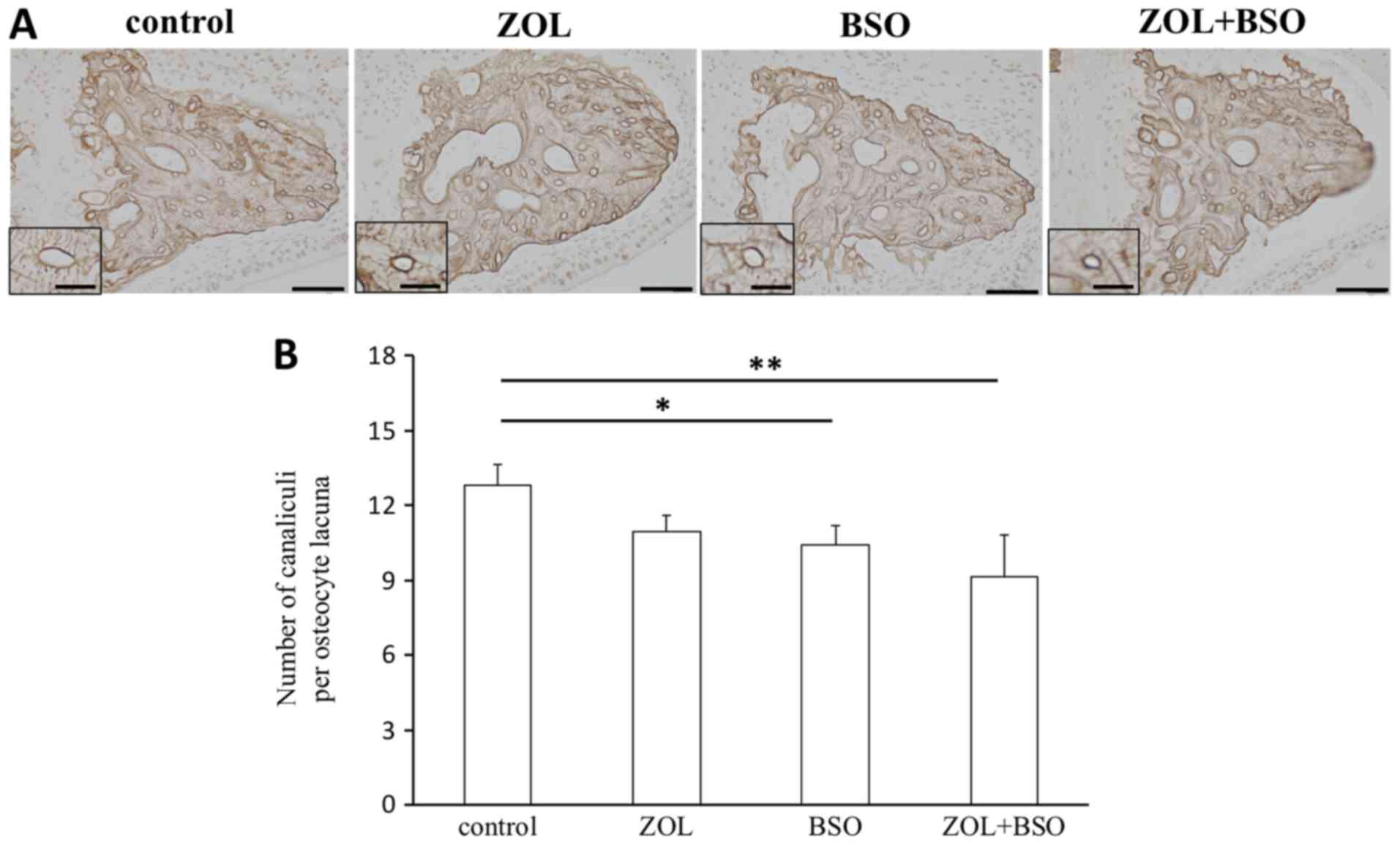
|
|
7
|
Urade M, Tanaka N, Furusawa K, Shimada J,
Shibata T, Kirita T, Yamamoto T, Ikebe T, Kitagawa Y and Fukuta J:
Nationwide survey for bisphosphonate-related osteonecrosis of the
jaws in Japan. J Oral Maxillofac Surg. 69:e364–e371. 2011.
View Article : Google Scholar : PubMed/NCBI
|
|
8
|
Doggrell SA: Clinical efficacy and safety
of zoledronic acid in prostate and breast cancer. Expert Rev
Anticancer Ther. 9:1211–1218. 2009. View Article : Google Scholar : PubMed/NCBI
|
|
9
|
Reid IR and Cornish J: Epidemiology and
pathogenesis of osteonecrosis of the jaw. Nat Rev Rheumatol.
8:90–96. 2011. View Article : Google Scholar : PubMed/NCBI
|
|
10
|
Kim JH, Park YB, Li Z, Shim JS, Moon HS,
Jung HS and Chung MK: Effect of alendronate on healing of
extraction sockets and healing around implants. Oral Dis.
17:705–711. 2011. View Article : Google Scholar : PubMed/NCBI
|
|
11
|
Howie RN, Borke JL, Kurago Z, Daoudi A,
Cray J, Zakhary IE, Brown TL, Raley JN, Tran LT, Messer R, et al: A
model for osteonecrosis of the jaw with zoledronate treatment
following repeated major trauma. PLoS One. 17:e01325202015.
View Article : Google Scholar
|
|
12
|
Poubel VLDN, Silva CAB, Mezzomo LAM, De
Luca Canto G and Rivero ERC: The risk of osteonecrosis on alveolar
healing after tooth extraction and systemic administration of
antiresorptive drugs in rodents: A systematic review. J
Craniomaxillofac Surg. 46:245–256. 2018. View Article : Google Scholar : PubMed/NCBI
|
|
13
|
Advisory T ask Force on
Bisphosphonate-Related Ostenonecrosis of the Jaws, American
Association of Oral and Maxillofacial Surgeons: American
Association of Oral and Maxillofacial Surgeons position paper on
bisphosphonate-related osteonecrosis of the jaws. J Oral Maxillofac
Surg. 65:369–376. 2007. View Article : Google Scholar : PubMed/NCBI
|
|
14
|
Sonis ST, Watkins BA, Lyng GD, Lerman MA
and Anderson KC: Bony changes in the jaws of rats treated with
zoledronic acid and dexamethasone before dental extractions mimic
bisphosphonate-related osteonecrosis in cancer patients. Oral
Oncol. 45:164–172. 2009. View Article : Google Scholar : PubMed/NCBI
|
|
15
|
Hokugo A, Christensen R, Chung EM, Sung
EC, Felsenfeld AL, Sayre JW, Garrett N, Adams JS and Nishimura I:
Increased prevalence of bisphosphonate-related osteonecrosis of the
jaw with vitamin D deficiency in rats. J Bone Miner Res.
25:1337–1349. 2010. View
Article : Google Scholar : PubMed/NCBI
|
|
16
|
Takaoka K, Yamamura M, Nishioka T, Abe T,
Tamaoka J, Segawa E, Shinohara M, Ueda H, Kishimoto H and Urade M:
Establishment of an animal model of bisphosphonate-related
osteonecrosis of the jaws in spontaneously diabetic torii rats.
PLoS One. 14:e01443552015. View Article : Google Scholar
|
|
17
|
Bamias A, Kastritis E, Bamia C,
Moulopoulos LA, Moulopoulos I, Bozasb G, Koutsoukou V, Gika D,
Anaqnostopoulos A, Papadimitriou C, et al: Osteonecrosis of the jaw
in cancer after treatment with bisphosphonates: Incidence and risk
factors. J Clin Oncol. 23:8580–8587. 2005. View Article : Google Scholar : PubMed/NCBI
|
|
18
|
Marx RE, Sawatari Y, Fortin M and Broumand
V: Bisphosphonate-induced exposed bone
(osteonecrosis/osteopetrosis) of the jaws: Risk factors,
recognition, prevention, and treatment. J Oral Maxillofac Surg.
63:1567–1575. 2005. View Article : Google Scholar : PubMed/NCBI
|
|
19
|
Jadu F, Lee L, Pharoah M, Reece D and Wang
L: A retrospective study assessing the incidence, risk factors and
comorbidities of pamidronate-related necrosis of the jaws in
multiple myeloma patients. Ann Oncol. 18:2015–2019. 2007.
View Article : Google Scholar : PubMed/NCBI
|
|
20
|
Almeida M, Han L, Martin-Millan M, Plotkin
LI, Stewart SA, Roberson PK, Kousteni S, O'Brien CA, Bellido T,
Parfitt AM, et al: Skeletal involution by age-associated oxidative
stress and its acceleration by loss of sex steroids. J Biol Chem.
282:27285–27297. 2007. View Article : Google Scholar : PubMed/NCBI
|
|
21
|
Almeida M, Ambrogini E, Han L, Manolagas
SC and Jilka RL: Increased lipid oxidation causes oxidative stress,
increased peroxisome proliferator-activated receptor-gamma
expression, and diminished pro-osteogenic Wnt signaling in the
skeleton. J Biol Chem. 284:27438–27448. 2009. View Article : Google Scholar : PubMed/NCBI
|
|
22
|
Nojiri H, Saita Y, Morikawa D, Kobayashi
K, Tsuda C, Miyazaki T, Saito M, Marumo K, Yonezawa I, Kaneko K, et
al: Cytoplasmic superoxide causes bone fragility owing to
low-turnover osteoporosis and impaired collagen cross-linking. J
Bone Miner Res. 26:2682–2694. 2011. View
Article : Google Scholar : PubMed/NCBI
|
|
23
|
Almeida M and O'Brien CA: Basic biology of
skeletal aging: Role of stress response pathways. J Gerontol Biol
Sci Med Sci. 68:1197–1208. 2013. View Article : Google Scholar
|
|
24
|
Domazetovic V, Marcucci G, Iantomasi T,
Brandi ML and Vincenzini MT: Oxidative stress in bone remodeling:
Role of antioxidants. Clin Cases Miner Bone Metab. 14:209–216.
2017. View Article : Google Scholar : PubMed/NCBI
|
|
25
|
Koçer G, Naziroğlu M, Çelik Ö, Önal L,
Özçelik D, Koçer M and Sönmez TT: Basic fibroblast growth factor
attenuates bisphosphonate-induced oxidative injury but decreases
zinc and copper levels in oral epithelium of rat. Biol Trace Elem
Res. 153:251–256. 2013. View Article : Google Scholar : PubMed/NCBI
|
|
26
|
Awodele O, Olayemi SO, Nwite JA and
Adeyemo TA: Investigation of the levels of oxidative stress
parameters in HIV and HIV-TB co-infected patients. J Infect Dev
Ctries. 6:79–85. 2012. View Article : Google Scholar : PubMed/NCBI
|
|
27
|
Lebreton F, van Schaik W, Sanguinetti M,
Posteraro B, Torelli R, Lee Bras F, Vemeuil N, Zhang X, Giard JC,
Dhalluin A, et al: AsrR is an oxidative stress sensing regulator
modulating Enterococcus faecium opportunistic traits, antimicrobial
resistance, and pathogenicity. PLoS Pathog. 8:e10028342012.
View Article : Google Scholar : PubMed/NCBI
|
|
28
|
McDevitt CA, Ogunniyi AD, Valkov E,
Lawrence MC, Kobe B, McEwan AG and Paton JC: A molecular mechanism
for bacterial susceptibility to zinc. PLoS Pathog. 7:e10023572011.
View Article : Google Scholar : PubMed/NCBI
|
|
29
|
Moye-Rowley WS: Transcription factors
regulating the response to oxidative stress in yeast. Antioxid
Redox Signal. 4:123–140. 2002. View Article : Google Scholar : PubMed/NCBI
|
|
30
|
Khandelwal VK, Mitrofan LM, Hyttinen JM,
Chaudhari KR, Buccione R, Kaarniranta K, Ravingerová T and
Mönkkönen J: Oxidative stress plays an important role in zoledronic
acid-induced autophagy. Physiol Res. 63 Suppl 4:S601–S612.
2014.PubMed/NCBI
|
|
31
|
Kuribayashi M, Fujioka M, Takahashi KA,
Arai Y, Ishida M, Goto T and Kubo T: Vitamin E prevents
steroid-induced osteonecrosis in rabbits. Acta Orthop. 81:154–160.
2010. View Article : Google Scholar
|
|
32
|
Ichiseki T, Matsumoto T, Nishino M,
Kaneuji A and Katsuda S: Oxidative stress and vascular permeability
in steroid-induced osteonecrosis model. J Orthop Sci. 9:509–515.
2004. View Article : Google Scholar : PubMed/NCBI
|
|
33
|
Ichiseki T, Kaneuji A, Katsuda S, Ueda Y,
Sugimori T and Matsumoto T: DNA oxidation injury in bone early
after steroid administration is involved in the pathogenesis of
steroid-induced osteonecrosis. Rheumatology (Oxford). 44:456–460.
2005. View Article : Google Scholar : PubMed/NCBI
|
|
34
|
Ichiseki T, Kaneuji A, Ueda Y, Nakagawa S,
Mikami T, Fukui K and Matsumoto T: Osteonecrosis development in a
novel rat model characterized by a single application of oxidative
stress. Arthritis Rheum. 63:2138–2141. 2011. View Article : Google Scholar : PubMed/NCBI
|
|
35
|
Kobayashi Y, Hiraga T, Ueda A, Wang L,
Matsumoto-Nakano M, Hata K, Yatani H and Yoneda T: Zoledronic acid
delays wound healing of the tooth extraction socket, inhibits oral
epithelial cell migration, and promotes proliferation and adhesion
to hydroxyapatite of oral bacteria, without causing osteonecrosis
of the jaw, in mice. J Bone Miner Metab. 28:165–175. 2010.
View Article : Google Scholar : PubMed/NCBI
|
|
36
|
Valavanidis A, Vlachogianni T and Fiotakis
C: 8-hydroxy-2′-deoxyguanosine (8-OHdG): A critical biomarker of
oxidative stress and carcinogenesis. J Environ Sci Health C Environ
Carcinog Ecotoxicol Rev. 27:120–139. 2009. View Article : Google Scholar : PubMed/NCBI
|
|
37
|
Jaiprakash A, Prasadam I, Feng JQ, Liu Y,
Crawford R and Xiao Y: Phenotypic characterization of
osteoarthritic osteocytes from the sclerotic zones: A possible
pathological role in subchondral bone sclerosis. Int J Biol Sci.
8:406–417. 2012. View Article : Google Scholar : PubMed/NCBI
|
|
38
|
Kawamoto T and Kawamoto K: Preparation of
thin frozen sections from nonfixed and undecalcified hard tissues
using Kawamoto's film method (2012). Methods Mol Biol.
1130:149–164. 2014. View Article : Google Scholar : PubMed/NCBI
|
|
39
|
Dempster DW, Compston JE, Drezner MK,
Glorieux FH, Kanis JA, Malluche H, Meunier PJ, Ott SM, Recker RR
and Parfitt AM: Standardized nomenclature, symbols and units for
bone histomorphometry: A 2012 update of the report of the ASBMR
Histomorphometry Nomenclature Committee. J Bone Miner Res. 28:2–17.
2013. View Article : Google Scholar : PubMed/NCBI
|
|
40
|
Huja SS and Beck FM: Bone remodeling in
maxilla, mandible, and femur of young dogs. Anat Rec (Hoboken).
291:1–5. 2008. View Article : Google Scholar : PubMed/NCBI
|
|
41
|
Rodan GA and Martin TJ: Therapeutic
approaches to bone diseases. Science. 289:1508–1514. 2000.
View Article : Google Scholar : PubMed/NCBI
|
|
42
|
Hughes DE, Wright KR, Uy HL, Sasaki A,
Yoneda T, Roodman GD, Mundy GR and Boyce BF: Bisphosphonates
promote apoptosis in murine osteoclasts in vitro and in vivo. J
Bone Miner Res. 10:1478–1487. 1995. View Article : Google Scholar : PubMed/NCBI
|
|
43
|
Selander KS, Mönkkönen J, Karhukorpi EK,
Härkönen P, Hannuniemi R and Väänänen HK: Characteristics of
clodronate-induced apoptosis in osteoclasts and macrophages. Mol
Pharmacol. 50:1127–1138. 1996.PubMed/NCBI
|
|
44
|
Hiroi-Furuya E, Kameda T, Hiura K, Mano H,
Miyazawa K, Nakamaru Y, Watanabe-Mano M, Okuda N, Shimada J,
Yamamoto Y, et al: Etidronate (EHDP) inhibits osteoclastic-bone
resorption, promotes apoptosis and disrupts actin rings in
isolate-mature osteoclasts. Calcif Tissue Int. 64:219–223. 1999.
View Article : Google Scholar : PubMed/NCBI
|
|
45
|
Santini D, Vespasiani Gentilucci U,
Vincenzi B, Picardi A, Vasaturo F, La Cesa A, Onori N, Scarpa S and
Tonini G: The antineoplastic role of bisphosphonates: From basic
research to clinical evidence. Ann Oncol. 14:1468–1476. 2003.
View Article : Google Scholar : PubMed/NCBI
|
|
46
|
Ruggiero SL, Dodson TB, Fantasia J,
Goodday R, Aghaloo T, Mehrotra B and O'Ryan F: American Association
of Oral and Maxillofacial Surgeons: American Association of Oral
and Maxillofacial Surgeons position paper on medication-related
osteonecrosis of the jaw-2014 update. J Oral Maxillofac Surg.
72:1938–1956. 2014. View Article : Google Scholar : PubMed/NCBI
|
|
47
|
Bonewald LF: The amazing osteocyte. J Bone
Miner Res. 26:229–238. 2011. View Article : Google Scholar : PubMed/NCBI
|
|
48
|
Busse B, Djonic D, Milovanovic P, Hahn M,
Püschel K, Ritchie RO, Djuric M and Amling M: Decrease in the
osteocyte lacunar density accompanied by hypermineralized lacunar
occlusion reveals failure and delay of remodeling in aged human
bone. Aging Cell. 9:1065–1075. 2010. View Article : Google Scholar : PubMed/NCBI
|
|
49
|
Dunstan CR, Somers NM and Evans RA:
Osteocyte death and hip fracture. Calcif Tissue Int. 53 Suppl
1:S113–S117. 1993. View Article : Google Scholar : PubMed/NCBI
|
|
50
|
Kobayashi K, Nojiri H, Saita Y, Morikawa
D, Ozawa Y, Watanabe K, Koike M, Asou Y, Shirasawa T, Yokote K, et
al: Mitochondrial superoxide in osteocytes perturbs canalicular
networks in the setting of age-related osteoporosis. Sci Rep.
5:91482015. View Article : Google Scholar : PubMed/NCBI
|