Introduction
Cervical cancer (CC) is the second most common
malignant cancer among women (1). CC
is characterized by high incidence and recurrence rates, as well as
high resistance to systemic therapies (2). Therefore, prognosis for patients with
CC remains relatively poor. Increasing evidence has demonstrated
that the human papillomavirus (HPV) is correlated with the
development of a high-grade precursor lesions and invasion in CC
(3). HPV can infect epithelial
cells, which remain active in cell-cycle progression and no longer
undergo apoptosis (4).
Studies have demonstrated that the Wnt signaling
pathway serves a key role in cell differentiation, proliferation,
migration and polarity (5,6). In addition, the Wnt signaling pathway
serves a key role in maintaining protein stability, subcellular
localization and transcriptional activity (7). In the progression of tumors, the
Wnt/β-catenin signaling pathway is an evolutionarily conserved and
versatile pathway (8). Aberrant
activation of the Wnt/β-catenin pathway can lead to abnormal
accumulation of β-catenin in the nucleus, which accelerates the
epithelial-mesenchymal transition (EMT) process (9,10). It is
therefore important to maintain appropriate Wnt signaling.
Gefitinib was one of the first-generation epidermal
growth factor receptor-tyrosine kinase inhibitors in clinical
trials, and is now widely used for the treatment of several types
of cancer (11,12). An increasing number of studies have
been designed to identify the efficacy and toxicity of gefitinib
(13,14). However, the underlying mechanism of
gefitinib in regulating CC progression remains unknown. Therefore,
the aim of the current study was to investigate the specific role
and underlying mechanism of gefitinib in CC using human CC cell
lines.
Materials and methods
Cell culture
Human cervical cancer cell lines HeLa and Siha were
obtained from the Institute of Life Sciences Cell Resource Center
(Shanghai, China). HeLa and Siha cells were cultured in minimal
essential medium (MEM, HyClone; GE Healthcare Life Sciences)
supplemented with 10% fetal bovine serum (HyClone; GE Healthcare
Life Sciences), penicillin (100 U/ml)-streptomycin (100 U/ml)
liquid (Thermo Fisher Scientific, Inc.) and 0.25 µg/ml amphotericin
B (Ameresco, Inc.). Cells were cultured at 37°C in a humidified
incubator containing 5% CO2.
Cell proliferation assay
HeLa and Siha cell proliferation was examined using
the cell counting kit-8 (CCK-8; Dojindo Molecular Technologies,
Inc.), according to the manufacturer's protocol. Exponentially
growing cells were seeded into 96-well plates at a density of
5×10³ cells/well in a final volume of 100 µl MEM and
cultured under normal conditions for 24 h at 37°C in a 5%
CO2-humidified incubator. Subsequently, different
concentrations (0.3125, 0.625, 1.25, 2.5, 5, 10, 20, 40 and 80
µmol/l) of gefitinib (cat. no. SML1657; Sigma-Aldrich; Merck KGaA;
Darmstadt, Germany) were added to each well, with DMSO used as the
vehicle control. After 48 h, 10 µl CCK-8 reagent was added to each
well and incubated for 1 h at 37°C. Cell proliferation was
calculated by measuring the absorbance at a wavelength of 450 nm
using a microplate reader (Bio-Rad Laboratories, Inc.). Growth
inhibition was calculated as a percentage of the untreated
controls. All experiments were performed in triplicate and the data
were expressed as the mean value ± standard deviation of five wells
per treatment. For each cell line, the half maximal inhibitory
concentration (IC50) was determined using the
four-parameter logistic model.
Cell cycle analysis
HeLa and Siha cells were seeded into six-well plates
at the density of 106 cells/well. Following treatment
with 10 µmol/l of gefitinib for 48 h, cells were harvested using
trypsin without EDTA, washed three times with ice-cold PBS and
fixed with 70% ethanol overnight at 4°C. Cells were subsequently
stained 25 µl propidium iodide with 10 µl RNase A at 37°C for 30
min in dark using the Cell cycle and apoptosis analysis kit (cat.
no. C1052; Beyotime Institute of Biotechnology, Haimen, China).
Cell cycle analysis was performed using a BD FACSCalibur system and
CellQuest pro software (version 2.0; BD Biosciences, Franklin
Lakes, NJ, USA). All experiments were performed in triplicate.
Flow cytometry evaluation of
apoptosis
Cell apoptosis was examined using the Annexin
V-fluorescein isothiocyanate (FITC)/propidium iodide (PI) Apoptosis
Detection kit (BD Biosciences). Following treatment with 10 µmol/l
of gefitinib for 48 h, cells were harvested using trypsin without
EDTA and washed three times with ice-cold PBS. Cells were
subsequently suspended at 1×10⁴ cells/ml in Annexin
V-Binding buffer and incubated with 5 µl Annexin V-FITC for 15 min
at 37°C in dark, followed by staining with 5 µl PI. Apoptotic cells
were immediately analyzed using a BD FACSCalibur system and
CellQuest pro software. All experiments were performed in
triplicate.
Dual-luciferase reporter assay
HeLa and Siha cells were seeded into 24-well plates
at a density of 1×10⁵ cells/well and incubated
overnight. After 24 h of culture, cells were co-transfected with
200 ng pTOP-Flash (Promega Corporation, Madison, WI, USA) or
pFOP-Flash reporter plasmids (Promega Corporation) and 200 ng
β-galactosidase (β-gal) using Lipofectamine® 2000
(Invitrogen; Thermo Fisher Scientific, Inc.) to monitor for
transfection efficiency. After transfection for 48 h, the activity
was measured. The TCF-responsive TOP-Flash reporter contains three
TCF binding sites, and the corresponding FOP-Flash contains three
mutated TCF sites (15). Cells were
treated with 10 µmol/l gefitinib for 4 h at 37°C and luciferase
activity was analyzed using a Dual-Luciferase Reporter assay system
(Promega Corporation), according to the manufacturer's protocol.
Luciferase activity was normalized for β-gal activity. The
experiment was performed in triplicate.
Western blot analysis
Total cellular protein was extracted from cells on
ice for 15 min using radioimmunoprecipitation assay buffer
(Beyotime Institute of Biotechnology) supplemented with fresh
proteinase inhibitor cocktail and phosphatase inhibitor
(Sigma-Aldrich; Merck KGaA). Samples were centrifuged at 11,000 × g
for 20 min at 4°C. Total protein was quantified using a
bicinchoninic acid assay (Sigma-Aldrich; Merck KGaA) and 20 µg
protein/well was separated via SDS-PAGE on a 10% gel. The
fractionated proteins were transferred onto polyvinylidene
difluoride membranes (EMD Millipore) and blocked for 1 h at room
temperature with 5% non-fat skimmed milk. The membranes were
incubated with primary antibodies, including E-cadherin (1:1,000;
cat. no. ab1416), Vimentin (1:1,000; cat. no. ab8978), GSK3β
(1:1,000; cat. no. ab93926), p-GSK3β (1:1,000; cat. no. ab131097),
β-catenin (1:1,000; cat. no. ab32572) and GAPDH (1:5,000; cat. no.
ab181602) overnight at 4°C (all from Abcam, Cambridge, UK).
Following primary antibody incubation, membranes were incubated
with horseradish peroxidase-conjugated secondary antibody,
horseradish peroxidase-conjugated goat anti-rabbit IgG (1:5,000;
cat. no. ZB-2306; OriGne Technologies, Inc.), for 1 h at room
temperature. Protein bands were visualized using a Western
Lightning® Chemiluminescence Reagent Plus according to
the manufacturer's protocol (cat. no. NEL105001EA; PerkinElmer,
Inc., Waltham, MA, USA). Protein expression was quantified using
ImageJ (version 1.8.0; National Institutes of Health, Bethesda, MD,
USA).
Immunofluorescence
HeLa and Siha cells were cultured in a six-well
plate with glass coverslips and following treatment with 10 µM
gefitinib at 37°C for 48 h, cells were fixed with 4%
paraformaldehyde for 30 min at room temperature. Cells were washed
three times with PBS for 5 min and blocked for 2 h at room
temperature with 8% bovine serum albumin (BSA; Sigma-Aldrich; Merck
KGaA). Subsequently, cells were incubated with primary antibodies
against E-cadherin (1:50; cat. no. ab1416) and vimentin (1:50; cat.
no. ab92547; both Abcam) in a humidified chamber overnight at 4°C.
Cells were washed three times with PBS. Following primary
incubation, cells were incubated with
tetramethylrhodamine-conjugated anti-rabbit IgG (1:500; cat. no.
ZDR5209; OriGene Technologies, Inc.) at room temperature for 30
min. Cell nuclei were counterstained with DAPI (1:1,000; cat. no.
C0060; Beijing Solarbio Science & Technology Co., Ltd.) for 20
min at room temperature. Cells were subsequently washed three times
with PBS in the dark and the coverslips were mounted with mounting
medium at room temperature for ~1 h in the dark. Fluorescence
intensity was observed under a fluorescence microscope
(magnification, ×40; XDS-500D; Shanghai Caikon Optical Instrument
Co., Ltd.).
Statistical analysis
Data were presented as the mean ± standard error of
the mean of at least three experiments. All statistical analyses
were performed using GraphPad Prism software (version 5.0; GraphPad
Software, Inc.). All experimental data were analyzed using the
unpaired Student's t-test or one-way analysis of variance followed
by Tukey's post hoc test. P<0.05 was considered to indicate a
statistically significant difference.
Results
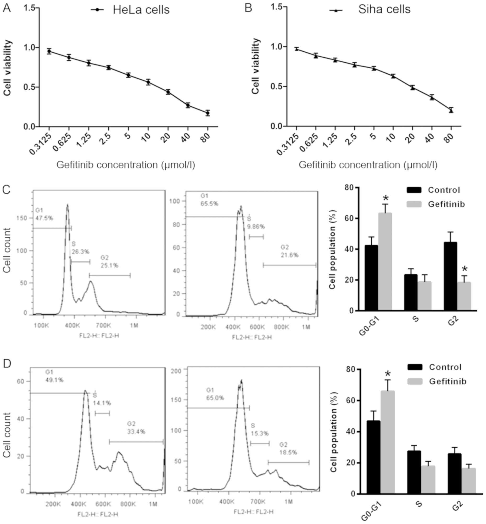
Gefitinib reduces proliferation and
induces cell cycle arrest in CC cells
To determine the effect of gefitinib on CC cell
growth, cell proliferation was examined in HeLa and Siha cells
following treatment with gefitinib. Treatment with gefitinib
exhibited strong cytotoxicity in HeLa cells [IC50,
16.19±0.26 µmol/l; 95% confidence interval (CI): 1.077–1.341;
Fig. 1A] and Siha cells
(IC50, 11.87±0.21 µmol/l, 95% CI: 1.003–1.146; Fig. 1B) compared with the control.
Furthermore, the effect of gefitinib on cell cycle distribution was
examined in HeLa and Siha cells. The number of cells in the
G0/G1 phase was significantly increased in
Hela and Siha cells following treatment with gefitinib compared
with the control (Fig. 1C and D),
which suggests that gefitinib may induce CC cell cycle arrest in
the G0/G1 phase.
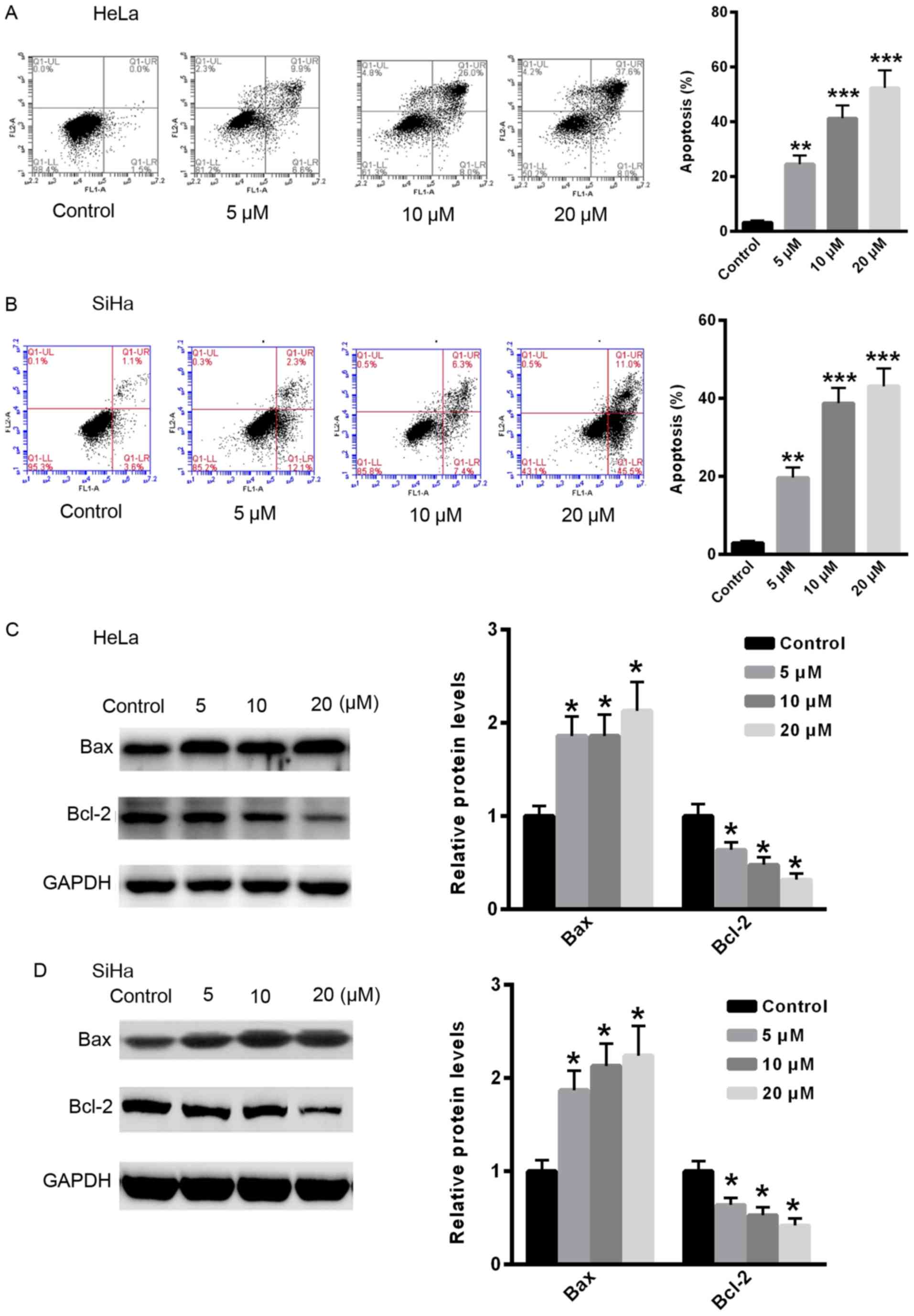
Gefitinib induces apoptosis in CC
cells
To examine the effect of gefitinib on CC cell
apoptosis, apoptosis was examined in HeLa and Siha cells following
treatment with gefitinib. Treatment with gefitinib significantly
induced apoptosis in CC cells in a dose-dependent manner compared
with the control (Fig. 2A and B).
Furthermore, the relative protein expression levels of
apoptosis-related proteins, Bax and Bcl-2, were examined. Following
treatment with gefitinib, the relative protein expression level of
Bcl-2 was significantly reduced, whereas the protein expression
level of Bax was significantly increased in HeLa and Siha cells
compared with the control (Fig. 2C and
D).
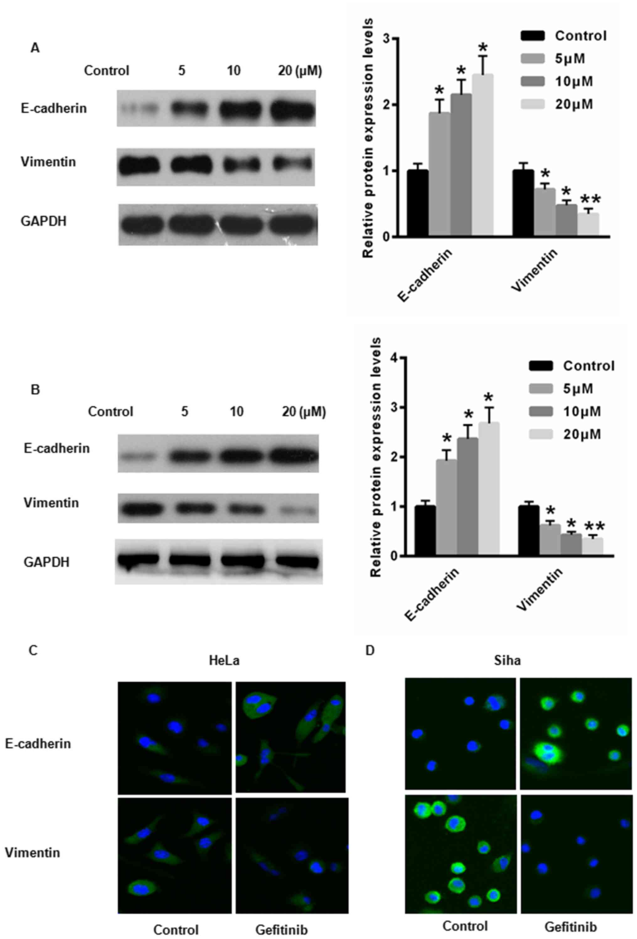
Gefitinib suppresses EMT in CC
cells
EMT is a key regulator of CC progression (16). Therefore, the expression levels of
E-cadherin, an epithelial cell marker, and vimentin, a mesenchymal
cell marker were examined in HeLa and Siha cells following
treatment with gefitinib. Following treatment with gefitinib, the
protein expression level of E-cadherin was significantly increased,
whereas the protein expression level of vimentin was significantly
reduced in HeLa and Siha cells compared with the control (Fig. 3A and B). Immunofluorescence
demonstrated increased E-cadherin expression and reduced vimentin
expression in HeLa and Siha cells following treatment with
gefitinib compared with the control (Fig. 3C and D. These results suggest that
gefitinib may suppress the EMT process in CC cells.
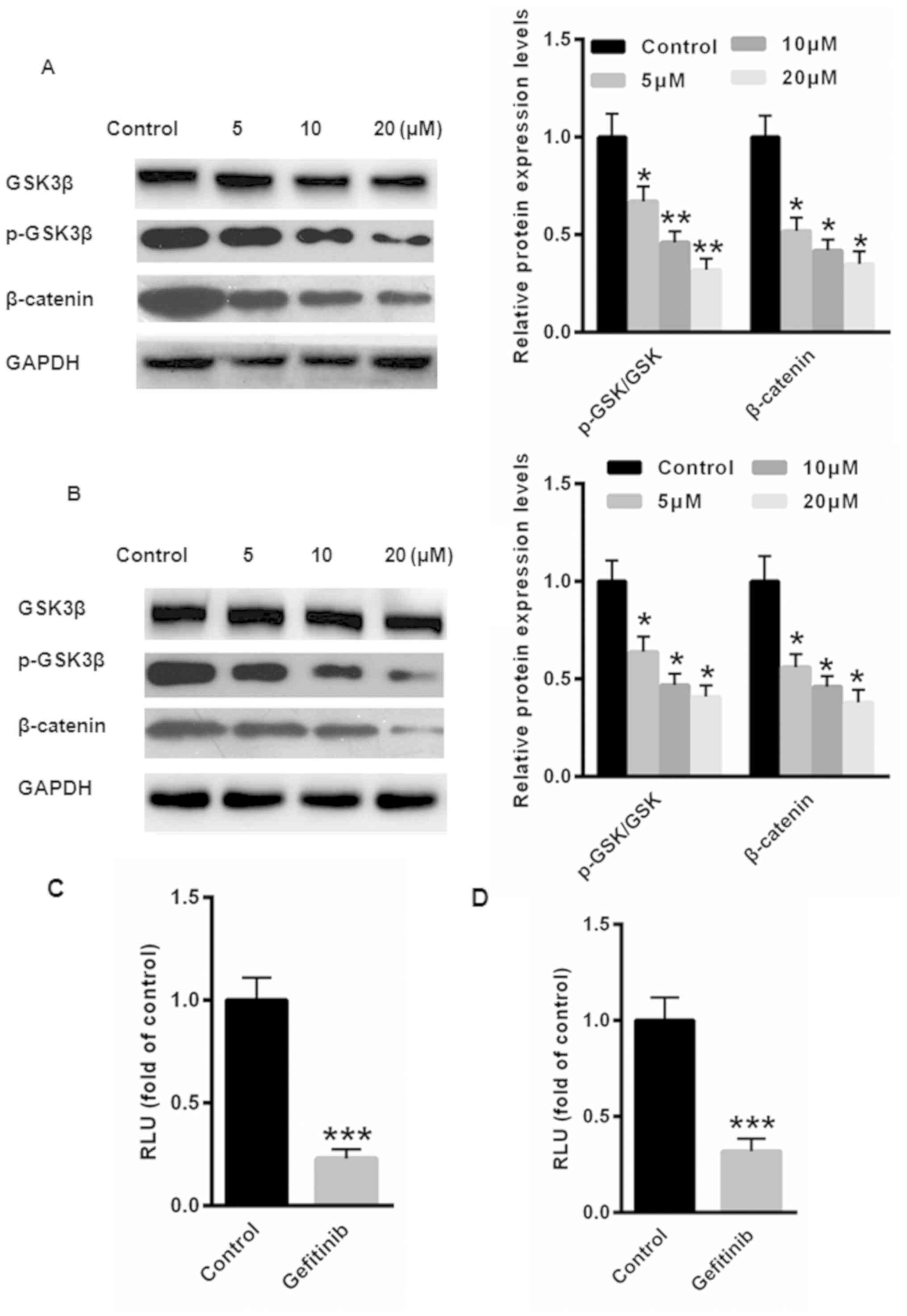
Gefitinib suppresses EMT via the
Wnt/β-catenin signaling pathway in CC cells
The Wnt/β-catenin signaling pathway is a major
contributor to CC tumorigenesis (17). GSK3β is a known negative regulator of
β-catenin. Therefore, the phosphorylation of GSK3β at Ser9, an
indicator of the activation state of GSK3β, was examined by western
blot analysis in HeLa and Siha cells following treatment with
gefitinib. The protein expression levels of p-GSK3β and β-catenin
and were significantly reduced in HeLa and Siha cells following
treatment with gefitinib compared with the control (Fig. 4A and B). Furthermore, luciferase
reporter assays demonstrated that activation of the Wnt/β-catenin
pathway was significantly suppressed following treatment with
gefitinib in HeLa and Siha cells compared with the control
(Fig. 4C and D). Taken together,
these results suggest that gefitinib may inactivate the
Wnt/β-catenin signaling pathway to inhibit EMT in CC cells.
Discussion
In recent years, significant advances have been made
in the diagnosis and treatment of CC, however, the overall 5-year
survival rate remains poor (18).
Currently, the most common treatment strategy for CC includes
surgery with platinum-based chemotherapy (19). Unfortunately, patients with advanced
CC relapse after primary treatment, and the majority of patients
succumb to recurrence and metastasis (20). It is therefore necessary to identify
novel therapeutic strategies for CC.
The in vitro and in vivo antitumor
activity of gefitinib has been reported in several types of human
cancer, including head and neck, colorectal, breast and lung cancer
(21–23). However, the effect of gefitinib in CC
remains unknown. In the current study, two CC cell lines, HeLa and
Siha, were used to investigate the effects of gefitinib. CCK-8
assays demonstrated that gefitinib exerted strong cytotoxicity in
HeLa and Siha cells. Flow cytometry was performed to examine cell
cycle progression and apoptosis in CC following treatment with
gefitinib. The current study demonstrated that treatment with
gefitinib enhanced the number of cells in the
G0/G1 phase and increased apoptosis in HeLa
and Siha cells. Furthermore, treatment with gefitinib reduced the
protein expression level of Bcl-2, and enhanced the protein
expression level of Bax. Taken together, these results suggest that
gefitinib may suppress CC cell proliferation and induce cell cycle
arrest and apoptosis.
To further investigate the underlying mechanism of
gefitinib in regulating CC progression, the EMT process was
examined in CC cells following treatment with gefitinib. In the
progression of CC, EMT is a key regulator that promotes cancer cell
proliferation and invasion (4). The
current study demonstrated that treatment with gefitinib suppressed
the EMT process by increasing the expression level of the
epithelial marker, E-cadherin, and decreasing the expression level
of the mesenchymal marker, vimentin. These results suggest that
gefitinib may suppress the EMT process in CC.
The canonical Wnt/β-catenin signaling pathway serves
an important role in EMT (25,26).
Abnormal activation of Wnt/β-catenin signaling is reported to
increase cancer cell proliferation, survival, differentiation and
EMT (27,28). The current study examined the
potential association between gefitinib and the Wnt/β-catenin
signaling pathway in CC cells. The current study demonstrated that
treatment with gefitinib decreased the protein expression levels of
p-GSK3β and β-catenin, which suggests that gefitinib may be a
potential novel therapeutic strategy in CC by suppressing the
Wnt/β-catenin signaling pathway and EMT to inhibit tumor metastasis
in CC cells.
In conclusion, the current study demonstrated that
gefitinib may suppress EMT during cell invasion and induce
apoptosis and cell cycle arrest by inhibiting the Wnt/β-catenin
signaling pathway.
Acknowledgements
Not applicable.
Funding
The current study was supported by a grant from
Puyang Oil Field General Hospital (grant no. 20160783).
Availability of data and materials
The datasets used and/or analyzed during the current
study are available from the corresponding author upon reasonable
request.
Authors' contributions
JZ performed the experiments and revised the
manuscript for important intellectual content. JY performed the
cell proliferation experiments and was involved in drafting the
manuscript. MY and LT designed the experiments, analyzed the data
and gave final approval of the version to be published. All authors
read and approved the final manuscript.
Ethics approval and consent to
participate
Not applicable.
Patient consent for publication
Not applicable.
Competing interests
The authors declare that they have no competing
interests.
References
|
1
|
Zhang R, Lu H, Lyu YY, Yang XM, Zhu LY,
Yang GD, Jiang PC, Re Y, Song WW, Wang JH, et al:
E6/E7-P53-POU2F1-CTHRC1 axis promotes cervical cancer metastasis
and activates Wnt/PCP pathway. Sci Rep. 7:447442017. View Article : Google Scholar : PubMed/NCBI
|
|
2
|
Bahrami A, Hasanzadeh M, ShahidSales S,
Yousefi Z, Kadkhodayan S, Farazestanian M, Joudi Mashhad M, Gharib
M, Mahdi Hassanian S and Avan A: Clinical significance and
prognosis value of Wnt signaling pathway in cervical cancer. J Cell
Biochem. 118:3028–3033. 2017. View Article : Google Scholar : PubMed/NCBI
|
|
3
|
Chung MT, Lai HC, Sytwu HK, Yan MD, Shih
YL, Chang CC, Yu MH, Liu HS, Chu DW and Lin YW: SFRP1 and SFRP2
suppress the transformation and invasion abilities of cervical
cancer cells through Wnt signal pathway. Gynecol Oncol.
112:646–653. 2009. View Article : Google Scholar : PubMed/NCBI
|
|
4
|
Cui N, Yang WT and Zheng PS: Slug inhibits
the proliferation and tumor formation of human cervical cancer
cells by up- regulating the p21/p27 proteins and down-regulating
the activity of the Wnt/β-catenin signaling pathway via the
trans-suppression Akt1/p-Akt1 expression. Oncotarget.
7:26152–26167. 2016. View Article : Google Scholar : PubMed/NCBI
|
|
5
|
Hua F, Liu S, Zhu L, Ma N, Jiang S and
Yang J: Highly expressed long non-coding RNA NNT-AS1 promotes cell
proliferation and invasion through Wnt/beta-catenin signaling
pathway in cervical cancer. Biomed Pharmacother. 92:1128–1134.
2017. View Article : Google Scholar : PubMed/NCBI
|
|
6
|
Kloth JN, Fleuren GJ, Oosting J, de
Menezes RX, Eilers PH, Kenter GG and Gorter A: Substantial changes
in gene expression of Wnt, MAPK and TNFalpha pathways induced by
TGF-beta1 in cervical cancer cell lines. Carcinogenesis.
26:1493–1502. 2005. View Article : Google Scholar : PubMed/NCBI
|
|
7
|
Kwan HT, Chan DW, Cai PC, Mak CS, Yung MM,
Leung TH, Wong OG, Cheung AN and Ngan HY: AMPK activators suppress
cervical cancer cell growth through inhibition of DVL3 mediated
Wnt/beta-catenin signaling activity. PLoS One. 8:e535972013.
View Article : Google Scholar : PubMed/NCBI
|
|
8
|
Lan K, Zhao Y, Fan Y, Ma B, Yang S, Liu Q,
Linghu H and Wang H: Sulfiredoxin may promote cervical cancer
metastasis via Wnt/β-catenin signaling pathway. Int J Mol Sci.
18:E9172017. View Article : Google Scholar : PubMed/NCBI
|
|
9
|
Lee J, Yoon YS and Chung JH: Epigenetic
silencing of the WNT antagonist DICKKOPF-1 in cervical cancer cell
lines. Gynecol Oncol. 109:270–274. 2008. View Article : Google Scholar : PubMed/NCBI
|
|
10
|
Li F, Wang T and Tang S: SOX14 promotes
proliferation and invasion of cervical cancer cells through
Wnt/β-catenin pathway. Int J Clin Exp Pathol. 8:1698–1704.
2015.PubMed/NCBI
|
|
11
|
Xu CR, Zhong WZ, Zhou Q, Zhang XC, Yang JJ
and Wu YL: Heterogeneity of the resistance to gefitinib treatment
in a non-small cell lung cancer patient with active epidermal
growth factor receptor mutation. Thorac Cancer. 8:51–53. 2017.
View Article : Google Scholar : PubMed/NCBI
|
|
12
|
Yang RF, Yu B, Zhang RQ, Wang XH, Li C,
Wang P, Zhang Y, Han B, Gao XX, Zhang L and Jiang ZM: Bevacizumab
and gefitinib enhanced whole-brain radiation therapy for brain
metastases due to non-small-cell lung cancer. Braz J Med Biol Res.
51:e60732017. View Article : Google Scholar : PubMed/NCBI
|
|
13
|
Yang XB, Chai XS, Wu WY, Long SQ, Deng H,
Pan ZQ, He WF, Zhou YS, Liao GY and Xiao SJ: Gefitinib plus Fuzheng
Kang'ai formula () in patients with advanced non-small cell lung
cancer with epidermal growth factor receptor mutation: A randomized
controlled trial. Chin J Integr Med. 24:734–740. 2018. View Article : Google Scholar : PubMed/NCBI
|
|
14
|
Yang Z, Hackshaw A, Feng Q, Fu X, Zhang Y,
Mao C and Tang J: Comparison of gefitinib, erlotinib and afatinib
in non-small cell lung cancer: A meta-analysis. Int J Cancer.
140:2805–2819. 2017. View Article : Google Scholar : PubMed/NCBI
|
|
15
|
Xu S and Gotlieb AI: Wnt3a/β-catenin
increases proliferation in heart valve interstitial cells.
Cardiovasc Pathol. 22:156–166. 2013. View Article : Google Scholar : PubMed/NCBI
|
|
16
|
Qureshi R, Arora H and Rizvi MA: EMT in
cervical cancer: Its role in tumour progression and response to
therapy. Cancer Lett. 356:321–331. 2015. View Article : Google Scholar : PubMed/NCBI
|
|
17
|
Xu S, Fan Y, Li D, Liu Y and Chen X:
Glycoprotein nonmetastatic melanoma protein B accelerates
tumorigenesis of cervical cancer in vitro by regulating the
Wnt/β-catenin pathway. Braz J Med Biol Res. 52:e75672018.
View Article : Google Scholar : PubMed/NCBI
|
|
18
|
Li S, Yang F, Wang M, Cao W and Yang Z:
miR-378 functions as an onco-miRNA by targeting the
ST7L/Wnt/β-catenin pathway in cervical cancer. Int J Mol Med.
40:1047–1056. 2017. View Article : Google Scholar : PubMed/NCBI
|
|
19
|
Liu P, Ma S, Liu H, Han H and Wang S: HCFU
inhibits cervical cancer cells growth and metastasis by
inactivating Wnt/β-catenin pathway. J Cell Biochem. Dec
12–2017.(Epub ahead of print) doi: 10.1002/jcb.26570.
|
|
20
|
Liu XF, Li XY, Zheng PS and Yang WT: DAX1
promotes cervical cancer cell growth and tumorigenicity through
activation of Wnt/β-catenin pathway via GSK3beta. Cell Death Dis.
9:3392018. View Article : Google Scholar : PubMed/NCBI
|
|
21
|
Hartmann S, Neckel N, Seher A, Mutzbauer
G, Brands RC, Linz C, Kübler AC and Müller-Richter UD: Erlotinib
and gefitinib responsiveness in head and neck cancer cell lines-a
comparing analysis with cetuximab. Clin Oral Investig. 20:759–769.
2016. View Article : Google Scholar : PubMed/NCBI
|
|
22
|
Li Q, Zhang D, Chen X, He L, Li T, Xu X
and Li M: Nuclear PKM2 contributes to gefitinib resistance via
upregulation of STAT3 activation in colorectal cancer. Sci Rep.
5:160822015. View Article : Google Scholar : PubMed/NCBI
|
|
23
|
Geng D, Sun D, Zhang L and Zhang W: The
therapy of gefitinib towards breast cancer partially through
reversing breast cancer biomarker arginine. Afr Health Sci.
15:594–597. 2015. View Article : Google Scholar : PubMed/NCBI
|
|
24
|
Jiang C, Xu R, Li XX, Wang YY, Liang WQ,
Zeng JD, Zhang SS, Xu XY, Yang Y, Zhang MY, et al: p53R2
overexpression in cervical cancer promotes AKT signaling and EMT,
and is correlated with tumor progression, metastasis and poor
prognosis. Cell Cycle. 16:1673–1682. 2017. View Article : Google Scholar : PubMed/NCBI
|
|
25
|
Fernando G, Paul F, Laura J, Alejandra AM,
Gabriela M and Alberto PL: Is the Wnt/β catenin signalling pathway
activated in Seminoma? An immunohistochemical study. J Cancer Res
Ther. 12:1075–1079. 2016. View Article : Google Scholar : PubMed/NCBI
|
|
26
|
Yang C, Du W and Yang D: Inhibition of
green tea polyphenol EGCG((−)-epigallocatechin-3-gallate) on the
proliferation of gastric cancer cells by suppressing canonical
wnt/β-catenin signalling pathway. Int J Food Sci Nutr. 67:818–827.
2016. View Article : Google Scholar : PubMed/NCBI
|
|
27
|
Mittag S, Valenta T, Weiske J, Bloch L,
Klingel S, Gradl D, Wetzel F, Chen Y, Petersen I, Basler K and
Huber O: A novel role for the tumour suppressor Nitrilase1
modulating the Wnt/β-catenin signalling pathway. Cell Discov.
2:150392016. View Article : Google Scholar : PubMed/NCBI
|
|
28
|
van Zuylen WJ, Rawlinson WD and Ford CE:
The Wnt pathway: A key network in cell signalling dysregulated by
viruses. Rev Med Virol. 26:340–355. 2016. View Article : Google Scholar : PubMed/NCBI
|