Introduction
Thyroid carcinoma is the most common malignant tumor
in the human neck, and its incidence is increasing markedly
worldwide (1). Papillary thyroid
cancer (PTC) is the most frequently observed thyroid carcinoma,
accounting for >90% of all thyroid carcinoma cases, and cervical
lymph node metastasis is very common in patients with PTC (2,3).
Cervical lymph node metastasis is an important risk factor for PTC
recurrence rate and survival rate reduction. The early detection of
cervical lymph node metastasis is therefore the key to improving
the cure rate and to reducing the recurrence of PTC (4,5).
Currently, ultrasound-guided fine-needle biopsy (US-guided FNAB) is
the most accurate and cost-effective method for evaluating cervical
lymph node metastasis (6). However,
6–8% of results obtained by US-guided FNAB are false negatives
(7). In addition, US-guided FNAB is
operator-dependent (8). Numerous
recent studies have demonstrated that fine-needle aspirate washout
thyroglobulin (FNA wash-out Tg) can also be used to identify
metastatic lymph nodes (9,10); however, the diagnostic threshold of
FNA wash-out Tg has yet to be established (10). Currently, the method of detecting
specific tumor molecular markers is having an increasing effect on
the diagnosis of malignancies.
Signal transducer and activator of transcription 3
(STAT3) is a cancer gene. STAT3 and its phosphorylated form,
pSTAT3, have pivotal roles in the processes of invasion and
metastasis in malignant tumors (11). Fibroblast growth factor 2 (FGF2) is a
growth factor that can promote angiogenesis (12). FGF2 is important in tumor
lymphangiogenesis and lymph node metastasis. Vascular endothelial
growth factor-C (VEGF-C) is involved in angiogenesis of a variety
of malignant tumors (13). Following
activation, STAT3 induces the expression of VEGF, thus promoting
tumor cell proliferation and angiogenesis.
The aim of the present study was to examine the
expression of STAT3, pSTAT3, FGF2 and VEGF-C in the cancerous and
adjacent normal thyroid tissues of patients with PTC using
immunohistochemistry and western blotting, and to explore the
association among STAT3, pSTAT3, FGF2 and VEGF-C.
Materials and methods
Patients
A total of 42 patients with PTC who were admitted to
the Second Hospital Affiliated to Hebei Medical University
(Shijiazhuang, China) between January 2012 and August 2013 were
enrolled in the present study. The patients included 33 females and
nine males, with an average age of 43.5 years (range, 20–65 years).
The diseases were pathologically confirmed following surgery. The
normal thyroid glands ≥2 cm away from the edge of the tumor in 20
patients, who were randomly selected from the 42 cases, were used
as controls. None of the patients had received anti-tumor therapy
before the surgery. Prior written and informed consent was obtained
from every patient and the study was approved by the Ethics Review
Board of the Second Hospital Affiliated to Hebei Medical
University.
Immunohistochemical staining
Immunohistochemistry was performed using a general
rat/rabbit immunohistochemical kit (Shanghai Gene Co., Ltd.,
Shanghai, China) using a two-step procedure. The specimens were
fixed with 4% paraformaldehyde for 48 h and embedded in paraffin.
The paraffin-embedded tissue was sliced continuously into 3–5-µm
sections, and the sections were deparaffinized and rehydrated.
Antigen retrieval was conductedby incubating with sodium citrate
for 20 min and endogenous peroxidase activity was blocked with 3%
hydrogen peroxide in methanol for 5 min at room temperature.
Non-specific binding was blocked by incubating the slides in 10%
goat serum for 15 min at room temperature. The sections were then
incubated overnight at 4°C with primary antibody. The primary
antibodies and dilution rates were as follows: Mouse anti-human
monoclonal STAT3 (1:200; cat. no. AO1236a; Abgent, San Diego, CA,
USA), rabbit anti-human polyclonal pSTAT3 (1:200; cat. no. AP3261a;
Abgent), rabbit anti-human polyclonal FGF2 (1:50; cat. no.
11234-1-AP; Proteintech, San Diego, CA, USA) and rabbit anti-human
monclonal VEGF-C (1:10; cat. no. AJ1813a Abgent). Following
incubation, the sections were placed at room temperature for 30 min
and incubated with biotinylated anti-mouse (1:100; Abcam,
Cambridge, UK) and anti-rabbit (1:100; Abcam, Cambridge, UK)
secondary antibodies at room temperature for 45 min. The slides
were developed using a 3,3′-diaminobenzidine kit (Shanghai
Gaochuang Chemical Technology Co., Ltd., Shanghai, China),
counterstained with hematoxylin, dehydrated through a graded
alcohol series and permeabilized in xylene. Images of the slides
were captured using a light microscope. The primary antibody was
replaced by phosphate-buffered saline in the blank controls. Image
analysis was carried out using Image-Pro software (Media
Cybernetics, Inc., Rockville, MD, USA). Measurements were performed
in triplicate for three separate sections of the tumor and averaged
to minimize heterogeneity within the tumors. Negative results were
defined as an absence of staining in the cytoplasm and staining of
<10% of the nuclei. A three-point scale, based on intensity, was
utilized in the scoring of cytoplasmic staining, as follows: 0,
negative; 1+, weakly positive; 2+, strongly positive. When staining
was present but not found in the cytoplasm, nuclear staining was
judged as positive when >10% of the nuclei showed positive
staining. For pSTAT3, the presence of any areas with >10% tumor
cells showing definitive nuclear staining resulted in a positive
score.
Western blot analyses
Tissue (100 mg) from each sample was collected and
homogenized with 1 ml lysis buffer. The specimens were then
centrifuged at 22,580 × g for 15 min at 4°C for the preparation of
the total protein. Proteins (100 µg/lane) were separated using
sodium dodecyl sulfate-polyacrylamide gel electrophoresis on
precast 10% polyacrylamide gels and transferred electrophoretically
to polyvinylidene difluoride membranes. The membranes were blocked
in 5% bovine serum albumin for 1 h at room temperature and then
probed overnight at 4°C with the appropriate primary antibodies, as
follows: Mouse anti-human monoclonal STAT3 (1:500;cat. no. AO1236a;
Abgent), rabbit anti-human polyclonal pSTAT3 (1:500; cat. no.
AP3261a; Abgent), rabbit anti-human polyclonal FGF2 (1:1,000; cat.
no. 11234-1-AP; Proteintech), rabbit anti-human monoclonal VEGF-C
(1:100; cat. no. AJ1813a Abgent) or rabbit anti-human polyclonal
β-actin (1:2,000; cat. no. ab8227; Abcam). The membranes were
subsequently incubated with secondary goat anti-rabbit
immunoglobulin G (1:5,000; Proteintech) at room temperature for 2
h. Target proteins were visualized using the enhanced
chemiluminescence detection system (Millipore, Billerica, MA, USA).
Images were obtained using a transmission scanner, with β-actin as
a reference protein. The image analysis was performed using ImageJ
software (US National Institutes of Health, Bethesda, MD, USA).
Statistical analysis
Statistical analyses were performed using SPSS
version 19.0 (IBM-SPSS, Armonk, NY, USA). χ2 tests were
performed to determine the statistical significance of any
association between the variables. Statistical significance was
defined as a P-value of <0.05.
Results
Differential expression of STAT3 and
pSTAT3 in the PTC and adjacent normal tissues
To determine the expression of STAT3 and pSTAT3 in
the PTC tissues of the 42 patients, immunohistochemical staining
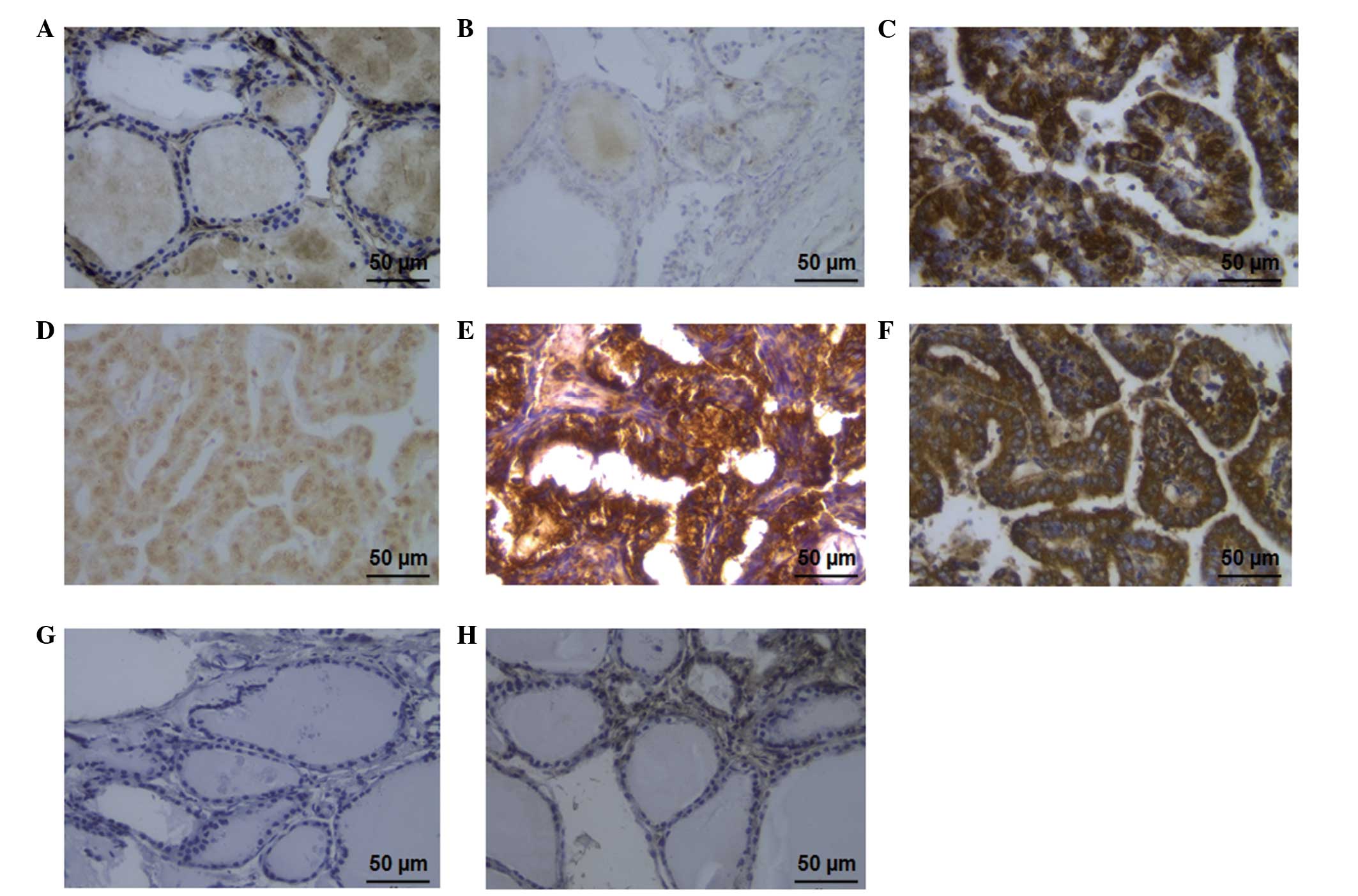
was performed, as shown in Fig. 1.
The adjacent normal tissues of 20 out of the 42 patients were also
examined to serve as the controls. The positive immunocytochemical
signal pattern for the STAT3 protein was indicated by the presence
of brownish yellow granules in the cytoplasm (Fig. 1A and C). pSTAT3 expression, which was
indicated by brownish yellow staining, was mainly located in the
nucleus (Fig. 1B and D). The
expression rates of STAT3 and pSTAT3 protein in the PTC tissues
were 76.2 and 42.9%, respectively; however, in the normal thyroid
tissues the expression rates were 40.0 and 0.0%, respectively
(Table I and Fig. 1). These results suggested that the
expression rates of STAT3 and pSTAT3 protein in the PTC tissues
were significantly different from those in the normal control
tissues.
 | Table I.Expression of STAT3, pSTAT3, FGF2 and
VEGF-C in the PTC and adjacent normal tissues, as determined by
immunohistochemical staining. |
Table I.
Expression of STAT3, pSTAT3, FGF2 and
VEGF-C in the PTC and adjacent normal tissues, as determined by
immunohistochemical staining.
| | Positive (n) |
|---|
|
|---|
| Tissue type | Negative (n) | 1+ | 2+ |
|---|
| Adjacent normal |
|
|
|
|
STAT3 | 12 | 8 | 0 |
|
pSTAT3 | 20 | 0 | 0 |
| FGF2 | 11 | 9 | 0 |
|
VEGF-C | 13 | 7 | 0 |
| PTC |
|
|
|
|
STAT3 | 10 | 12 | 20 |
|
pSTAT3 | 24 | 7 | 11 |
| FGF2 | 8 | 19 | 15 |
|
VEGF-C | 11 | 13 | 18 |
FGF2 is an upstream cytokine that regulates the
STAT3 signal transduction pathway. VEGF-C serves as a downstream
protein regulator of STAT3 and promotes lymphangiogenesis by
activating the VEGF receptor 3 (VEGFR-3). To confirm the above
results, the expression of FGF2 and VEGF-C in the PTC and adjacent
tissues was detected (Fig. 1). The
expression rate of FGF2 in the PTC tissues was 81.0%, compared with
45.0% in the adjacent normal tissues, suggesting a significantly
higher rate in the PTC tissues than that in the normal group
(P<0.05) (Table I and Fig. 1E and G). The positive
immunocytochemical signal pattern for the VEGF-C protein was the
presence of brownish yellow granules in the cytoplasm. The
expression rate in the PTC group was 73.8% (Fig. 1F), compared with 35.0% in the
adjacent normal tissues (Fig. 1H);
therefore, the VEGF-C protein levels in the PTC tissues were
significantly higher than those in the normal tissues (P<0.05).
In summary, the expression levels of STAT3 and pSTAT3 were
significantly increased in the PTC tissues when compared with those
in the adjacent normal tissues.
As shown in Table
II, STAT3, pSTAT3, FGF2 and VEGF-C expression was not
associated with the patient age, gender, tumor size or number of
lesions (P>0.05); however, notable correlation was found between
the expression of these proteins and cancer development, such as
lymph node metastasis (P<0.05), which is consistent with the
results in Fig. 1.
| Table II.Expression levels of STAT3, pSTAT3,
FGF2, and VEGF-C are associated with clinical pathological
parameters. |
Table II.
Expression levels of STAT3, pSTAT3,
FGF2, and VEGF-C are associated with clinical pathological
parameters.
| | STAT3 | pSTAT3 | FGF2 | VEGF-C |
|---|
|
|
|
|
|---|
| —Parameter | n | (-) | (+) | P-value | (-) | (+) | P-value | (-) | (+) | P-value | (-) | (+) | P-value |
|---|
| Age (years) |
|
|
|
|
|
|
|
|
|
|
|
|
|
|
<45 | 22 | 6 | 16 | >0.05 | 15 | 7 | >0.05 | 6 | 16 | >0.05 | 6 | 16 | >0.05 |
|
≥45 | 20 | 4 | 15 |
| 9 | 11 |
| 2 | 18 |
| 5 | 15 |
|
| Gender |
|
|
|
|
|
|
|
|
|
|
|
|
|
|
Male | 9 | 2 | 7 | >0.05 | 7 | 2 | >0.05 | 3 | 6 | >0.05 | 2 | 7 | >0.05 |
|
Female | 33 | 8 | 25 |
| 17 | 16 |
| 5 | 28 |
| 9 | 24 |
|
| Tumor size
(cm) |
|
|
|
|
|
|
|
|
|
|
|
|
|
| ≤1 | 8 | 5 | 3 | >0.05 | 6 | 2 | >0.05 | 5 | 3 | >0.05 | 3 | 5 | >0.05 |
|
>1 | 34 | 5 | 29 |
| 18 | 16 |
| 3 | 31 |
| 8 | 26 |
|
| Lymph node |
|
|
|
|
|
|
|
|
|
|
|
|
|
| Without
metastasis | 22 | 8 | 14 | <0.05 | 16 | 6 | <0.05 | 7 | 15 | <0.05 | 9 | 13 | <0.05 |
|
Metastasis | 20 | 2 | 18 |
| 8 | 12 |
| 1 | 19 |
| 2 | 18 |
|
| Number of
lesions |
|
|
|
|
|
|
|
|
|
|
|
|
|
|
Single | 32 | 7 | 25 | >0.05 | 19 | 13 | >0.05 | 6 | 26 | >0.05 | 8 | 24 | >0.05 |
|
Multiple | 10 | 3 | 7 |
| 5 | 5 |
| 2 | 8 |
| 2 | 8 |
|
Western blot analyses of STAT3 and
pSTAT3 expression in the PTC and adjacent normal tissues
To investigate whether the STAT3 and pSTAT3 proteins
exhibited differential expression in the PTC and adjacent normal
tissues, the total proteins were extracted and subjected to western
blot analysis, with the cellular β-actin protein serving as a
loading control. Since FGF2 is an upstream cytokine regulating the
STAT3 signal transduction pathway and VEGF-C is a downstream
protein for the regulation of STAT3, the expression of these
proteins was also detected using western blot analyses in this
study. Representative blots are shown in Fig. 2A. The mean normalized optical density
(OD) of the protein bands relative to the OD of the β-actin band
from each protein was calculated, and statistical analysis was
performed (Fig. 2B).
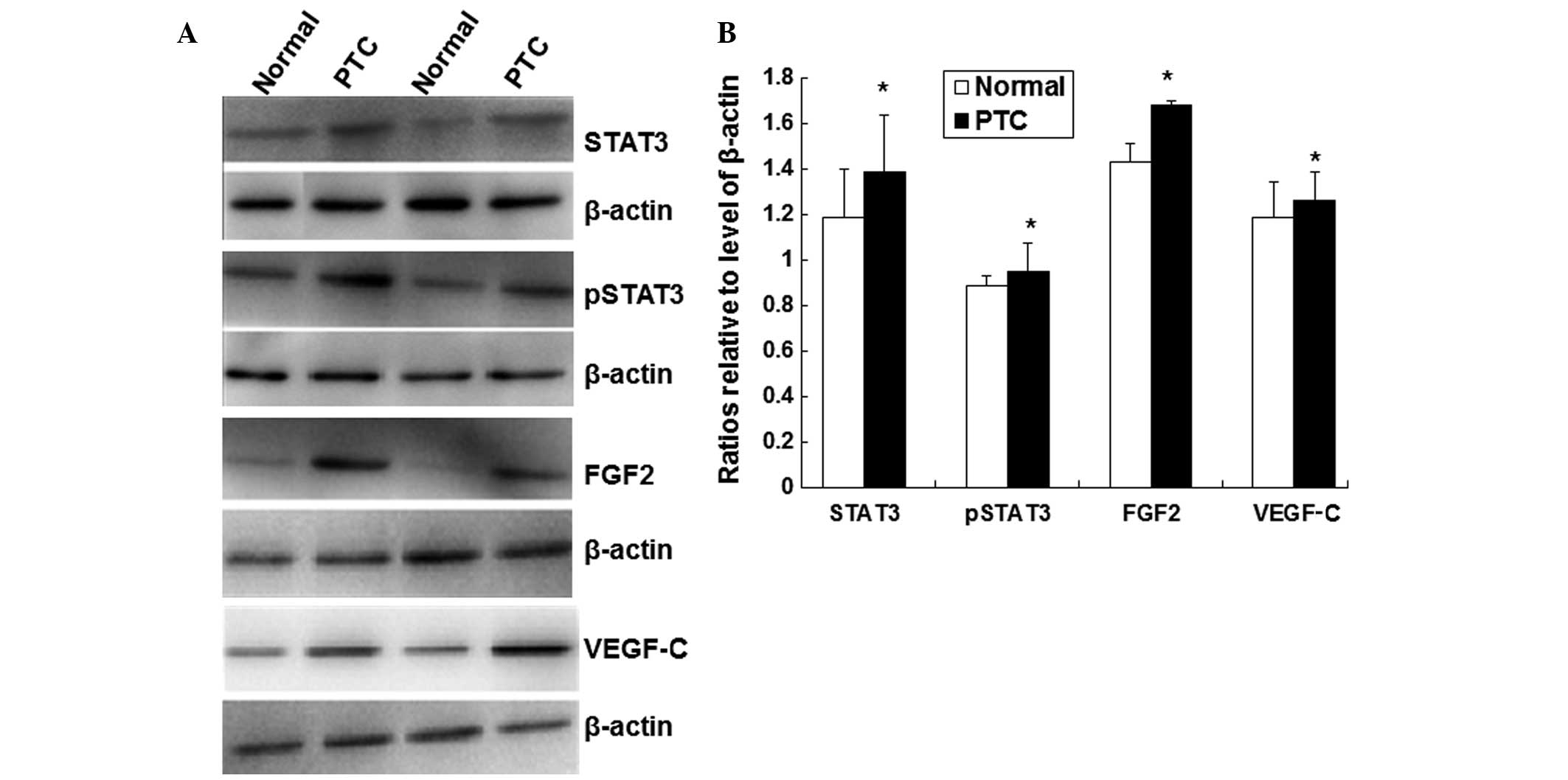 | Figure 2.Expression of STAT3, pSTAT3, FGF2 and
VEGF-C proteins as determined by western blotting. (A) Tissue (100
mg) from each sample was collected and homogenized with 1 ml lysis
buffer. Proteins (100 µg/lane) were separated using sodium dodecyl
sulfate-polyacrylamide gel electrophoresis on precast 10%
polyacrylamide gels and transferred electrophoretically to
polyvinylidene difluoride membranes. The following primary
antibodies were used: Rabbit anti-human STAT3 (1:500), rabbit
anti-human pSTAT3 (1:500), rabbit anti-human FGF2 (1:1,000), rabbit
anti-human VEGF-C (1:100) or rabbit anti-human β-actin. The
membranes were then incubated with secondary goat anti-rabbit
antibody (1:5,000) at room temperature for 2 h. Target proteins
were visualized using the enhanced chemiluminescence detection
system, and images were obtained using a transmission scanner, with
β-actin as a reference protein. (B) Histograms showing the mean
normalized OD of the detected protein bands relative to the OD of
the β-actin band. Error bars show the standard error of the mean.
*P<0.05 when compared with the normal controls. STAT3, signal
transducer and activator of transcription 3; pSTAT3, phosphorylated
STAT3; FGF2, fibroblast growth factor 2; VEGF-C, vascular
endothelial growth factor-C; PTC, papillary thyroid cancer; OD,
optical density. |
As shown in Fig. 2A and
B, the mean levels of STAT3 and pSTAT3 proteins, as well as the
levels of FGF2 and VEGF-C, were significantly increased in the PTC
tissues in comparison with the levels in the adjacent normal
tissues (P<0.05). These results suggest that the increased
levels of STAT3 and pSTAT3 may be associated with the development
of PTC.
Through Pearson correlation analysis it was found
that the expression of STAT3 and pSTAT3 exhibited a linear
correlation in the PTC tissues (r=0.662, P<0.05). Furthermore,
the expression of STAT3 and VEGF-C showed a linear correlation
(r=0.733, P<0.05). These results are consistent with the finding
that increased levels of STAT3 and pSTAT3 may be associated with
the development of PTC.
Discussion
Cervical lymph node metastasis is an important risk
factor that can increase the PTC recurrence rate and reduce the
survival rate (3). According to
statistics, 20–90% of patients with PTC have already developed
cervical lymph node metastasis before they are diagnosed (14). Cervical lymph node metastasis mostly
occurs in the VI region. Between 28 and 33% of cervical lymph node
metastasis cases are not found by preoperative radiological
examination and surgical treatment but are confirmed following
prophylactic VI cleaning (15). At
present, there is no ideal method to determine whether a patient
exhibits lymph node metastasis prior to surgery, and potential
lymph node metastasis or micrometastasis can be hard to find in
preoperative routine examination. An improvement in the
preoperative diagnosis rate for cervical lymph node metastasis is
therefore of vital significance and could reduce the repeated
surgery rate, avoid the patient being subjected to excessive
surgery and improve the survival rate. Equally, improved methods of
preoperative diagnosis would be applicable to patients who are
difficult to diagnose using preoperative fine-needle aspiration
biopsy (FNA). At present, the method of detecting specific tumor
molecular markers is playing an increasingly important role in the
diagnosis of malignant tumors. The ability to determine whether a
patient with thyroid carcinoma exhibits cervical lymph node
metastasis through the detection of the aforementioned molecular
markers is likely to provide an important contribution to the
improvement of the surgical treatment of thyroid cancer.
Janus protein tyrosine kinase (JAK)]-STAT pathways
are important intracellular signal transduction pathways that are
associated with tumor occurrence, development and metastasis
(16,17). The JAK-STAT3 pathway can be activated
by a variety of extracellular signal proteins, including cytokines,
epidermal growth factor and non-receptor tyrosine kinases. JAKs are
activated by the binding of the ligand to the receptor. The JAKs
then phosphorylate tyrosine residues on the receptor with enhanced
kinase activity, and sites are created for interactions with
proteins containing phosphotyrosine-binding domains (18). STAT3 proteins, which have
phosphotyrosine residue-binding domains, are recruited to the
receptors where they are also tyrosine-phosphorylated. The
phosphorylated STAT3 proteins then translocate into the cell
nucleus and bind to target genes (19). This affects a series of associated
basic cell functions, including factor activation, protein
expression, tumor progression and metastasis.
STAT3 expression increases in a variety of malignant
tumor types, such as breast, pancreatic, lung and ovarian cancer
and melanoma, and plays a crucial role in tumorigenesis, tumor
angiogenesis, invasion and metastasis (20–22). The
STAT3 signal transduction pathway may perform a vital role in the
tumor microenvironment of immune tolerance. According to early
studies, STAT3 can inhibit the differentiation and maturation of
dendritic cells, induce T-cell immune tolerance and promote the
secretion of certain immunosuppressive cytokines, such as
transforming growth factor-β, interleukin (IL)-10, IL-6 and VEGF,
thereby promoting tumor growth (23–26).
Chen et al (27) found that
the expression of STAT3 and pSTAT3 in breast cancer tissues was
significantly higher than that in paracancerous tissues, and that
the expression level of STAT3 was closely associated with axillary
lymph node metastasis. It was therefore proposed in the same study
that STAT3 could be used as a predictor of breast cancer
postoperative survival rate. In a study by Bednarek et al
(28), it was found that STAT3 could
significantly inhibit the growth of the bladder cancer cell line
T24 and induce apoptosis through RNA interference silencing. There
are, however, different viewpoints: A previous study (29) showed that the expression of pSTAT3 in
tumor cells was inversely correlated with tumor diameter, vascular
invasion and distant metastasis. Furthermore, pSTAT3 can inhibit
tumor growth, and the pSTAT3 expression level of inpatients with
pulmonary metastasis and bone metastasis of thyroid carcinoma has
been found to be lower than that of those without metastasis
(29).
The results of the present study showed that STAT3
was expressed in the cytoplasm of the PTC and adjacent normal
tissues. The expression of pSTAT3 was solely found in the nuclei of
the PTC tissues. The expression of STAT3 and pSTAT3 in the PTC
tissues was significantly higher than that in the paracancerous
tissues, and the expression levels in the PTC with lymph node
metastasis group were significantly higher than those in the PTC
without lymph node metastasis group. The trends of the two were
accordant. This showed that STAT3 and pSTAT3 play important roles
in promoting the occurrence, development and lymph node metastasis
of PTC. The expression levels were not found to be associated with
age, gender, tumor size or the number of lesions.
PTC tumor cells induce local blood vessel and
lymphatic regeneration, and then promote cervical lymph node
metastasis. FGF2, a type of multipotent cell growth factor, is an
important upstream cytokine regulator of the STAT3 signal
transduction pathway that is widely distributed in a variety of
tissues and organs derived from the mesoderm and neuroectoderm, as
well as in tumor tissue. FGF2 activates target cell FGFRs
predominantly by autocrine or paracrine means, causes a series of
intracellular signal transduction pathways and plays an important
role in promoting tumor angiogenesis, lymphangiogenesis and lymph
node metastasis (30–33).
VEGF-C is the only currently known lymph-specific
angiogenic factor. VEGF-C can combine with VEGFR-3 and induce the
phosphorylation of VEGFR-3, which causes lymphatic endothelial cell
proliferation. VEGF-C, as a downstream protein of regulator STAT3,
plays an important role in tumor angiogenesis. The VEGF-C
expression level has been shown to be positively associated with
blood vessel density and lymph node metastasis in tumor tissues
(13,34,35). In
a study by Karaca et al (36)
it was reported that the expression levels of serum VEGF in
patients with thyroid carcinoma were significantly increased; VEGF
can therefore be used as a predictor of cervical lymph node
metastasis and local recurrence of PTC. The preoperative serum
VEGF-C level can be used as a preoperative diagnostic predictor
irrespective of whether a patient exhibits cervical lymph node
metastasis (37). Furthermore, the
level of VEGF-C is closely associated with tumor size, lymph node
metastasis and distant metastasis.
The results of the present study showed that the
expression of FGF2 and VEGF-C in the PTC group was higher than that
in the adjacent normal tissues. Additionally, the expression levels
in the PTC with lymph node metastasis group were higher than those
in the PTC without lymph node metastasis group. This observation is
consistent with the results of former studies (37,38),
indicating that FGF2 and VEGF-C may promote local tissue
angiogenesis and lymphangiogenesis in PTC, thus promoting PTC
formation, local infiltration and lymph node metastasis; however,
there is no correlation in patients with PTC between FGF2 and
VEGF-C expression and age, gender, tumor size or the number of
lesions.
The results of the present correlation analysis
showed that STAT3 and pSTAT3 expression was higher in PTC tissues
and was positively correlated with FGF2 and VEGF-C expression. This
indicates that STAT3 and pSTAT3 enhance lymphatic and vascular
density and play a significant role in the local invasion and lymph
node metastasis associated with PTC. The detection of STAT3 and
pSTAT3 is simple and applicable to patients who cannot be diagnosed
using preoperative FNA. In conclusion, STAT3 and pSTAT3, two novel
immunohistochemical markers, may represent a valid adjunctive tool
for screening PTC with lymph node metastasis.
Acknowledgements
This study was supported by the Hebei Province
Science and Technology Support Program (grant no. 10246140D) of
China.
References
|
1
|
Kent WD, Hall SF, Isolato PA, Houlden RL,
George RL and Groome PA: Increased incidence of differentiated
thyroid carcinoma and detection of subclinical disease. CMAJ.
177:1357–1361. 2007. View Article : Google Scholar : PubMed/NCBI
|
|
2
|
Alvarado R, Sywak MS, Delbridge L and Sidu
SB: Central lymph node dissection as a secondary procedure for
papillary thyroid cancer: Is there added morbidity? Surgery.
145:514–518. 2009. View Article : Google Scholar : PubMed/NCBI
|
|
3
|
Lee YS, Lim YS, Lee JC, Wang SG, Kim IJ
and Lee BJ: Clinical implication of the number of central lymph
node metastasis in papillary thyroid carcinoma: preliminary report.
World J Surg. 34:2558–2563. 2010. View Article : Google Scholar : PubMed/NCBI
|
|
4
|
Wan H, Zhang B, Liu S, et al: Preliminary
study of patterns of level IIb lymph node metastasis in papillary
thyroid carcinoma. Zhonghua Er Bai Yan Hou Tou Jing Wai Ke Za Zhi.
49:27–30. 2014.[(In Chinese)].
|
|
5
|
Eun YG, Lee YC and Kwon KH: Predictive
factors of contralateral paratracheal lymph node metastasis in
papillary thyroid cancer: prospective multicenter study.
Otolaryngol Head Neck Surg. 150:210–215. 2014. View Article : Google Scholar : PubMed/NCBI
|
|
6
|
Cooper DS, Doherty GM, Haugen BR, et al:
American Thyroid Association Guidelines Taskforce: Management
guidelines for patients with thyroid nodules and differentiated
thyroid cancer. Thyroid. 16:109–142. 2006. View Article : Google Scholar : PubMed/NCBI
|
|
7
|
Frasoldati A, Toschi E, Zini M, et al:
Role of thyroglobulin measurement in fine-needle aspiration
biopsies of cervical lymph nodes in patients with differentiated
thyroid cancer. Thyroid. 9:105–111. 1999. View Article : Google Scholar : PubMed/NCBI
|
|
8
|
Choi JS, Kim J, Kwak JY, Kim MJ, Chang HS
and Kim EK: Preoperative staging of papillary thyroid carcinoma:
comparison of ultrasound imaging and CT. AJR Am J Roentgenol.
193:871–878. 2009. View Article : Google Scholar : PubMed/NCBI
|
|
9
|
Kim MJ, Kim EK, Kim BM, et al:
Thyroglobulin measurement in fine-needle aspirate washouts: the
criteria for neck node dissection for patients with thyroid cancer.
Clin Endocrinol (Oxf). 70:145–151. 2009. View Article : Google Scholar : PubMed/NCBI
|
|
10
|
Pacini F, Fugazzola L, Lippi F, et al:
Detection of thyroglobulin in fine needle aspirates of nonthyroidal
neck masses: a clue to the diagnosis of metastatic differentiated
thyroid cancer. J Clin Endocrinol Metab. 74:1401–1404. 1992.
View Article : Google Scholar : PubMed/NCBI
|
|
11
|
Zhang Z, Bai Y, Li P, Zhao J, Wang Y, Sun
L and Tang J: Relationship between activated STAT3 protein and
epithelial-mesenchymal transition in papillary thyroid carcinonma.
Lin Chung Er Bi Yan Hou Tou Jing Wai Ke Za Zhi. 27:1265–1268.
2013.[(In Chinese)]. PubMed/NCBI
|
|
12
|
Slattery ML, John EM, Stern MC, et al:
Associations with growth factor genes (FGF1, FGF2, PDGFB, FGFR2,
NRG2, EGF, ERBB2) with breast cancer risk and survival: the Breast
Cancer Health Disparities Study. Breast Cancer Res Treat.
140:587–601. 2013. View Article : Google Scholar : PubMed/NCBI
|
|
13
|
Jebreel A, England J, Bedford K, Murphy J,
Karsai L and Atkin S: Vascular endothelial growth factor (VEGF),
VEGF receptors expression and microvascular density in benign and
malignant thyroid diseases. Int J Exp Pathol. 88:271–277. 2007.
View Article : Google Scholar : PubMed/NCBI
|
|
14
|
Dralle H and Machens A: Surgical
approaches in thyroid cancer and lymph-node metastases. Best Pract
Res Clin Endocrinol Metab. 22:971–987. 2008. View Article : Google Scholar : PubMed/NCBI
|
|
15
|
Sakorafas GH, Sampanis D and Safioleas M:
Cervical lymph node dissection in papillary thyroid cancer: current
trends, persisting controversies, and unclarified uncertainties.
Surg Oncol. 19:e57–e70. 2010. View Article : Google Scholar : PubMed/NCBI
|
|
16
|
Bournazou E and Bromberg J: Targeting the
tumor microenvironment: JAK-STAT3 signaling. JAKSTAT.
2:e238282013.PubMed/NCBI
|
|
17
|
Voskas D, Ling LS and Woodgett JR: Signals
controlling un-differentiated states in embryonic stem and cancer
cells: role of the phosphatidylinositol 3′ kinase pathway. J Cell
Physiol. 229:1312–1322. 2014. View Article : Google Scholar : PubMed/NCBI
|
|
18
|
Zouein FA, Duhé RJ and Booz GW: JAKs go
nuclear: emerging role of nuclear JAK1 and JAK2 in gene expression
and cell growth. Growth Factors. 29:245–252. 2011. View Article : Google Scholar : PubMed/NCBI
|
|
19
|
Kamran MZ, Patil P and Gude RP: Role of
STAT3 in cancer metastasis and translational advances. Biomed Res
Int. 2013:4218212013. View Article : Google Scholar : PubMed/NCBI
|
|
20
|
Feng Y, Ke C, Tang Q, et al: Metformin
promotes autophagy and apoptosis in esophageal squamous cell
carcinoma by downregulating Stat3 signaling. Cell Death Dis.
5:e10882014. View Article : Google Scholar : PubMed/NCBI
|
|
21
|
Pandurangan AK and Esa NM: Signal
transducer and activator of transcription 3 - a promising target in
colitis-associated cancer. Asian Pac J Cancer Prev. 15:551–560.
2014. View Article : Google Scholar : PubMed/NCBI
|
|
22
|
Zhang J, Gill A, Atmore B, Johns A,
Delbridge L, Lai R and McMullen T: Upregulation of the signal
transducers and activators of transcription 3 (STAT3) pathway in
lymphatic metastases of papillary thyroid cancer. Int J Clin Exp
Pathol. 4:356–362. 2011.PubMed/NCBI
|
|
23
|
Kawakami Y, Yaguchi T, Sumimoto H, et al:
Improvement of cancer immunotherapy by combing molecular targeted
therapy. Front Oncol. 3:1362013. View Article : Google Scholar : PubMed/NCBI
|
|
24
|
Gu L, Talati P, Vogiatzi P, et al:
Pharmacological suppression of JAK1/2 by JAK1/2 inhibitor AZD1480
potently inhibits IL-6-induced experimental prostate cancer
metastases formation. Mol Cancer Ther. 13:1246–1258. 2014.
View Article : Google Scholar : PubMed/NCBI
|
|
25
|
Farren MR, Carlson LM, Netherby CS, et al:
Tumor-induced STAT3 signaling in myeloid cells impairs dendritic
cell generation by decreasing PKCβII abundance. Sci Signal.
7:ra162014. View Article : Google Scholar : PubMed/NCBI
|
|
26
|
Iwata-Kajihara T, Sumimoto H, Kawamura N,
et al: Enhanced cancer immunotherapy using STAT3-depleted dendritic
cells with high Th1-inducing ability and resistance to cancer
cell-derived inhibitory factors. J Immunol. 187:27–36. 2011.
View Article : Google Scholar : PubMed/NCBI
|
|
27
|
Chen Y, Wang J, Wang X, et al: STAT3, a
poor survival predicator, is associated with lymph node metastasis
from breast cancer. J Breast Cancer. 16:40–49. 2013. View Article : Google Scholar : PubMed/NCBI
|
|
28
|
Bednarek I, Sypniewski D, Gawlik N,
Galilejczyk A and Goraus K: The efficiency of silencing expression
of the gene coding STAT3 transcriptional factor and susceptibility
of bladder cancer cells to apoptosis. Contemp Oncol (Pozn).
16:316–321. 2012.PubMed/NCBI
|
|
29
|
Couto JP, Daly L, Almeida A, et al: STAT3
negatively regulates thyroid tumorigenesis. Proc Natl Acad Sci USA.
109:E2361–E2370. 2012. View Article : Google Scholar : PubMed/NCBI
|
|
30
|
Dong L, Li Y, Cao J, et al: FGF2 regulates
melanocytes viability through the STAT3-transactivated PAX3
transcription. Cell Death Differ. 19:616–622. 2012. View Article : Google Scholar : PubMed/NCBI
|
|
31
|
Coleman SJ, Chioni AM, Ghallab M, et al:
Nuclear translocation of FGFR1 and FGF2 in pancreatic stellate
cells facilitates pancreatic cancer cell invasion. EMBO Mol Med.
6:467–481. 2014. View Article : Google Scholar : PubMed/NCBI
|
|
32
|
Lee HJ, Seo AN, Park SY, et al: Low
prognostic implication of fibroblast growth factor family
activation in triple-negative breast cancer subsets. Ann Surg
Oncol. 21:1561–1568. 2014. View Article : Google Scholar : PubMed/NCBI
|
|
33
|
Meng QH, Xu E, Hildebrandt MA, et al:
Genetic variants in the fibroblast growth factor pathway as
potential markers of ovarian cancer risk, therapeutic response, and
clinical outcome. Clin Chem. 60:222–232. 2014. View Article : Google Scholar : PubMed/NCBI
|
|
34
|
Huang C, Huang R, Chang W, et al: The
expression and clinical significance of pSTAT3, VEGF and VEGF-C in
pancreatic adenocarcinoma. Neoplasma. 59:52–61. 2012. View Article : Google Scholar : PubMed/NCBI
|
|
35
|
Yu XM, Lo CY, Chan WF, Lam KY, Leung P and
Luk JM: Increased expression of vascular endothelial growth factor
C in papillary thyroid carcinoma correlates with cervical lymph
node metastases. Clin Cancer Res. 11:8063–8069. 2005. View Article : Google Scholar : PubMed/NCBI
|
|
36
|
Karaca Z, Tanriverdi F, Unluhizarci K, et
al: VEGFR1 expression is related to lymph node metastasis and serum
VEGF may be a marker of progression in the follow-up of patients
with differentiated thyroid carcinoma. Eur J Endocrinol.
164:277–284. 2011. View Article : Google Scholar : PubMed/NCBI
|
|
37
|
Hai-yun S, Deng-ting C and Shou-yi D:
Preoperative and postoperative serum VEGF-C levels and its clinical
significance. Xi Bao Yu Fen Zi Mian Yi Xue Za Zhi. 26:903–904.
2010.[(In Chinese)]. PubMed/NCBI
|
|
38
|
Emoto N, Onose H, Sugihara H, Minami S,
Shimizu K and Wakabayashi I: Fibroblast growth factor-2 free from
extracellular matrix is increased in papillary thyroid carcinomas
and Graves' thyroids. Thyroid. 8:491–497. 1998. View Article : Google Scholar : PubMed/NCBI
|