Introduction
It is well known that oxidative stress occurs as a
result of excessive reactive oxygen species (ROS), such as singlet
oxygen, superoxide anions, and hydrogen peroxide
(H2O2). These ROS cause irreversible damage
to cellular components, including lipids, proteins, DNA, and other
macromolecules, which has been linked to cell death (1,2).
ROS are also known to contribute to various pathological
conditions, including cancer; inflammatory, neurodegenerative and
cardiovascular diseases; and aging (3–5).
Therefore, the human body has various defense systems against
oxidative stress that are surpassed in extensive damage (6,7).
These mechanisms use antioxidant enzymes or antioxidant compounds.
Among these systems, the nuclear transcription factor erythroid
2-related factor 2 (Nrf2) signaling pathway has recently attracted
interest as a candidate for protection from oxidative damage as it
is involved in cellular antioxidant defenses (8,9).
Nrf2 is an essential transcription factor that regulates a number
of detoxifying and antioxidant defense genes by binding to
antioxidant responsive elements (AREs) (10,11). Under quiescent conditions,
Nrf2-dependent transcription is suppressed by the negative
regulator Kelch-like ECH-associated protein 1 (Keap1), which
facilitates the degradation of Nrf2 through ubiquitinated
proteasomal degradation (12,13). Upon stimulation, Nrf2 escapes
Keap1-mediated repression, translocates into the nucleus, and
subsequently binds to AREs present in the promoter regions of an
array of genes, including heme oxygenase-1 (HO-1) and
NAD(P)H:quinone oxidoreductase 1 (NQO1), involved in cellular
antioxidant defense (14,15). HO-1 can catalyze the degradation
of heme, resulting in the formation of the antioxidant bilirubin
when biliverdin reductase is present (16,17). NQO1, a cytosolic flavoprotein,
facilitates the detoxification and excretion of endogenous and
exogenous chemicals via a reduction reaction that converts quinones
to hydroquinones, limiting the subsequent generation of ROS
(18,19).
Esculetin (6,7-dihydroxycoumarin) is a phenolic
compound and derivative of coumarin (20,21). This compound is found in many
plants, such as Artemisia capillaris, Ceratostigma
willmottianum and Citrus limonia that have been used in
traditional Oriental herbal medicine for many decades (22,23). Esculetin has been reported to have
diverse pharmacological actions, including anti-neurotoxicity
(24,25), anti-angiogenesis (26), anti-inflammatory (27,28), and antitumor activities (29–32). More recently, esculetin has been
shown to have beneficial effects on cellular oxidative stress
(33–35). However, the inhibitory mechanisms
of esculetin vis-à-vis the beneficial effect of esculetin against
oxidative stress have not been fully studied to date. Therefore, in
the present study, we evaluated the protective effects of esculetin
on H2O2-induced oxidative stress and whether
esculetin can activate Nrf2 signaling in mouse-derived C2C12
myoblasts.
Materials and methods
Reagents and antibodies
Esculetin (6,7-dihydroxycoumarin) was purchased from
Sigma Chemical Co. (St. Louis, MO, USA), dissolved in dimethyl
sulfoxide (DMSO, vehicle), and adjusted to final concentrations
using complete culture medium. The final DMSO concentration was
<0.1% in all experiments. Dulbecco's modified Eagle's medium
(DMEM), fetal bovine serum (FBS), and other tissue culture reagents
were obtained from WelGENE Inc. (Daegu, Korea).
H2O2,
3-(4,5-dimethylthiazol-2-yl)-2,5-diphenyltetrazolium bromide (MTT),
N-acetyl-L-cysteine (NAC), 4′,6-diamidino-2- phenylindole
(DAPI), propidium iodide (PI) and zinc protoporphyrin IX (ZnPP)
were also purchased from Sigma Chemical Co. PD98059, an ERK
inhibitor, was purchased from Calbiochem, Inc. (San Diego, CA,
USA). 2′,7′-Dichlorofluorescein diacetate (DCFDA) and an Annexin
V-fluorescein isothiocyanate (FITC) apoptosis detection kit were
purchased from Molecular Probes, Inc. (Eugene, OR, USA) and R&D
Systems Inc. (Minneapolis, MN, USA), respectively. Primary
antibodies (Table I) were
purchased from Santa Cruz Biotechnology, Inc. (Dallas, TX, USA),
Cell Signaling Technology, Inc. (Danvers, MA, USA) and Abcam, Inc.
(Cambridge, MA, USA). An enhanced chemiluminescence (ECL) kit and
horseradish (HRP)-conjugated secondary antibodies were obtained
from Amersham Life Science (Arlington Heights, IL, USA). All other
chemicals not specifically mentioned here were purchased from Sigma
Chemical Co.
 | Table IAntibodies used in the present
study. |
Table I
Antibodies used in the present
study.
| Antibody | Origin | Company | Catalogue no. |
|---|
| Actin | Mouse
monoclonal | Santa Cruz
Biotechnology, Inc. | SC-47778 |
| p-γH2AX | Rabbit
monoclonal | Cell Signaling
Technology, Inc. | 9718 |
| γH2AX | Rabbit
monoclonal | Cell Signaling
Technology, Inc. | 7631 |
| PARP | Rabbit
polyclonal | Santa Cruz
Biotechnology, Inc. | SC-7150 |
| p-Nrf2 | Rabbit
monoclonal | Abcam, Inc. | ab76026 |
| Nrf2 | Rabbit
polyclonal | Santa Cruz
Biotechnology, Inc. | SC-13032 |
| Keap1 | Goat
polyclonal | Santa Cruz
Biotechnology, Inc. | SC-15246 |
| HO-1 | Rabbit
polyclonal | Santa Cruz
Biotechnology, Inc. | SC-10789 |
| NQO1 | Goat
polyclonal | Santa Cruz
Biotechnology, Inc. | SC-16464 |
| p-ERK | Mouse
monoclonal | Cell Signaling
Technology, Inc. | 9106 |
| ERK | Rabbit
polyclonal | Santa Cruz
Biotechnology, Inc. | SC-154 |
| p-JNK | Mouse
monoclonal | Cell Signaling
Technology, Inc. | 9255 |
| JNK | Rabbit
monoclonal | Cell Signaling
Technology, Inc. | 9252 |
| p-p38 MAPK | Rabbit
monoclonal | Cell Signaling
Technology, Inc. | 9211 |
| p38 MAPK | Rabbit
polyclonal | Santa Cruz
Biotechnology, Inc. | SC-535 |
Cell culture and viability assay
C2C12 myoblasts obtained from the American Type
Culture Collection (Manassas, VA, USA) were cultured in DMEM
supplemented with 10% heat-inactivated FBS and 100 µg/ml of
penicillin/streptomycin antibiotics in a humidified atmosphere
containing 5% CO2 and 95% air at 37°C. The cells were
pre-treated with various concentrations (0–5 µM) of
esculetin for 1 h and then incubated with or without 1 mM
H2O2 for 6 h in the absence or presence of 5
mM NAC or 50 µM PD98059. To measure cell viability, the
cells were maintained with MTT at a final concentration of 0.5
mg/ml for 3 h, and the formazan that formed was dissolved in DMSO.
Optical density was measured at 540 nm using an enzyme-linked
immunosorbent assay (ELISA) plate reader (Dynatech MR-7000;
Dynatech Laboratories, Chantilly, VA, USA). The optical density of
the formazan formed in the control (untreated) cells was used to
represent 100% viability (36).
Measurement of ROS generation
To measure ROS levels, the cells were washed twice
with phosphate-buffered saline (PBS) and lysed with 1% Triton X-100
in PBS for 10 min at 37°C. The cells were pre-treated with 5
µM esculetin for 1 h and then incubated with or without 1 mM
H2O2 for 6 h in the absence or presence of 5
mM NAC or 50 µM PD98059. The cells were then stained with 10
µM DCFDA for 20 min at room temperature in the dark. The
green fluorescence of DCF was recorded at 515 nm using a flow
cytometer, and 10,000 events were counted per sample. The results
are expressed as the percentage of increase relative to the
non-treated cells.
Comet assay (single-cell gel
electrophoresis assay)
A comet assay was performed to detect DNA migrating
from single cells in the gel, following a previously described
method (37). Briefly, the cells
were exposed to 1 mM H2O2 for 6 h in the
presence and absence of 5 µM esculetin for 1 h. The cells
were suspended in 1% low melting point agarose and aliquoted onto
glass microscope slides. The slides were placed in single rows and
electrophoresed at 30 V (1 V/cm) and 300 mA for 20 min to draw
negatively charged DNA toward the anode. Finally, the slides were
washed with 0.4 M Tris (pH 7.5) at 4°C and stained with 20
µg/ml PI. The slides were examined under a fluorescence
microscope (Carl Zeiss AG, Oberkochen, Germany), and the resulting
images were analyzed.
Western blot analysis
The cells were pre-treated with 5 µM
esculetin for 1 h and then incubated with or without 1 mM
H2O2 for 6 h, or pre-treated with or without
50 µM PD98059 for 1 h and then treated with 5 µM
esculetin for 4 h. The cells were harvested, washed with PBS, and
lysed on ice for 30 min in lysis buffer (20 mM sucrose, 1 mM
ethylenediaminetetraacetic acid, 20 µM Tris-HCl, pH 7.2, 1
mM dithiothreitol, 10 mM KCl, 1.5 mM MgCl2, and 5
µg/ml aprotinin). Subsequently, an equal amount of protein
for each sample was separated by sodium dodecyl sulfate
(SDS)-polyacrylamide gel electrophoresis and transferred to
polyvinylidene fluoride membranes (Schleicher & Schuell, Inc.,
Keene, NH, USA). The membranes were blocked with 5% skim milk and
then incubated overnight at 4°C with desired primary antibodies.
The membranes were further incubated with corresponding
HRP-conjugated secondary antibodies for 2 h at room temperature.
The proteins of interest were visualized using an ECL detection
system.
Detection of nuclear morphological
changes
The cells were pre-treated with 5 µM
esculetin for 1 h and then incubated with or without 1 mM
H2O2 for 6 h. The detection of chromatin
condensation and nuclear fragmentation in the nuclei of apoptotic
cells was performed using DAPI staining. The cells were harvested,
washed with PBS twice, and fixed with 3.7% paraformaldehyde in PBS
for 10 min at 25°C. The fixed cells were washed with PBS and
stained with 1 mg/ml DAPI solution for 10 min. The cells were then
washed twice with PBS and observed under a fluorescence microscope
(38).
Flow cytometric detection of
apoptosis
The cells were pre-treated for 1 h with the
indicated concentrations of esculetin and then incubated for 6 h
with or without 1 mM H2O2 in the absence or
presence of 50 µM PD98059. The rate of apoptosis was
determined using an Annexin V-FITC apoptosis detection kit. After
treatment with the agents, the cells in each sample were stained
with Annexin V-FITC and PI in accordance with the manufacturer's
instructions. After a 15-min incubation at room temperature in the
dark, the degree of apoptosis was quantified as a percentage of the
Annexin V-positive and PI-negative (Annexin
V+/PI− cells) cells using a flow cytometer
(Becton Dickinson, San Jose, CA, USA) (39).
Statistical analysis
Unless specified otherwise, data are expressed as
the means ± standard deviation (SD) of at least three independent
experiments. A one-way analysis of variance (SPSS version 12.0
software) and a Scheffe's test were used to determine the
significance of differences between groups. P<0.05 was
considered to indicate a statistically significant difference.
Results
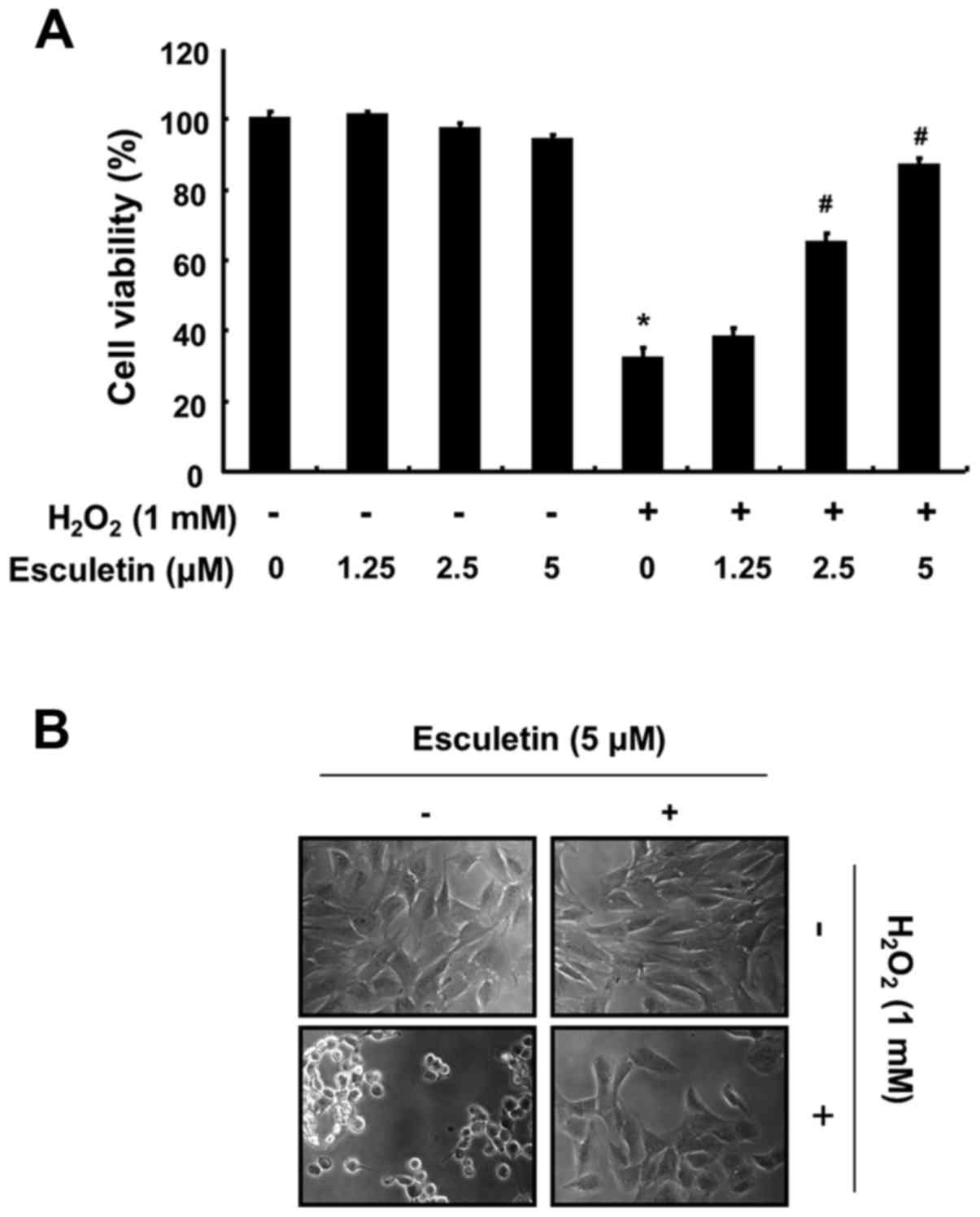
Effects of esculetin on
H2O2-induced cytotoxicity in C2C12 cells
To determine the protective effects of esculetin
against H2O2-induced cytotoxicity, C2C12
cells were pre-treated with various concentrations of esculetin
(1.25–5 µM) for 1 h and then exposed to
H2O2 (1 mM) for a further 6 h. The
concentrations of esculetin that did not have any measurable
adverse effects on the cells were selected (data not shown). As
shown in Fig. 1A, exposure to
H2O2 alone significantly reduced cell
viability (more than 60%) when measured using the MTT assay,
whereas the H2O2-induced reduction in cell
viability was prevented by pre-treatment with esculetin in a
concentration-dependent manner. In addition,
H2O2 stimulation significantly induced
morphological changes, including extensive cytosolic vacuolization
and the presence of irregular cell membrane buds, which were
effectively attenuated by esculetin pre-treatment (Fig. 1B).
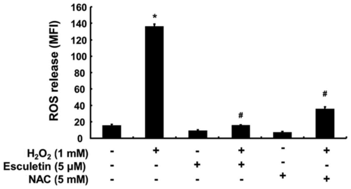
Inhibition of
H2O2-induced ROS generation by esculetin in
C2C12 cells
The intracellular ROS generation was monitored to
investigate whether esculetin can prevent
H2O2-induced ROS generation. The results of
the flow cytometric analysis using DCFDA as a fluorescence probe
demonstrated that the intensity of the DCF-liberated fluorescent
signal from the H2O2-exposed cells was
significantly increased; however, the signal was markedly reduced
in the presence of 5 µM esculetin (Fig. 2). As a positive control, the ROS
scavenger NAC at 5 mM also markedly attenuated
H2O2-induced ROS generation, indicating that
esculetin scavenged H2O2-induced ROS
accumulation.
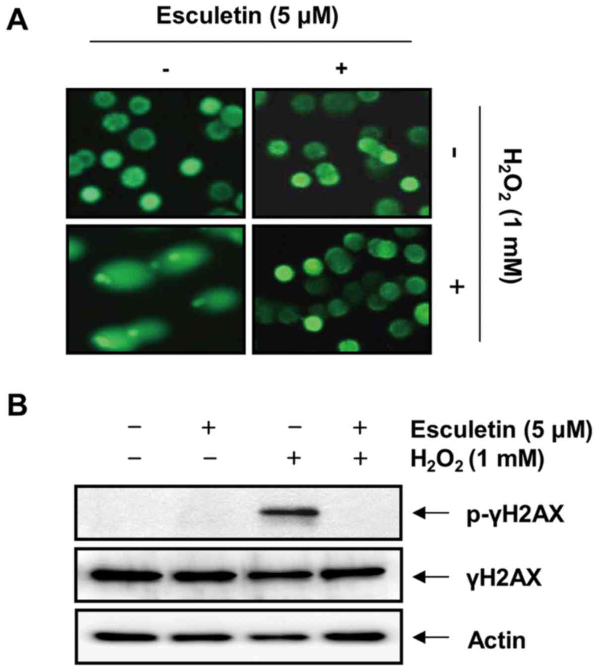
Esculetin protects C2C12 cells from
H2O2-induced DNA damage
We then examined the effects of esculetin on
H2O2-mediated DNA damage in C2C12 cells.
Fig. 3A shows the results of the
comet assay performed to evaluate the protective effect of
esculetin against H2O2-induced DNA damage.
Exposure to H2O2 leads to the loss of
membrane integrity; therefore, the fragmented DNA appeared outside
the cell as comet-like structures; however, this adverse effect was
markedly inhibited by esculetin. In addition, treatment of the
C2C12 cells with H2O2 upregulated the level
of the phosphorylated histone variant H2AX at serine 139 (p-γH2AX),
a sensitive marker of DNA double-strand breaks (40) (Fig.
3B). However, pre-treatment with esculetin significantly
decreased H2O2-induced p-γH2AX
expression.
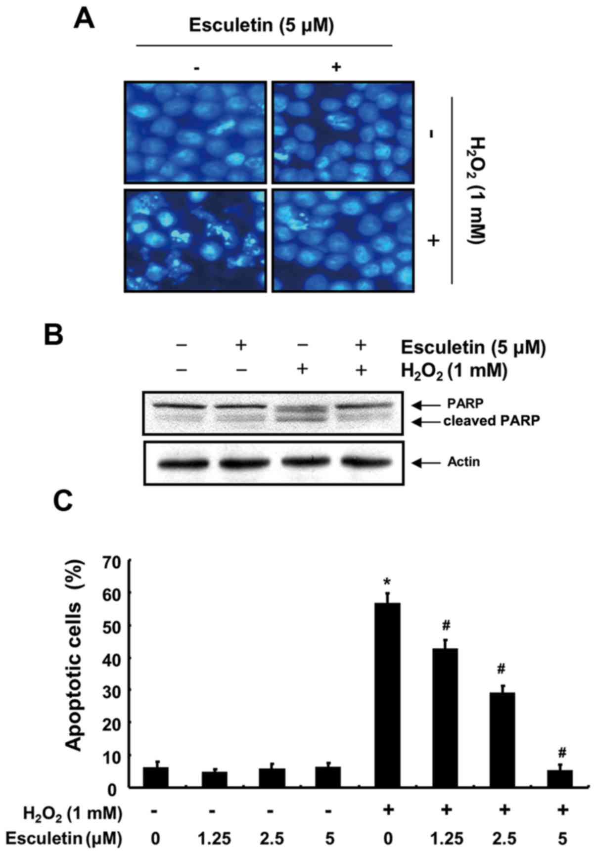
Inhibition of the
H2O2-induced apoptosis of C2C12 cells by
esculetin
To evaluate the potential effect of esculetin on
H2O2-induced C2C12 cell apoptosis, we
examined apoptotic features by measuring chromatin condensation in
the nuclei, poly(ADP ribose) polymerase (PARP) cleavage, and
Annexin V-positive cells. DAPI staining revealed increased nuclei
with chromatin condensation and the formation of apoptotic bodies,
characteristic morphological changes of apoptosis, in cells
cultured with 1 mM H2O2. However, the control
and esculetin (5 µM)-treated groups showed few apoptotic
cells, and pre-treatment of the cells with esculetin significantly
abrogated the H2O2-induced apoptotic
characteristics (Fig. 4A). The
results of western blot analysis also indicated a marked increase
in the level of cleaved PARP, an apoptotic marker protein, in the
H2O2-treated cells compared with the control
group, and treatment with esculetin significantly decreased the
levels of cleaved PARP (Fig. 4B).
Furthermore, the results of flow cytometric analysis revealed an
increase in the percentage of Annexin-positive C2C12 cells exposed
to H2O2 compared with the cells in the
control group. By contrast, treatment of the cells with esculetin
prior to exposure to H2O2 strongly protected
the C2C12 cells against apoptosis in a concentration-dependent
manner (Fig. 4C). These results
clearly indicated that esculetin inhibited
H2O2-induced apoptotic signaling in the
H2O2-exposed C2C12 cells.
Induction of Nrf2 and NQO1 expression by
esculetin in C2C12 cells
To determine whether the protective effects of
esculetin against H2O2-induced oxidative
stress and apoptosis result from the induction of the expression of
antioxidant genes, such as HO-1 and NQO1, and their transcription
factor Nrf2, western blot analysis was performed. As shown in
Fig. 5, the esculetin-treated
cells exhibited a significant increase in the protein levels of
NQO1 compared to these levels in the control group; however, no
changes were observed in the levels of HO-1. Furthermore, esculetin
enhanced the phosphorylated levels of endogenous Nrf2 in a
time-dependent manner without affecting their total steady-state
levels and obtained greatest induction at 5 µM after 4 h. By
contrast, esculetin reduced the levels of Keap1 under the same
conditions.
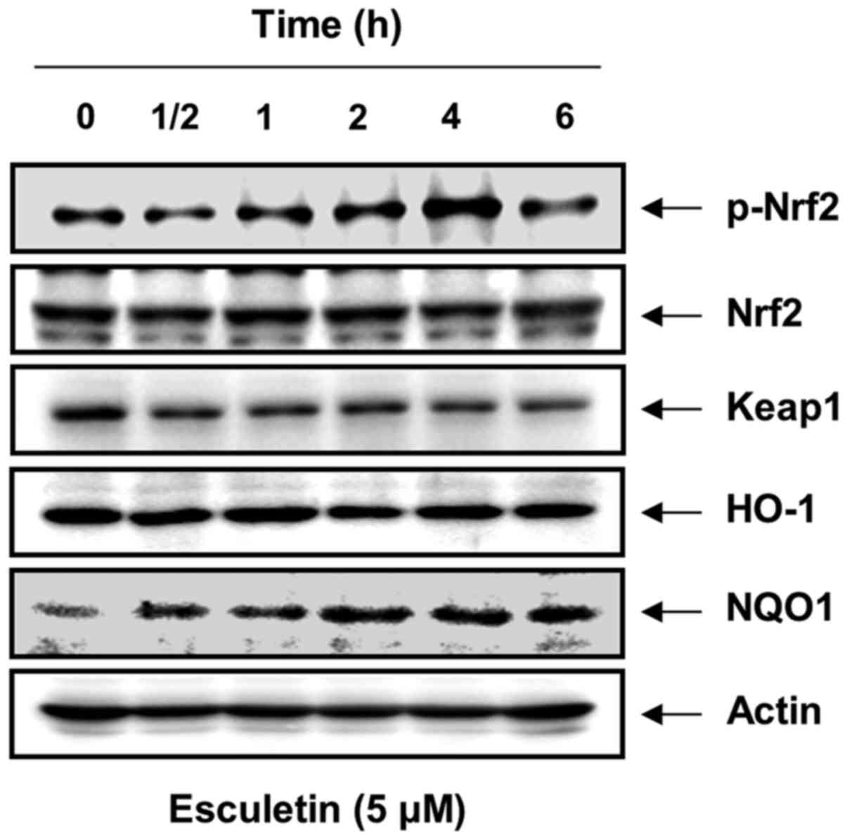 | Figure 5Effects of esculetin on the
expression of Nrf2, Keap1, HO-1, and NQO1 in C2C12 cells. The cells
were incubated with 5 µM escu-letin for the indicated time
periods. Cellular proteins were separated on SDS-polyacrylamide
gels and then transferred onto membranes. The membranes were probed
with specific antibodies against Nrf2, p-Nrf2, Keap1, HO-1, and
NQO1. Proteins were visualized using an ECL detection system. Actin
was used as an internal control. Nrf2, nuclear factor erythroid
2-related factor 2; Keap1, Kelch-like ECH-associated protein 1;
HO-1, heme oxy-genase-1; NQO1, NAD(P)H:quinone oxidoreductase 1;
SDS, sodium dodecyl sulfate; ECL, enhanced chemiluminescence. |
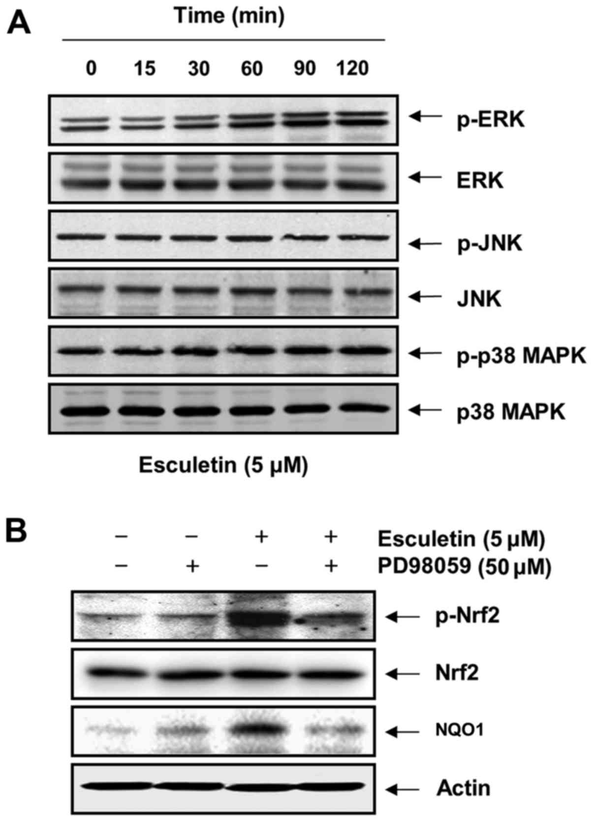
Phosphorylation of Nrf2 by esculetin
through the activation of extracellular signal-regulated kinase
(ERK) in C2C12 cells
To investigate whether Nrf2 phosphorylation by
esculetin in C2C12 cells is affected by the activation of MAPKs as
upstream signaling mediators, we assessed the phosphorylated forms
of ERK, JNK and p38 MAPK. As shown in Fig. 6A, although the total protein
levels of ERK did not show notable changes, esculetin markedly
increased the phosphorylation of ERK within 1 h of treatment, while
the phosphorylation levels of JNK and p38 MAPK remained unaltered.
The dependence of the phosphorylation of Nrf2 challenged with
esculetin upon the activation of ERK was confirmed using a specific
inhibitor of ERK, PD98059. For this experiment, the C2C12 cells
were pre-treated with 50 µM PD98059 for 1 h and then treated
with esculetin for 4 h. We found that treatment with PD98059
effectively reduced the esculetin-induced phosphorylation of Nrf2,
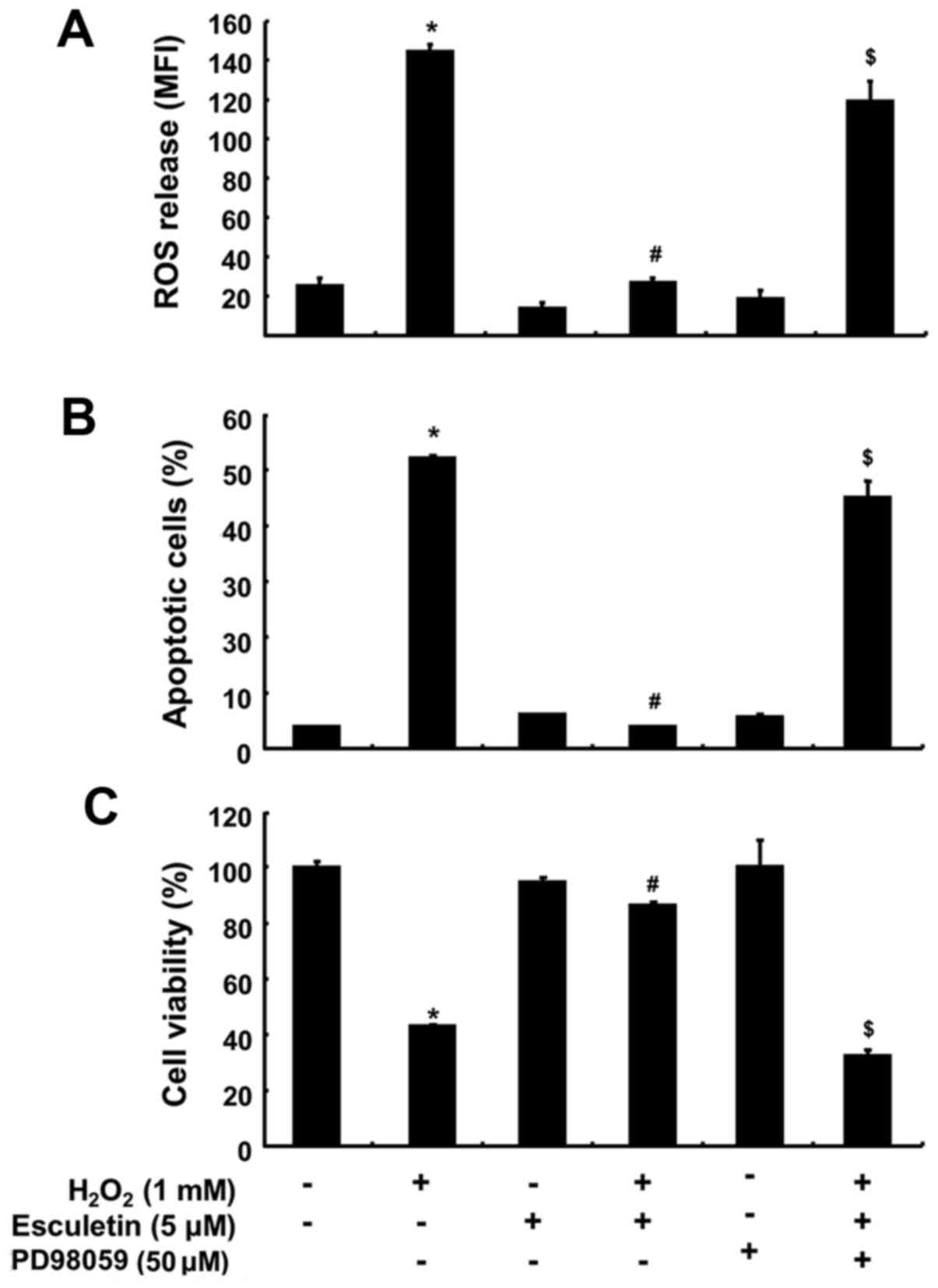
with a resulting decrease in the expression of NQO1 (Fig. 6B). In addition, co-pre-treatment
with PD98059 and esculetin prior to exposure to
H2O2 markedly abrogated the protective
effects of esculetin against H2O2-induced ROS
generation and apoptosis as well as growth reduction (Fig. 7).
Discussion
Many recent studies have reported that natural
compounds have broad protective effects against oxidative stress.
Moreover, the removal of excess ROS or the suppression of their
generation by antioxidants may be effective in preventing oxidative
DNA damage and cell death (6,7).
In this study, esculetin showed intracellular ROS scavenging
activities and provided cytoprotection against oxidative stress in
C2C12 cells (Figs. 1 and 2), suggesting that it may be involved in
the activation of antioxidant enzymes. Our data also demonstrated
that esculetin effectively protected C2C12 cells from
H2O2-mediated DNA damage and apoptosis
(Figs. 3 and 4).
Accumulating evidence indicates that the
transcription factor Nrf2 may serve as a critical regulator of the
cellular antioxidant response to protect against oxidative
stress-induced DNA damage and apoptosis (9,10).
When stimulated by inducers, Nrf2 is released from Keap1, leading
to phosphorylation of Nrf2, which is a critical process in the
nuclear translocation of Nrf2 (41–43). In the nucleus, Nrf2 dimerizes with
other cofactors and binds AREs to induce detoxification enzymes and
antioxidant proteins in response to a number of stimuli, including
oxidative stress (11,15). Therefore, we selected phase-2
anti-oxidative enzymes such as HO-1 and NQO1 and ascertained
whether they would be regulated via the Nrf2 signaling pathway in
H2O2-induced C2C12 cell damage and
esculetin-mediated cytoprotection. The results demonstrated that
esculetin induced the phosphorylation of Nrf2 and the expression of
NQO1 but not HO-1, along with the downregulation of Keap1
expression (Fig. 5), which is
consistent with a previous study (24). These results indicate that
esculetin may stimulate Nrf2 activation by enhancing Nrf2
phosphorylation and reducing Keap1 at the same time, in other words
by increasing the ratio of Nrf2/Keap1.
Several reports have suggested that the MAPK
signaling pathway is a central regulatory pathway for Nrf2
phosphorylation and nuclear translocation associated with inducible
expression of antioxidant enzymes (42,43). To further identify the signaling
pathways affected by esculetin that enhance Nrf2 phosphorylation
and NQO1 expression, we investigated the effects of esculetin on
three MAPK cascades. Immunoblotting data indicated that
phosphorylation of ERK occurred at 30 min after esculetin treatment
and was sustained for up to 120 min, while JNK and ERK were not
affected (Fig. 6A). Moreover,
esculetin induced the phosphorylation of Nrf2 and NQO1 expression
was markedly suppressed by PD98059, a specific inhibitor of ERK
(Fig. 6B). These observations
suggest that ERK appears to play a major role and upregulated Nrf2
phosphorylation in the induction of downstream NQO1 expression in
esculetin-treated C2C12 cells. In parallel with these observations,
we also found that blockage of ERK activation with PD98059 markedly
abrogated the protective effects of esculetin against
H2O2-induced ROS generation, apoptosis, and
inhibition of the growth of the C2C12 cells (Fig. 7). The data provide positive
evidence that the ERK signaling pathway is involved in the
esculetin-mediated activation of Nrf2 and upregulation of NQO1;
therefore, regulation of the Nrf2/NQO1 pathway can reduce
H2O2-induced oxidative damage in C2C12
cells.
Taken together, the present results demonstrated
that esculetin exhibits potent cytoprotective effects against cell
toxicity resulting from exposure to H2O2 via
scavenging ROS. Moreover, the phosphorylation of Nrf2 and
upregulation of NQO1 via ERK signaling are critical for protection
against H2O2-induced oxidative stress.
Although further research and clinical trials are needed to further
elucidate the molecular mechanisms detected herein, the findings of
our study suggest that esculetin has potential therapeutic value as
an antioxidant agent.
Acknowledgments
This research was supported by Basic Science
Research Program through the National Research Foundation of Korea
(NRF) grant funded by the Korea government (nos.
2015R1A2A1A10051603 and 2015R1A2A2A01004633).
References
|
1
|
Lyakhovich A and Graifer D:
Mitochondria-mediated oxidative stress: Old target for new drugs.
Curr Med Chem. 22:3040–3053. 2015. View Article : Google Scholar : PubMed/NCBI
|
|
2
|
Ermakov AV, Konkova MS, Kostyuk SV,
Izevskaya VL, Baranova A and Veiko NN: Oxidized extracellular DNA
as a stress signal in human cells. Oxid Med Cell Longev.
2013:6497472013. View Article : Google Scholar : PubMed/NCBI
|
|
3
|
Dai DF, Chiao YA, Marcinek DJ, Szeto HH
and Rabinovitch PS: Mitochondrial oxidative stress in aging and
healthspan. Longev Healthspan. 3:62014. View Article : Google Scholar : PubMed/NCBI
|
|
4
|
Brieger K, Schiavone S, Miller FJ Jr and
Krause KH: Reactive oxygen species: From health to disease. Swiss
Med Wkly. 142:w136592012.PubMed/NCBI
|
|
5
|
Mena S, Ortega A and Estrela JM: Oxidative
stress in environmental-induced carcinogenesis. Mutat Res.
674:36–44. 2009. View Article : Google Scholar
|
|
6
|
Rajendran P, Nandakumar N, Rengarajan T,
Palaniswami R, Gnanadhas EN, Lakshminarasaiah U, Gopas J and
Nishigaki I: Antioxidants and human diseases. Clin Chim Acta.
436:332–347. 2014. View Article : Google Scholar : PubMed/NCBI
|
|
7
|
Yang HY and Lee TH: Antioxidant enzymes as
redox-based biomarkers: A brief review. BMB Rep. 48:200–208. 2015.
View Article : Google Scholar : PubMed/NCBI
|
|
8
|
Silva-Palacios A, Königsberg M and Zazueta
C: Nrf2 signaling and redox homeostasis in the aging heart: A
potential target to prevent cardiovascular diseases? Ageing Res
Rev. 26:81–95. 2016. View Article : Google Scholar : PubMed/NCBI
|
|
9
|
Huang Y, Li W, Su ZY and Kong AN: The
complexity of the Nrf2 pathway: Beyond the antioxidant response. J
Nutr Biochem. 26:1401–1413. 2015. View Article : Google Scholar : PubMed/NCBI
|
|
10
|
Gan L and Johnson JA: Oxidative damage and
the Nrf2-ARE pathway in neurodegenerative diseases. Biochim Biophys
Acta. 1842:1208–1218. 2014. View Article : Google Scholar : PubMed/NCBI
|
|
11
|
Suzuki T and Yamamoto M: Molecular basis
of the Keap1-Nrf2 system. Free Radic Biol Med. 88:93–100. 2015.
View Article : Google Scholar : PubMed/NCBI
|
|
12
|
O'Connell MA and Hayes JD: The Keap1/Nrf2
pathway in health and disease: From the bench to the clinic.
Biochem Soc Trans. 43:687–689. 2015. View Article : Google Scholar : PubMed/NCBI
|
|
13
|
Jaramillo MC and Zhang DD: The emerging
role of the Nrf2-Keap1 signaling pathway in cancer. Genes Dev.
27:2179–2191. 2013. View Article : Google Scholar : PubMed/NCBI
|
|
14
|
Murakami S and Motohashi H: Roles of Nrf2
in cell proliferation and differentiation. Free Radic Biol Med.
88:168–178. 2015. View Article : Google Scholar : PubMed/NCBI
|
|
15
|
Stefanson AL and Bakovic M: Dietary
regulation of Keap1/Nrf2/ARE pathway: Focus on plant-derived
compounds and trace minerals. Nutrients. 6:3777–3801. 2014.
View Article : Google Scholar : PubMed/NCBI
|
|
16
|
Mancuso C and Barone E: The heme
oxygenase/biliverdin reductase pathway in drug research and
development. Curr Drug Metab. 10:579–594. 2009. View Article : Google Scholar : PubMed/NCBI
|
|
17
|
Wegiel B, Nemeth Z, Correa-Costa M, Bulmer
AC and Otterbein LE: Heme oxygenase-1: A metabolic Nike. Antioxid
Redox Signal. 20:1709–1722. 2014. View Article : Google Scholar :
|
|
18
|
Baulig A, Garlatti M, Bonvallot V,
Marchand A, Barouki R, Marano F and Baeza-Squiban A: Involvement of
reactive oxygen species in the metabolic pathways triggered by
diesel exhaust particles in human airway epithelial cells. Am J
Physiol Lung Cell Mol Physiol. 285:L671–L679. 2003. View Article : Google Scholar : PubMed/NCBI
|
|
19
|
Piao MS, Choi JY, Lee DH, Yun SJ, Lee JB
and Lee SC: Differentiation-dependent expression of NADP(H):quinone
oxidoreductase-1 via NF-E2 related factor-2 activation in human
epidermal keratinocytes. J Dermatol Sci. 62:147–153. 2011.
View Article : Google Scholar : PubMed/NCBI
|
|
20
|
Fylaktakidou KC, Hadjipavlou-Litina DJ,
Litinas KE and Nicolaides DN: Natural and synthetic coumarin
derivatives with anti-inflammatory/antioxidant activities. Curr
Pharm Des. 10:3813–3833. 2004. View Article : Google Scholar
|
|
21
|
Lacy A and O'Kennedy R: Studies on
coumarins and coumarin-related compounds to determine their
therapeutic role in the treatment of cancer. Curr Pharm Des.
10:3797–3811. 2004. View Article : Google Scholar : PubMed/NCBI
|
|
22
|
Chang WS, Lin CC, Chuang SC and Chiang HC:
Superoxide anion scavenging effect of coumarins. Am J Chin Med.
24:11–17. 1996. View Article : Google Scholar : PubMed/NCBI
|
|
23
|
Hu J, Zhu Q, Bai S and Jia Z: New
eudesmane sesquiterpene and other constituents from Artemisia
mongolica. Planta Med. 62:477–478. 1996. View Article : Google Scholar : PubMed/NCBI
|
|
24
|
Subramaniam SR and Ellis EM:
Esculetin-induced protection of human hepatoma HepG2 cells against
hydrogen peroxide is associated with the Nrf2-dependent induction
of the NAD(P)H: quinone oxidoreductase 1 gene. Toxicol Appl
Pharmacol. 250:130–136. 2011. View Article : Google Scholar
|
|
25
|
Lee CR, Shin EJ, Kim HC, Choi YS, Shin T
and Wie MB: Esculetin inhibits N-methyl-D-aspartate neurotoxicity
via glutathione preservation in primary cortical cultures. Lab Anim
Res. 27:259–263. 2011. View Article : Google Scholar : PubMed/NCBI
|
|
26
|
Park SL, Won SY, Song JH, Lee SY, Kim WJ
and Moon SK: Esculetin inhibits VEGF-induced angiogenesis both in
vitro and in vivo. Am J Chin Med. 44:61–76. 2016. View Article : Google Scholar : PubMed/NCBI
|
|
27
|
Zhu L, Nang C, Luo F, Pan H, Zhang K, Liu
J, Zhou R, Gao J, Chang X, He H, et al: Esculetin attenuates
lipopolysaccharide (LPS)-induced neuroinflammatory processes and
depressive-like behavior in mice. Physiol Behav. 163:184–192. 2016.
View Article : Google Scholar : PubMed/NCBI
|
|
28
|
Hong SH, Jeong HK, Han MH, Park C and Choi
YH: Esculetin suppresses lipopolysaccharide-induced inflammatory
mediators and cytokines by inhibiting nuclear factor-κB
translocation in RAW 264.7 macrophages. Mol Med Rep. 10:3241–3246.
2014.PubMed/NCBI
|
|
29
|
Jeon YJ, Cho JH, Lee SY, Choi YH, Park H,
Jung S, Shim JH and Chae JI: Esculetin induces apoptosis through
EGFR/PI3K/Akt signaling pathway and nucleophosmin relocalization. J
Cell Biochem. 117:1210–1221. 2016. View Article : Google Scholar
|
|
30
|
Lee SY, Lim TG, Chen H, Jung SK, Lee HJ,
Lee MH, Kim DJ, Shin A, Lee KW, Bode AM, et al: Esculetin
suppresses proliferation of human colon cancer cells by directly
targeting β-catenin. Cancer Prev Res (Phila). 6:1356–1364. 2013.
View Article : Google Scholar
|
|
31
|
Park C, Jin CY, Kim GY, Choi IW, Kwon TK,
Choi BT, Lee SJ, Lee WH and Choi YH: Induction of apoptosis by
esculetin in human leukemia U937 cells through activation of JNK
and ERK. Toxicol Appl Pharmacol. 227:219–228. 2008. View Article : Google Scholar
|
|
32
|
Park C, Jin CY, Kwon HJ, Hwang HJ, Kim GY,
Choi IW, Kwon TK, Kim BW, Kim WJ and Choi YH: Induction of
apoptosis by esculetin in human leukemia U937 cells: Roles of Bcl-2
and extracellular-regulated kinase signaling. Toxicol In Vitro.
24:486–494. 2010. View Article : Google Scholar
|
|
33
|
Sulakhiya K, Keshavlal GP, Bezbaruah BB,
Dwivedi S, Gurjar SS, Munde N, Jangra A, Lahkar M and Gogoi R:
Lipopolysaccharide induced anxiety- and depressive-like behaviour
in mice are prevented by chronic pre-treatment of esculetin.
Neurosci Lett. 611:106–111. 2016. View Article : Google Scholar
|
|
34
|
Kim JH, Jeong MS, Kim DY, Her S and Wie
MB: Zinc oxide nanoparticles induce lipoxygenase-mediated apoptosis
and necrosis in human neuroblastoma SH-SY5Y cells. Neurochem Int.
90:204–214. 2015. View Article : Google Scholar : PubMed/NCBI
|
|
35
|
Prabakaran D and Ashokkumar N: Protective
effect of esculetin on hyperglycemia-mediated oxidative damage in
the hepatic and renal tissues of experimental diabetic rats.
Biochimie. 95:366–373. 2013. View Article : Google Scholar
|
|
36
|
Hong SH, Sim MJ and Kim YC:
Melanogenesis-promoting effects of Rhynchosia nulubilis and
Rhynchosia volubilis ethanol extracts in melan-a cells. Toxicol
Res. 32:141–147. 2016. View Article : Google Scholar : PubMed/NCBI
|
|
37
|
Gunasekarana V, Raj GV and Chand P: A
comprehensive review on clinical applications of comet assay. J
Clin Diagn Res. 9:GE01–GE05. 2015.PubMed/NCBI
|
|
38
|
Zhao X, Sun P, Qian Y and Suo H: D.
candidum has in vitro anticancer effects in HCT-116 cancer cells
and exerts in vivo anti-metastatic effects in mice. Nutr Res Pract.
8:487–493. 2014. View Article : Google Scholar : PubMed/NCBI
|
|
39
|
Kwon T, Rho JK, Lee JC, Park YH, Shin HJ,
Cho S, Kang YK, Kim Y, Yoon DY and Yu DY: An important role for
peroxiredoxin II in survival of A549 lung cancer cells resistant to
gefitinib. Exp Mol Med. 47:e1652015. View Article : Google Scholar : PubMed/NCBI
|
|
40
|
Rogakou EP, Pilch DR, Orr AH, Ivanova VS
and Bonner WM: DNA double-stranded breaks induce histone H2AX
phosphorylation on serine 139. J Biol Chem. 273:5858–5868. 1998.
View Article : Google Scholar : PubMed/NCBI
|
|
41
|
Venugopal R and Jaiswal AK: Nrf1 and Nrf2
positively and c-Fos and Fra1 negatively regulate the human
antioxidant response element-mediated expression of NAD(P)H:quinone
oxidoreductase1 gene. Proc Natl Acad Sci USA. 93:14960–14965. 1996.
View Article : Google Scholar : PubMed/NCBI
|
|
42
|
Kweon MH, Adhami VM, Lee JS and Mukhtar H:
Constitutive overexpression of Nrf2-dependent heme oxygenase-1 in
A549 cells contributes to resistance to apoptosis induced by
epigallocatechin 3-gallate. J Biol Chem. 281:33761–33772. 2006.
View Article : Google Scholar : PubMed/NCBI
|
|
43
|
Surh YJ, Kundu JK and Na HK: Nrf2 as a
master redox switch in turning on the cellular signaling involved
in the induction of cytoprotective genes by some chemopreventive
phytochemicals. Planta Med. 74:1526–1539. 2008. View Article : Google Scholar : PubMed/NCBI
|