Introduction
Colorectal cancer (CRC) is the fourth leading cause
of cancer-related mortality worldwide, accounting for over 600,000
deaths annually (1). Although
surgical resection is the most powerful treatment for the complete
cure of CRC, a local or distant tumor recurrence sometimes develops
even after a curative resection of the primary tumor is achieved.
Therefore, the identification of reliable criteria and/or novel
biomarkers for predicting a risk of recurrence is needed to
identify the patients who should receive postoperative adjuvant
chemotherapy. Recent clinical trials and basic research have
designed to investigate biomolecules available as prognostic
factors (2). However, none of them
have been incorporated into the clinical practice to assess
patients with CRC.
The trefoil factor (TFF) proteins (TFF1, TFF2 and
TFF3) are characterized by the presence of at least one
40-aminoacid protein domain with three conserved disulfide bonds,
which is termed the trefoil motif (3,4).
TFF1 and TFF2 are usually expressed in the stomach and duodenal
epithelium, respectively. TFF3 is predominantly expressed in the
goblet cells of the small intestine and colon (4). TFFs are rapidly and coordinately
secreted from mucus-secreting cells when the gastrointestinal
mucosa suffers mechanical and/or chemical damage, and they respond
to the mucosal injury (4,5). Recent studies suggest that TFFs are
involved in the development and progression of various types of
cancer, including breast, lung, prostate, stomach and colon cancer
(6–15). In general, TFF1 seems to function
as a stomach-specific tumor suppressor gene (16,17),
whereas TFF2 and TFF3 are thought to augment tumor progression by
increasing tumor invasion and metastasis (16,18).
However, the more precise role of TFFs in human cancer remains
unclear. There are few reports on studies that investigated the
clinical significance of TFFs expression using excised primary CRC
specimens (19). In the present
study, we evaluated the mRNA expression levels of TFF1, TFF2 and
TFF3 in excised CRC specimens and assessed the correlation between
TFF expression and the clinicopathological findings in CRC
patients.
Materials and methods
Patients and tissue samples
A total of 154 consecutive patients with CRC who
underwent surgical resection of the primary tumor between January
2005 and December 2007 at the Saga University Hospital were
enrolled in the present study. Twenty patients with distant
metastasis who underwent a palliative resection of the tumor to
release the disease-related symptoms were also included. All
patients were histologically diagnosed with CRC, and none of them
had received chemotherapy and/or radiotherapy before surgery. The
clinicopathological findings are described according to the
criteria of the TNM Classification System of Malignant Tumors 7th
edition (20). Adjuvant
chemotherapy was added after surgery in 47 patients. Among them, 40
patients received oral tegafur-uracil (UFT) plus leucovorin, 6
patients received S-1, and 1 patient received intravenous
5-fluorouracil (5-FU) with leucovorin for at least 6 months. The
median follow-up period after surgery was 59.3 months (range,
2.0–88.7 months). Informed consent for the use of tissue specimens
was obtained from all patients, and the study protocol was approved
by the Ethics Committee of Saga University, Faculty of
Medicine.
Total RNA isolation and real-time
RT-PCR
At surgery, all samples of normal and cancer tissue
obtained from excised specimens were immediately flash frozen in
liquid nitrogen and stored at −80°C until use. Total RNA was
extracted from each tumor and the corresponding normal tissue
specimens with the RNeasy total RNA reagent set (Qiagen, Venlo, The
Netherlands). For each sample, 1 μg RNA was converted into cDNA
using a ReverTra Ace (Toyobo, Osaka, Japan) reverse transcription
(RT) reaction kit. The cDNA was used as a template for the
polymerase chain reaction (PCR). Real-time quantitative RT-PCR was
performed with the LightCycler™ instrument system (Roche
Diagnostics, Mannheim, Germany) using the LightCycler-FastStart DNA
Master Plus SYBR-Green kit (Roche Diagnostics) according to the
manufacturer’s instructions. The primers were designed as follows:
TFF1 sense, 5′-TTG TGG TTT TCC TGG TGT CA-3′ and antisense, 5′-GGG
ACG TCG ATG GTA TTA GG-3′; TFF2 sense, 5′-AGC AAG AGT CGG ATC AGT
GC-3′ and antisense, 5′-AGA AGC AGC ACT TCC GAG AG-3′; TFF3 sense,
5′-CAA GCA AAC AAT CCA GAG CA-3′ and antisense, 5′-CTC AGG ACT CGC
TTC ATG GT-3′. All experiments were carried out in triplicate, and
the mean values were calculated.
Immunohistochemistry
The paraffin-embedded sections were incubated with
anti-TFF3 antibody [Human TFF3 monoclonal antibody (M01), clone
3D9, 1:500; Abnova Corp., Taipei, Taiwan] overnight at 4°C and with
the corresponding secondary antibody for 30 min at room
temperature. The slides were then washed in phosphate-buffered
saline (PBS), followed by incubation with a DAB
(3,3′-diaminobenzidine) substrate kit (Nichirei Co., Tokyo, Japan).
Tumors with either cytoplasmic or both cytoplasmic and membranous
staining were considered to be positive for TFF3, and the level of
staining was scored according to a comprehensive scoring formula
previously described (21).
Briefly, the intensity of staining was scored as follows: zero, no
staining; 1, weak staining; 2, moderate staining; and 3, strong
staining. The extent of staining was scored as follows: zero, no
positively stained cells; 1, <33% of the tumor cells had
positive staining; 2, 33–67% positively stained tumor cells; and 3,
>67% tumor cells with positive staining. Tumors with a total
score higher than six were considered to have high TFF3
expression.
Statistical analysis
Statistical analyses were performed using the
computer software SPSS 15.0J for windows program (SPSS, Inc.,
Chicago, IL, USA). Comparisons of clinical variables between the
two groups were performed with the Mann-Whitney U-tests.
Comparisons of categorical data between the two groups were
performed with Chi-square tests. A cut-off value for the TFF3
expression status was determined using a receiver-operator
characteristic (ROC) curve. The survival curves were generated
using the Kaplan-Meier method, and statistical differences were
compared using log-rank tests. Both univariate and multivariate
analyses for survival were performed using Cox’s proportional
hazards model. P-values <0.05 were considered statistically
significant.
Results
Patient characteristics
The characteristics of the patients and their TFF
expression levels are summarized in Table I. The 154 patients included 85
(55.2%) males and 69 (44.8%) females, ranging in age from 35 to 89
years (median, 69 years). Among them, 115 patients (74.7%) were
diagnosed with colon cancer, while the remaining 39 (25.3%) were
determined to have rectal cancer. Lymph node metastasis was found
in 73 patients (47.4%), and distant metastasis was observed in 20
patients (13.0%).
 | Table IRelationship between TFF family
expression and clinicopathological findings. |
Table I
Relationship between TFF family
expression and clinicopathological findings.
| TFF1 expression | TFF2 expression | TFF3 expression |
|---|
|
|
|
|
|---|
| Characteristics | Low (n=77) | High (n=77) | P-value | Low (n=77) | High (n=77) | P-value | Low (n=77) | High (n=77) | P-value |
|---|
| Age (mean) | 68.1±10.0 | 68.3±12.0 | 0.924 | 66.8±11.1 | 69.5±10.7 | 0.129 | 67.7±10.3 | 68.7±11.6 | 0.583 |
| Gender |
| Male | 43 | 42 | 0.871 | 43 | 42 | 0.871 | 42 | 43 | 0.871 |
| Female | 34 | 35 | | 34 | 35 | | 35 | 34 | |
| Location |
| Colon | 56 | 59 | 0.578 | 55 | 60 | 0.354 | 51 | 64 | 0.016 |
| Rectum | 21 | 18 | | 22 | 17 | | 26 | 13 | |
| Histology |
| Well | 30 | 31 | 0.869 | 34 | 27 | 0.249 | 32 | 29 | 0.621 |
| Others | 47 | 46 | | 43 | 50 | | 45 | 48 | |
| T |
| 1/2 | 18 | 18 | 1.000 | 18 | 18 | 1.000 | 18 | 18 | 1.000 |
| 3/4 | 59 | 59 | | 59 | 59 | | 59 | 59 | |
| N |
| Negative | 42 | 39 | 0.628 | 43 | 38 | 0.420 | 42 | 39 | 0.628 |
| Positive | 35 | 38 | | 34 | 39 | | 35 | 38 | |
| ly |
| Negative | 28 | 34 | 0.324 | 32 | 30 | 0.742 | 32 | 30 | 0.742 |
| Positive | 49 | 43 | | 45 | 47 | | 45 | 47 | |
| v |
| Negative | 52 | 52 | 1.000 | 53 | 51 | 0.731 | 51 | 53 | 0.731 |
| Positive | 25 | 25 | | 24 | 26 | | 26 | 24 | |
| Stage |
| 0/I/II | 41 | 36 | 0.420 | 42 | 35 | 0.259 | 42 | 35 | 0.259 |
| III/IV | 36 | 41 | | 35 | 42 | | 35 | 42 | |
| Distant
metastasis |
| Negative | 71 | 63 | 0.055 | 68 | 66 | 0.632 | 72 | 62 | 0.017 |
| Positive | 6 | 14 | | 9 | 11 | | 5 | 15 | |
Relationship between TFF expression and
the clinicopathological findings in CRC patients
The expression status of each patient’s TFF was
classified into a high- or low-expression group. For the purpose of
the statistical analysis, the median of this series was used as a
cut-off value to distinguish tumors with high or low TFF
expression. As shown in Table I,
neither TFF1 nor TFF2 expression was associated with the tumor
histology, depth of cancer invasion, lymph node metastasis,
lymphatic invasion or vascular invasion. Conversely, expression
status of TFF3 was significantly associated with the location of
the disease and the presence of distant metastasis (P=0.016 and
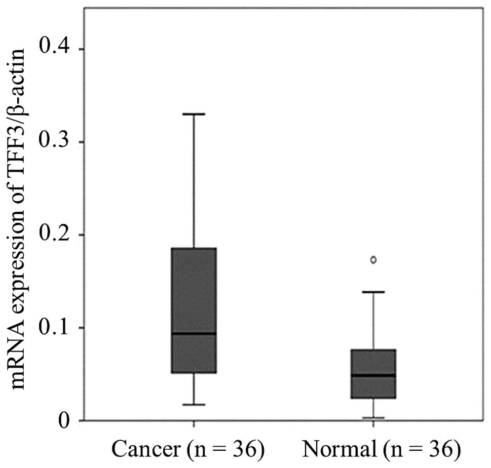
0.017, respectively). In addition, the expression level of TFF3 in
cancer tissue was significantly higher than that of the
corresponding normal tissue (P=0.001) (Fig. 1).
Immunohistochemical staining for
TFF3
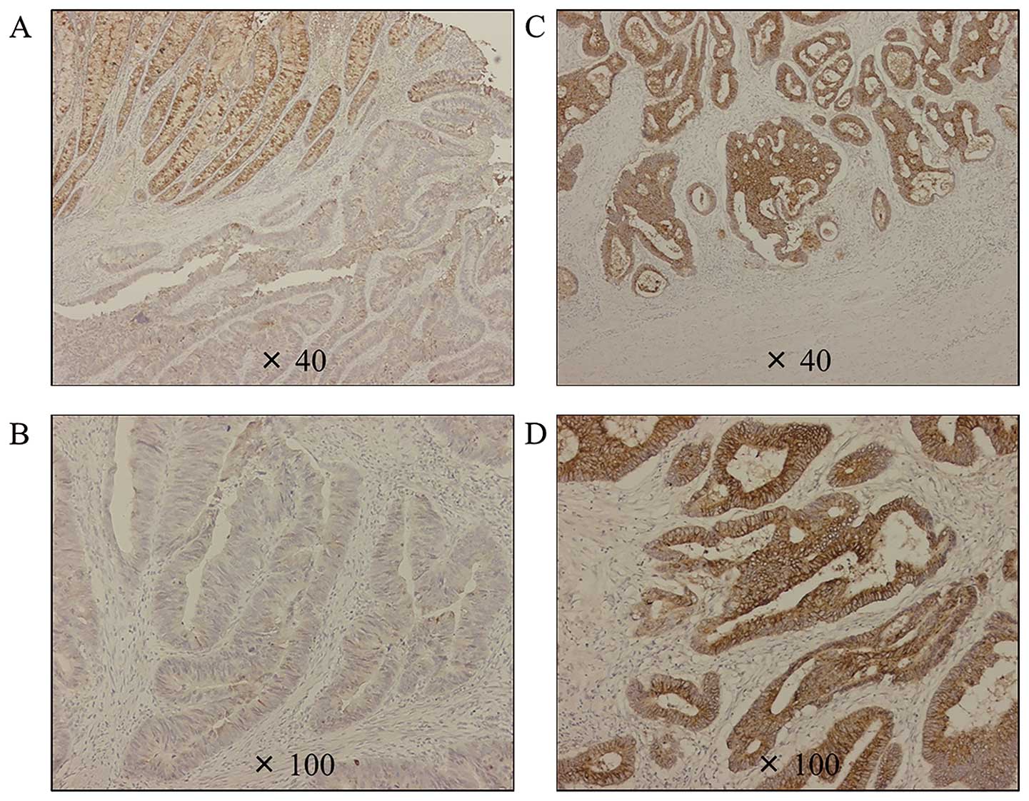
The immunohistochemical staining for TFF3 was
predominantly distributed in the cytoplasm of cancer cells.
Fig. 2A and B indicates low TFF3
expression, while Fig. 2C and D
depicts high TFF3 expression. The immunohistochemical staining
status of TFF3 was significantly correlated with the corresponding
mRNA expression level (data not shown).
Definition of the cut-off value for TFF3
expression status
The TFF3 expression status was re-classified into
positive- and negative-expression groups using the appropriate
cut-off value that was determined by a ROC curve generated
according to the presence of distant metastasis. The cut-off value
was determined to be 0.134 with 60.0% sensitivity and 73.1%
specificity [area under the curve (AUC) 0.662, 95% CI 0.531–0.793,
P=0.020]. Forty-eight patients were classified as belonging to the
positive expression group, while the remaining 106 patients were
included in the negative expression group.
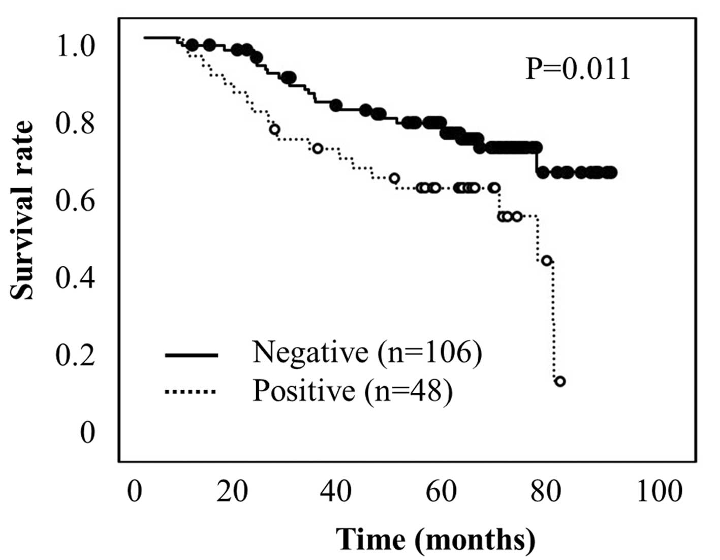
Kaplan-Meier survival analysis according
to TFF3 expression
The Kaplan-Meier survival analysis with log-rank
tests revealed a strong correlation between TFF3 expression and
disease-specific survival (Fig.
3). The patients with positive TFF3 expression (n=48) had a
significantly shorter survival than those who were negative for
TFF3 (P=0.011).
Univariate and multivariate analyses for
disease-specific survival in 154 CRC patients
As summarized in Table
II, tumor histology [hazard ratio (HR)=2.827, P=0.004), cancer
invasion depth (HR=5.464, P=0.005), regional lymph node metastasis
(HR=4.167, P<0.001), lymphatic invasion (HR=4.255, P<0.001),
vascular invasion (HR=2.825, P<0.001), and the positive
expression of TFF3 (HR=2.103, P=0.013) were significantly
associated with poor survival according to the univariate analysis.
In addition, the multivariate analysis of the variables that were
significant according to the univariate analysis was carried out
using Cox’s proportional hazards model (Table II). The results demonstrated that
positive lymph node metastasis (HR=2.681, P=0.011), vascular
invasion (HR=1.905, P=0.035), and the positive expression of TFF3
(HR=2.226, P=0.009) were independently associated with poor
survival in CRC patients.
| Table IIStatistical analysis of
disease-specific survival of colorectal patients (n=154). |
Table II
Statistical analysis of
disease-specific survival of colorectal patients (n=154).
| Univariate
analysis | Multivariate
analysis |
|---|
|
|
|
|---|
|
Characteristics | HR | 95% CI | P-value | HR | 95% CI | P-value |
|---|
| Age (≤69 vs. >69
years) | 0.568 | 0.076–4.237 | 0.581 | | | |
| Gender (male vs.
female) | 0.767 | 0.424–1.388 | 0.381 | | | |
| Location (colon vs.
rectum) | 1.419 | 0.701–2.872 | 0.331 | | | |
| Histology (well vs.
others) | 2.827 | 1.401–5.705 | 0.004 | 1.779 | 0.865–3.655 | 0.117 |
| T (1/2 vs.
3/4) | 5.464 | 1.689–17.544 | 0.005 | 0.417 | 0.112–1.209 | 0.160 |
| N (negative vs.
positive) | 4.167 | 2.119–8.197 |
<0.001 | 2.681 | 1.259–5.714 | 0.011 |
| ly (negative vs.
positive) | 4.255 | 1.894–9.524 |
<0.001 | 0.625 | 0.250–1.497 | 0.309 |
| v (negative vs.
positive) | 2.825 | 1.580–5.051 |
<0.001 | 1.905 | 1.046–3.460 | 0.035 |
| TFF3 (negative vs.
positive) | 2.103 | 1.173–3.771 | 0.013 | 2.226 | 1.221–4.057 | 0.009 |
The prognostic impact of TFF3 for early
recurrence after curative surgery
Of the total 154 patients 134 did not have any
findings of metastasis at surgery. However, 36 of the 134 patients
exhibited positive TFF3 expression, and 7 of the 36 patients
(19.4%) developed recurrent disease within one year. On the
contrary, local or distant tumor recurrence was observed in only 7
of 98 (7.1%) patients with negative TFF3 expression. The frequency
of early recurrence within one year after curative surgery was
significantly different between TFF3-positive and -negative
patients (P=0.039).
Discussion
This is the first report demonstrating that positive
TFF3 expression could be a prognostic marker for recurrence in CRC
patients who underwent curative resection by using the excised
specimens. The TFF proteins are widely expressed in the
gastrointestinal epithelium, and they are well known for their
protective and healing effects after mucosal damage in the
gastrointestinal tract (4,5). TFFs protect cells from apoptotic
death and stimulate cell migration to achieve their role in
epithelial tissue (22,23). Although most studies have focused
on their effects following mucosal injury, recent evidence has
indicated that TFFs contribute to the oncogenic transformation,
growth and metastasis of human solid tumors (21,24–26).
However, to the best of our knowledge, the role of TFF expression
in CRC has never been investigated in excised specimens. The
present study evaluated the expression status of TFFs in a large
cohort of samples from patients with CRC. Our results demonstrate
the clinical significance of TFFs and highlight their potential as
therapeutic targets in CRC patients.
TFF1 was initially described as an
estrogen-inducible gene in the hormone-sensitive breast cancer cell
line MCF-7 (27). However, a later
observation that TFF1 knockout mice develop gastric adenomas and
adenocarcinomas indicated the role of TFF1 as a tumor suppressor in
the stomach (16). Our present
study also demonstrated that loss of TFF1 expression in gastric
cancer contributed to tumor invasion and was associated with poor
survival (17). TFF1 is absent in
normal colon tissue but is induced at high levels in CRCs (28). Rodrigues et al (29) demonstrated that forced TFF1
expression promotes adenoma-carcinoma transition in human colon
epithelial cells by using premalignant adenoma cells. In this
series, TFF1 expression status was not associated with
clinicopathological findings such as tumor histology, cancer
invasion depth and lymph node metastasis (Table I). One previous study reported that
TFF1 does not have any clinicopathological significance in CRCs
(30). Taken together, the
existing evidence seems to indicate that TFF1 is involved in the
development of CRC rather than its progression.
The role of TFF2 in cancer biology remains unclear.
Kosriwong et al (31)
showed that positive TFF2 immunostaining in cholangiocarcinoma was
markedly increased compared with normal and precancerous tissues,
thus, suggesting that it plays an important role in tumor
progression. In contrast, another study demonstrated that
Helicobacter pylori infection promotes hypermethylation of
the TFF2 promoter and silencing of TFF2 expression during gastric
carcinogenesis, indicating that it acts as a tumor-suppressor gene
(32). Therefore, the role of TFF2
in human malignancies has been controversial so far and
relationship between TFF2 expression and the clinical parameters of
CRCs unknown. Similarly, in our data, clinical values of TFF2 in
patients with CRC are still obscure.
TFF3 is upregulated in most human malignancies,
including CRC. Yio et al (12) demonstrated that TFF3 contributes to
the aggressive behavior of colon cancer cells in a rat model. Rat
colon cancer cells that natively express TFF3 showed enhanced
migration and invasion and less apoptosis compared with a sister
cell line that does not express TFF3. In cooperation with vascular
endothelial growth factor (VEGF), TFF3 activates the signal
transducer and activator of transcription (STAT) 3 signaling
pathway, which promotes cellular invasion and tumor growth in human
colon cancer (33). In the present
study, high TFF3 expression was significantly associated with the
presence of distant metastasis, including liver metastasis, lung
metastasis, para-aortic lymph node metastasis and peritoneal
dissemination (Table I), thus,
indicating that TFF3 promotes the aggressive behavior of CRC. In
addition, TFF3 expression was significantly higher in cancer
tissues compared to the corresponding normal mucosa both at the
mRNA level (P=0.001; Fig. 1) and
protein level (Fig. 2). The
survival analysis demonstrated that patients with positive TFF3
expression showed significantly worse survival than those who were
negative for TFF3 (P=0.011; Fig.
3). Furthermore, the prognostic value of TFF3 in CRC patients
was identified as an independent prognostic factor in a
multivariate analysis (P=0.009; Table
II). Moreover, one recent study demonstrated a relationship
between TFF3 expression and lymph node metastasis and concluded
that TFF3 expression might play a role in promoting lymph node
metastases of CRCs (19). Another
group found that TFF3 was frequently expressed in both primary
colon cancer and liver metastases but not in normal liver tissue
(13). Taken together, the results
suggest that TFF3 is associated with patient survival and the
metastatic potential of CRC. Thus, TFF3 might be a possible
prognostic marker for disease recurrence after curative surgery. We
performed an additional statistical analysis that included the 134
patients without metastatic disease at the time of surgery. Among
the 134 CRC patients who received curative surgery, patients with
positive TFF3 expression exhibited a significantly higher
recurrence frequency within one year in compared with those with
negative TFF3. Although further studies of additional cohorts are
needed, our results clearly demonstrate the prognostic potential of
TFF3 for early CRC recurrence in a clinical setting.
In conclusion, positive TFF3 expression is an
independent poor-prognostic factor in CRC patients, and may be a
useful marker for early recurrence.
References
|
1
|
Ferlay J, Parkin DM and Steliarova-Foucher
E: Estimates of cancer incidence and mortality in Europe in 2008.
Eur J Cancer. 46:765–781. 2010. View Article : Google Scholar : PubMed/NCBI
|
|
2
|
Bacolod MD and Barany F: Molecular
profiling of colon tumors: the search for clinically relevant
biomarkers of progression, prognosis, therapeutics, and
predisposition. Ann Surg Oncol. 18:3694–3700. 2011. View Article : Google Scholar : PubMed/NCBI
|
|
3
|
Wong WM, Poulsom R and Wright NA: Trefoil
peptides. Gut. 44:890–895. 1999. View Article : Google Scholar : PubMed/NCBI
|
|
4
|
Hoffmann W and Jagla W: Cell type specific
expression of secretory TFF peptides: colocalization with mucins
and synthesis in the brain. Int Rev Cytol. 213:147–181. 2002.
View Article : Google Scholar : PubMed/NCBI
|
|
5
|
Taupin D and Podolsky DK: Trefoil factors:
initiators of mucosal healing. Nat Rev Mol Cell Biol. 4:721–732.
2003. View
Article : Google Scholar : PubMed/NCBI
|
|
6
|
Poulsom R, Hanby AM, Lalani EN, Hauser F,
Hoffmann W and Stamp GW: Intestinal trefoil factor (TFF 3) and pS2
(TFF 1), but not spasmolytic polypeptide (TFF 2) mRNAs are
co-expressed in normal, hyperplastic, and neoplastic human breast
epithelium. J Pathol. 183:30–38. 1997. View Article : Google Scholar : PubMed/NCBI
|
|
7
|
Buache E, Etique N, Alpy F, et al:
Deficiency in trefoil factor 1 (TFF1) increases tumorigenicity of
human breast cancer cells and mammary tumor development in
TFF1-knockout mice. Oncogene. 30:3261–3273. 2011. View Article : Google Scholar : PubMed/NCBI
|
|
8
|
Qu Y, Yang Y, Ma D and Xiao W: Increased
trefoil factor 3 levels in the serum of patients with three major
histological subtypes of lung cancer. Oncol Rep. 27:1277–1283.
2012.PubMed/NCBI
|
|
9
|
Bougen NM, Amiry N, Yuan Y, et al: Trefoil
factor 1 suppression of E-cadherin enhances prostate carcinoma cell
invasiveness and metastasis. Cancer Lett. 332:19–29. 2013.
View Article : Google Scholar
|
|
10
|
Kirikoshi H and Katoh M: Expression of
TFF1, TFF2 and TFF3 in gastric cancer. Int J Oncol. 21:655–659.
2002.PubMed/NCBI
|
|
11
|
Leung WK, Yu J, Chan FK, et al: Expression
of trefoil peptides (TFF1, TFF2, and TFF3) in gastric carcinomas,
intestinal metaplasia, and non-neoplastic gastric tissues. J
Pathol. 197:582–588. 2002. View Article : Google Scholar : PubMed/NCBI
|
|
12
|
Yio X, Zhang JY, Babyatsky M, et al:
Trefoil factor family-3 is associated with aggressive behavior of
colon cancer cells. Clin Exp Metastasis. 22:157–165. 2005.
View Article : Google Scholar : PubMed/NCBI
|
|
13
|
Babyatsky M, Lin J, Yio X, et al: Trefoil
factor-3 expression in human colon cancer liver metastasis. Clin
Exp Metastasis. 26:143–151. 2009. View Article : Google Scholar
|
|
14
|
Efstathiou JA, Noda M, Rowan A, et al:
Intestinal trefoil factor controls the expression of the
adenomatous polyposis coli-catenin and the E-cadherin-catenin
complexes in human colon carcinoma cells. Proc Natl Acad Sci USA.
95:3122–3127. 1998. View Article : Google Scholar : PubMed/NCBI
|
|
15
|
Taupin D, Ooi K, Yeomans N and Giraud A:
Conserved expression of intestinal trefoil factor in the human
colonic adenoma-carcinoma sequence. Lab Invest. 75:25–32.
1996.PubMed/NCBI
|
|
16
|
Lefebvre O, Chenard MP, Masson R, et al:
Gastric mucosa abnormalities and tumorigenesis in mice lacking the
pS2 trefoil protein. Science. 274:259–262. 1996. View Article : Google Scholar : PubMed/NCBI
|
|
17
|
Tanaka T, Nakamura J, Kitajima Y, et al:
Loss of trefoil factor 1 is regulated by DNA methylation and is an
independent predictive factor for poor survival in advanced gastric
cancer. Int J Oncol. 42:894–902. 2013.PubMed/NCBI
|
|
18
|
Tebbutt NC, Giraud AS, Inglese M, et al:
Reciprocal regulation of gastrointestinal homeostasis by SHP2 and
STAT-mediated trefoil gene activation in gp130 mutant mice. Nat
Med. 8:1089–1097. 2002. View
Article : Google Scholar : PubMed/NCBI
|
|
19
|
Huang YG, Li YF, Wang LP and Zhang Y:
Aberrant expression of trefoil factor 3 is associated with
colorectal carcinoma metastasis. J Cancer Res Ther. 9:376–380.
2013. View Article : Google Scholar : PubMed/NCBI
|
|
20
|
Sobin LH, Gospodarowicz MK and Wittekind
C: TNM Classification of Malignant Tumours. 7th Edition.
Wiley-Blackwell; Oxford: 2009
|
|
21
|
May FE and Westley BR: Expression of human
intestinal trefoil factor in malignant cells and its regulation by
oestrogen in breast cancer cells. J Pathol. 182:404–413. 1997.
View Article : Google Scholar : PubMed/NCBI
|
|
22
|
Hoffmann W: Trefoil factors TFF (trefoil
factor family) peptide-triggered signals promoting mucosal
restitution. Cell Mol Life Sci. 62:2932–2938. 2005. View Article : Google Scholar : PubMed/NCBI
|
|
23
|
Thim L and May FE: Structure of mammalian
trefoil factors and functional insights. Cell Mol Life Sci.
62:2956–2973. 2005. View Article : Google Scholar : PubMed/NCBI
|
|
24
|
Henry JA, Bennett MK, Piggott NH, Levett
DL, May FE and Westley BR: Expression of the pNR-2/pS2 protein in
diverse human epithelial tumours. Br J Cancer. 64:677–682. 1991.
View Article : Google Scholar : PubMed/NCBI
|
|
25
|
Chan VY, Chan MW, Leung WK, Leung PS, Sung
JJ and Chan FK: Intestinal trefoil factor promotes invasion in
non-tumorigenic Rat-2 fibroblast cell. Regul Pept. 127:87–94. 2005.
View Article : Google Scholar : PubMed/NCBI
|
|
26
|
Dhar DK, Wang TC, Tabara H, et al:
Expression of trefoil factor family members correlates with patient
prognosis and neoangiogenesis. Clin Cancer Res. 11:6472–6478. 2005.
View Article : Google Scholar : PubMed/NCBI
|
|
27
|
Masiakowski P, Breathnach R, Bloch J,
Gannon F, Krust A and Chambon P: Cloning of cDNA sequences of
hormone-regulated genes from the MCF-7 human breast cancer cell
line. Nucleic Acids Res. 10:7895–7903. 1982. View Article : Google Scholar : PubMed/NCBI
|
|
28
|
Emami S, Rodrigues S, Rodrigue CM, et al:
Trefoil factor family (TFF) peptides and cancer progression.
Peptides. 25:885–898. 2004. View Article : Google Scholar : PubMed/NCBI
|
|
29
|
Rodrigues S, Rodrigue CM, Attoub S, et al:
Induction of the adenoma-carcinoma progression and Cdc25A-B
phosphatases by the trefoil factor TFF1 in human colon epithelial
cells. Oncogene. 25:6628–6636. 2006. View Article : Google Scholar : PubMed/NCBI
|
|
30
|
Vizoso FJ, Fagilde MC, Corte MD, et al:
Cytosolic levels of an estrogen-induced breast cancer-associated
peptide (TFF1/pS2) in colorectal cancer: clinical significance and
relationship with steroid receptors. Int J Biol Markers.
18:301–310. 2003.
|
|
31
|
Kosriwong K, Menheniott TR, Giraud AS,
Jearanaikoon P, Sripa B and Limpaiboon T: Trefoil factors: tumor
progression markers and mitogens via EGFR/MAPK activation in
cholangiocarcinoma. World J Gastroenterol. 17:1631–1641. 2011.
View Article : Google Scholar : PubMed/NCBI
|
|
32
|
Peterson AJ, Menheniott TR, O’Connor L, et
al: Helicobacter pylori infection promotes methylation and
silencing of trefoil factor 2, leading to gastric tumor development
in mice and humans. Gastroenterology. 139:2005–2017. 2010.
View Article : Google Scholar : PubMed/NCBI
|
|
33
|
Rivat C, Rodrigues S, Bruyneel E, et al:
Implication of STAT3 signaling in human colonic cancer cells during
intestinal trefoil factor 3 (TFF3) - and vascular endothelial
growth factor-mediated cellular invasion and tumor growth. Cancer
Res. 65:195–202. 2005.PubMed/NCBI
|