Introduction
Glycosylation of human malignant lymphoma cells is
known to be closely associated with the patient clinical outcome
(1,2). We speculated that cell surface
glycans may play an important role in lymphoma cell behavior. In a
recent study we showed that loss of cell surface Peanut agglutinin
(PNA) reactive oligosaccharides is closely associated with a poor
prognosis of patients with Burkitt's lymphoma (3). Cell surface O-linked oligosaccharides
play an important role in cell adhesion (4–6) or
galectin-induced cell death (7).
We also previously showed that loss of cell surface
L-PHA reactive oligosaccharides or ConA reactive oligosaccharides
is also closely associated with a poor prognosis of patients with
Burkitt's lymphoma (3). Several
studies have suggested that remodeling of the cell surface N-linked
oligosaccharides of various cell lines can regulate cell adhesive
properties to the extracellular matrix (ECM) (8,9). We
previously reported that alteration of N-linked oligosaccharides by
treatment of cells with the glycosylation inhibitor swainsonine
(SW) resulted in enhancement of lymphoma cell adhesion to
galectin-1 (10).
In the present study we analyzed whether treatment
with the O-glycosylation inhibitor BZ, the N-glycosylation
inhibitors, swainsonine (SW) or TM, or with an enzyme that cleaves
sialic acid, neuraminidase, might influence adhesion to the ECM of
cells of a Burkitt's lymphoma and an anaplastic large cell lymphoma
cell line. The biological significance of cell surface
oligosaccharides associated with β1-integrin mediated lymphoma cell
adhesion to the ECM is discussed.
Materials and methods
Cell lines
The HBL-8 cell line was established in our
laboratory from a patient who had Burkitt's lymphoma (10). The human anaplastic large cell
lymphoma cell line, H-ALCL, was also established in our laboratory
from a patient. HBL-8 cells (clone, 3G3) and H-ALCL cells were
grown in RPMI-1640 culture medium containing 15% fetal calf serum,
under 5% CO2 at 37ºC. Flow cytometric analysis indicated
that the H-ALCL cell line expresses the galectin-1 receptors,
CD45RA (leukocyte common antigen, LCA) and CD45RO (UCHL-1) (data
not shown). The use of these two cell lines that were derived from
patients was approved by the Bioethics Committee of Fukushima
Medical University. The informed consent was obtained by disclosure
of research concept on Web site Home Page in Fukushima Medical
University.
Reagents
The biotinylated lectins, Arachis hypogaea
(PNA; BA-2301-2), Phaseolus vulgaris (L-PHA; BA-1801-2),
Canavalia ensiformis (ConA; BA-1104-5 and Helix
Pomatia (HPA; BA-3601-1) were purchased from EY Laboratories
(San Mateo, CA, USA). Anti-VLA-1 antibody, clone 5E8D9, was from
Upstate Biotechnology (NY, USA). Anti-VLA-2 antibody, clone AK-7,
and anti-VLA-3 antibody, clone C3 II.1 were from BD Pharmingen
(USA). Anti-VLA-4 antibody, clone HP2/1, was from Immunotech, a
Beckman Coulter Co. (France). Anti-VLA-5 antibody, clone NKI-SAM-1,
was from Chemicon International (USA). Anti-CD45 antibody
(leukocyte common antigen, LCA) was from Nichirei, H0408,
Japan.
Flow cytometric analysis
In brief, 5×10⁵ cells of the HBL-8 3G3
cloned cell line were suspended in 100 μl phosphate-buffered saline
(PBS), and incubated with 5 μl biotinylated lectins or anti-VLA
monoclonal antibodies at 4ºC for 20 min and, then washed twice with
PBS. The cells were then incubated with 5 μl avidin-FITC (Vector
Laboratories, Inc., Burlingame, CA, USA) at 4ºC for 20 min or with
5 μl fluorescein conjugated anti-mouse immunoglobulin (#AMI 4408,
BioSource International Inc., CA, USA) at 4ºC for 20 min, and were
subsequently washed twice with PBS, following which fluorescence
intensity was analyzed using a FACScan.
For inhibition of O-linked oligosaccharides,
5×10⁶ HBL-8 3G3 cloned cells were incubated at 37ºC in
20 ml RPMI-1640 containing 15% FCS with or without 2 mM BZ for 48 h
before flow cytometric analysis using biotinylated HPA lectin. For
inhibition of N-glycans, 1×10⁷ HBL-8 3G3 cloned cells
were incubated at 37ºC in 20 ml RPMI-1640 containing 15% FCS with
or without 0.1 μg/ml SW or with or without 1.0 μg/ml TM for 24 h
before flow cytometric analysis using biotinylated L-PHA, ConA or
PNA lectins.
Cell adhesion assay
The 96-well tissue culture plates were coated with
the matrix protein fibronectin (4305-FN, R&D Systems, USA: 0.5,
1.0 and 1.5 μg/well), human recombinant galectin-1 (10 μg/well,
ATGP0385, ATGen Co. Ltd., USA) and galectin-3 (2 μg/well, PROSPEC,
CYT-606, Funakoshi, Japan), and were dried at room temperature
overnight. Each well was filled with 100 μl PBS solution and the
PBS was then removed by aspiration. Each well was filled with
RPMI-1640 culture medium containing 15% BSA and 15% FCS, and was
cultured at 37ºC for 60 min. After aspiration of the medium, HBL-8
or H-ALCL cells (100 μl from the cell density at 1×10⁶/2
ml) were added to each well and were incubated at 37ºC for 1 or 2
h. After aspiration of the medium, PBS solution was added to each
well and the PBS was then aspirated to remove non-adhered cells.
Subsequently, 100 μl of 3.7% formaldehyde was added to each well to
fix the adhered cells, and was incubated at RT for 40 min. After
aspiration of the formaldehyde, 100 μl of 0.1% crystal violet was
added to each well and the plates were incubated at RT for 40 min.
After aspiration of the crystal violet, 200 μl of PBS was added to
each well and then removed by aspiration. Subsequently, 100 μl of
10% acetic acid was added to each well and the absorbance at
570–655 or 570 nm was determined using an ELISA plate reader
(iMark™, Microplate Reader, Bio-Rad, Hercules, CA, USA) (10). The capacity of the cells to adhere
to the extracellular matrix was evaluated by analysis of the
absorbance in tested wells.
To confirm the β1-integrin-dependency of cell
adhesion, inhibition assays were performed by pre-incubation of the
cells with 2 μg isotype control Ab/100 μl PBS for 1 h (purified
mouse IgG, κ isotype, cat no. 555746, BD Pharmingen, Japan), 2 μg
anti-VLA-4 antibody/100 μl PBS for 1 h (HP2/1, no. 0764,
Immunotech, A Coulter Co.), 2 μg ant-VLA-5 antibody/100 μl PBS for
1 h (MAB1986, Chemicon), 1 μg/μl Fibronectin CS-1 peptide for 1 h
(GWB-B6016B, GenWay Biotech. Inc. San Diego, CA, USA) or with 1
μg/μl Fibronectin RGD peptide for 1 h (A8052; Sigma, Japan) prior
to the fibronectin or galectin adhesion assay.
For inhibition of O-linked oligosaccharides,
5×10⁶ HBL-8 3G3 cloned cells were incubated at 37ºC in
20 ml RPMI-1640 containing 15% FCS with or without 2 mM BZ for 48 h
before the adhesion assay. For inhibition of N-glycans,
1×10⁷ HBL-8 3G3 cloned cells were incubated at 37ºC in
20 ml RPMI-1640 containing 15% FCS with or without 0.1 μg/ml SW for
24 h before the adhesion assay; or 5×10⁶ HBL-8 3G3
cloned cells were incubated at 37ºC in 20 ml RPMI-1640 containing
15% FCS with or without 1.0 μg/ml TM for 24 h before the adhesion
assay.
To analyze the role of cell surface sialic acid in
cell adhesion to galectin, H-ALCL cells were pre-treated with or
without 0.2 U/ml neuraminidase from Arthrobacter ureafaciens
(no. 10269611001, Roche, Germany) at 37ºC for 30 min before the
adhesion/invasion assay. To analyze the geta-galactose dependency,
the cell adhesion assay to galectins was performed with 0.1 M
β-lactose, and in the control, with 0.1 M sucrose.
Invasion assay
The invasion assay (haptotaxis) was performed as
reported (12) with several
modifications. The 24-well culture plate was filled with 600 μl the
culture medium RPMI-1640 containing 15% BSA 15% FCS. The lower
surfaces of the membranes of transwell chamber, chemotaxicell
(Krabo, Japan) witrh 8-μm pore membrane were coated with 10 μl
galectin-1, galectin-3 or galectin-8 and dried at RT. Then coated
chemotaxicells were inserted into each well. In total 100 μl of
3×10⁶/ml H-ALCL cells was inserted into each
chemotaxicell and incubated at 37ºC for 24 h. After incubation, the
invaded cells in the lower surfaces of each well were counted by
trypan-blue exclusion methods. The cell count was performed using
triplicate wells (n=3). To evaluation of effect of AZA1, the cells
were pre-incubated with 25 μM AZA1, for 1 h.
Statistical analysis
The p-values in the experimental results were
calculated based on Student's t-test. p<0.05 was considered
statistically significant. Data analysis was performed using
Microsoft Office Excel 2007.
Results
Cell surface expression of VLA
integrins
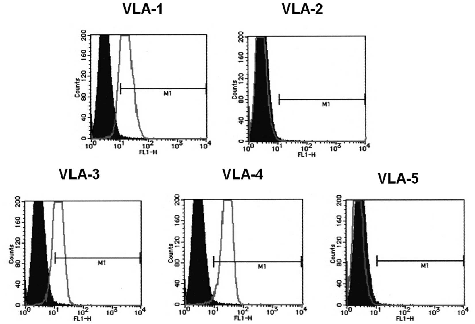
Flow cytometric analysis indicated that the
integrins VLA-1, -3 and -4 were expressed on the cell surface of
HBL-8 3G3 cloned cells, but that VLA-2 and -5 were not expressed
(Fig. 1). Previous
immunohistochemical findings showed that the H-ALCL cells express
VLA-5, but not VLA-4 (11).
Effect of alteration of cell surface O-
and N-glycans on cell lectin reactivity
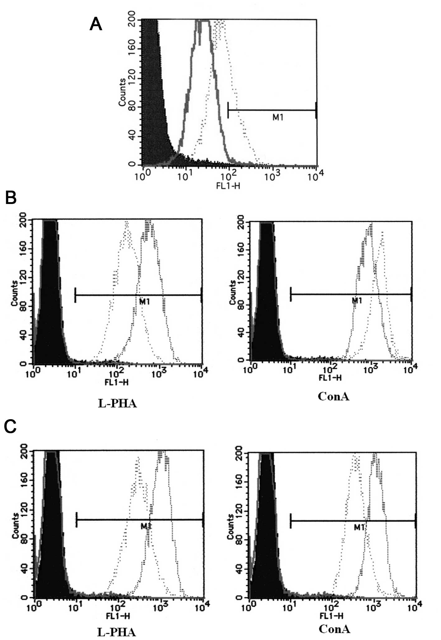
Flow cytometric analysis of HBL-8 3G3 cells
indicated that the HPA lectin reactivity of the cells increased
after inhibition of O-glycosylation with BZ treatment (Fig. 2A). Following inhibition of
N-glycosylation with SW treatment the L-PHA reactivity of the cells
decreased and the ConA reactivity of the cells increased (Fig. 2B). On the other hand after
inhibition of N-glycosylation with TM treatment both the L-PHA
reactivity and the ConA reactivity of the cells decreased (Fig. 2C). Our recent report showed that
L-PHA reactivity of H-ALCL cells also decreased after TM treatment
(11). These data show that
inhibition of O- and N-glycosylation specifically altered cell
surface lectin reactivity.
Effect of alteration of cell surface
glycosylation on cell adhesion to fibronectin
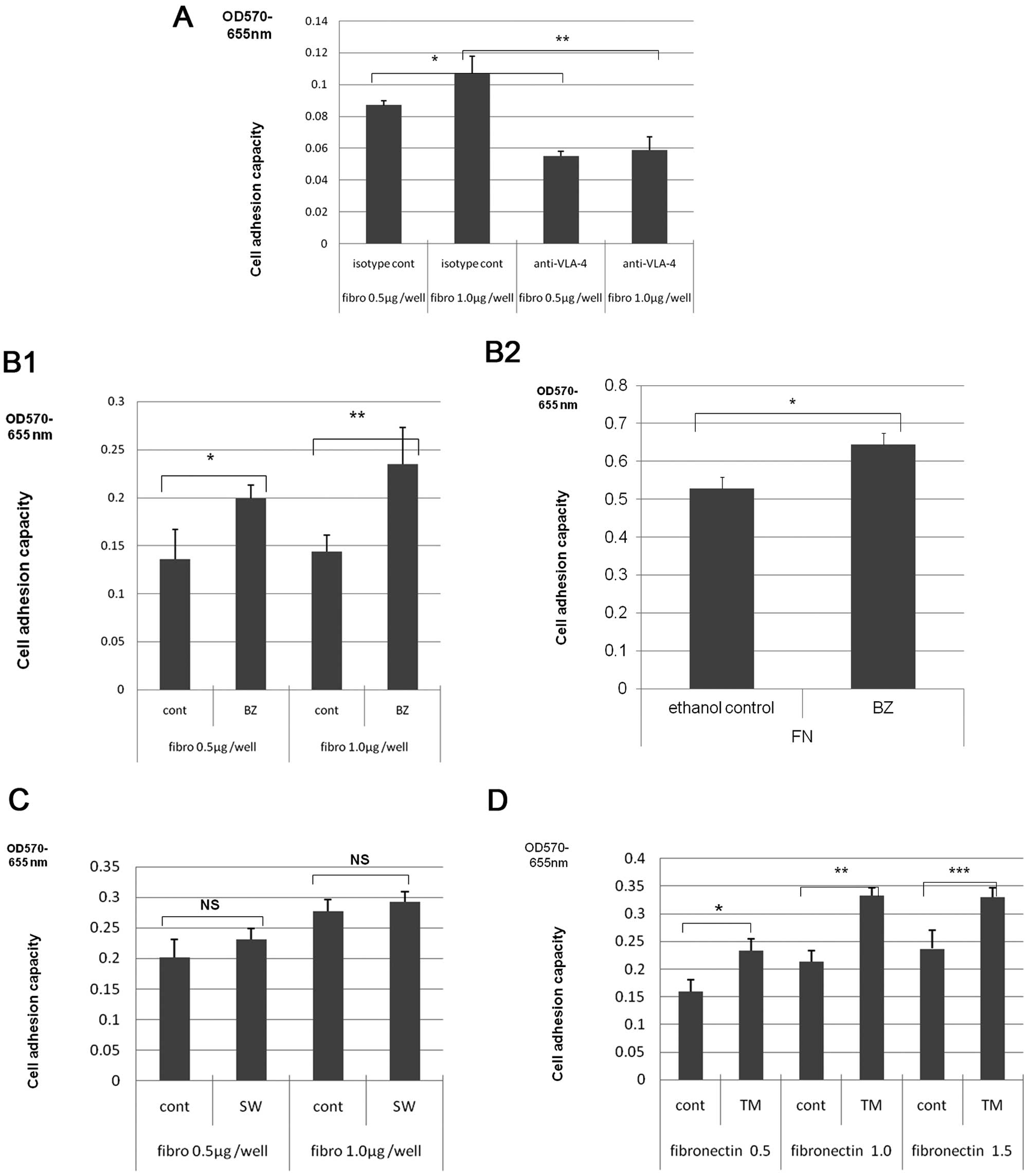
A fibronectin-coated cell adhesion assay showed that
HBL-8 lymphoma cells adhered to fibronectin. Pre-treatment of the
cells with anti-VLA-4 or control antibodies indicated that this
adhesion was mediated by the integrin VLA-4 (Fig. 3A). The number of HBL-8 3G3 or
H-ALCL cells that adhered to fibronectin increased following BZ
treatment compared to the number of adhered cells in the absence of
BZ treatment [Fig. 3B-1 (HBL-8 3G3)
and B-2 (H-ALCL)]. There was no significant difference in the
number of HBL-8 3G3 cells that adhered to fibronectin between
non-treated and SW-treated cells (Fig.
3C). On the other hand treatment with TM enhanced HBL-8 3G3
cell adhesion to fibronectin compared to non-treated cells
(Fig. 3D). Thus, alteration of
cell surface glycosylation modulates cell adhesion to fibronectin.
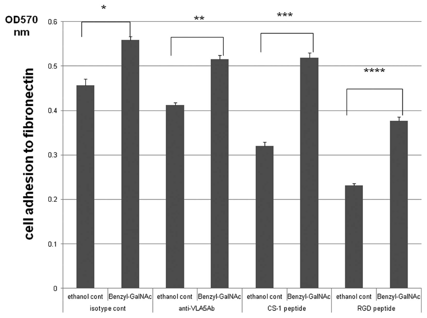
The enhancement of H-ALCL adhesion to fibronectin by BZ was not
mediated by the integrin VLA-5, or by CS-1 or RGD protein sequences
(Fig. 4).
Effect of alteration of cell surface
glycosylation on H-ALCL cell adhesion to galectins
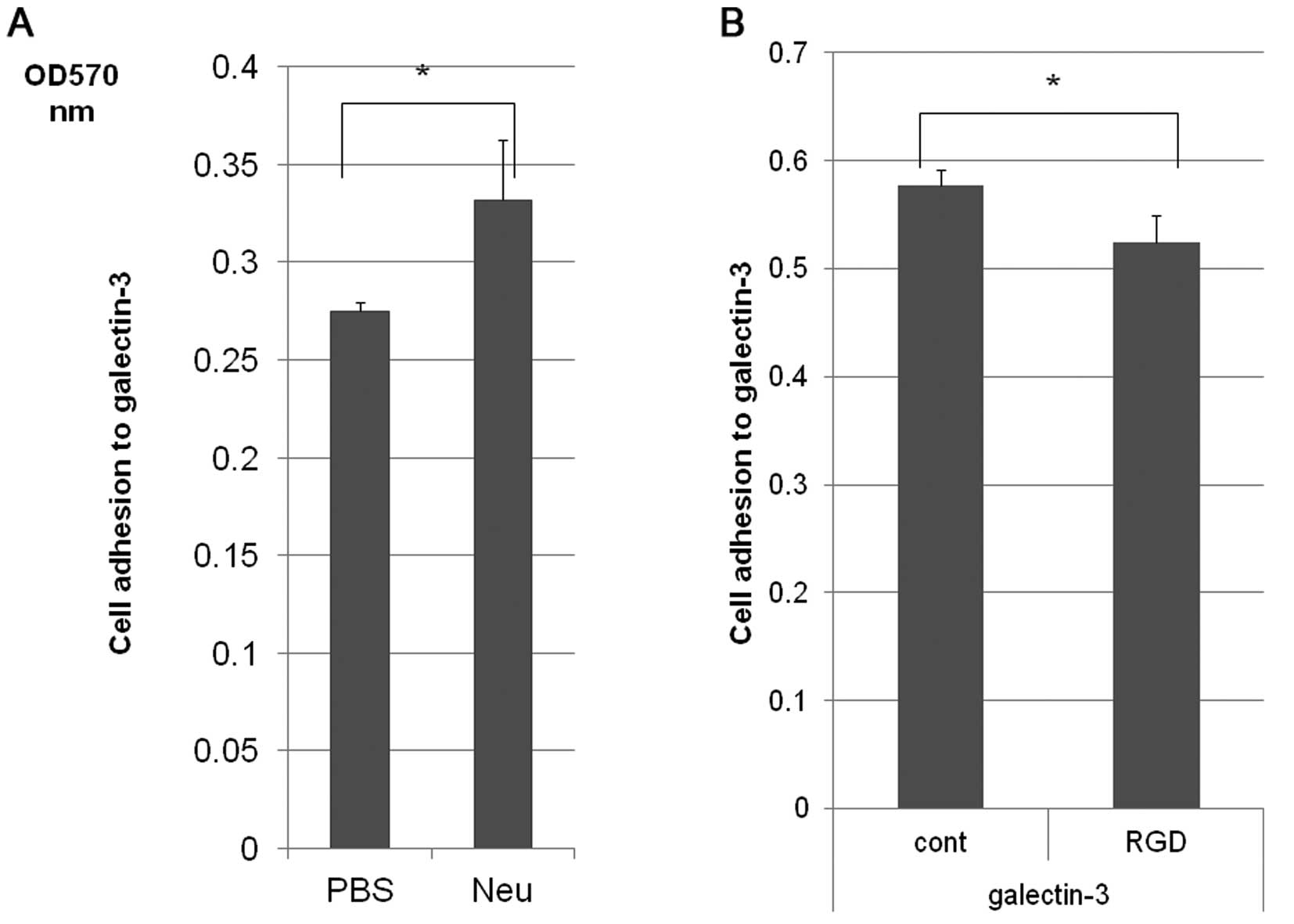
H-ALCL cells also adhered to galectin-3 and this
adhesion to galectin-3 was enhanced by cleavage of cell surface
sialic acids by pre-treatment with neuraminidase (Fig. 5A). Furthermore, H-ALCL cell
adhesion to galectin-3 was inhibited by RGD peptide pre-treatment
(Fig. 5B), with serum-free medium.
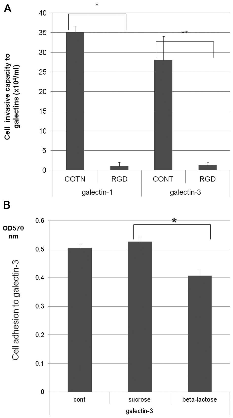
The ability of H-ALCL cells to invade galectin-1 and galectin-3 was
markedly inhibited by pre-incubation with the RGD peptide,
indicating that this invasion was dependent on the RGD sequence
(Fig. 6A). Further inhibition
experiments showed that inhibition of H-ALCL cell adhesion to
galectin-3 by β-lactose was much higher compared to inhibition by
sucrose suggesting that H-ALCL cell adhesion to galectin-3 might be
mediated by β-galactose on the cell surface (Fig. 6B).
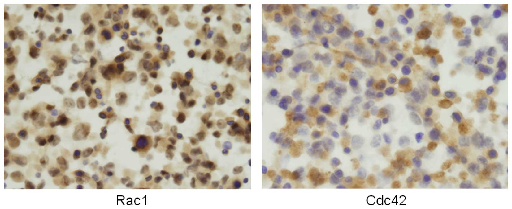
Potential involvement of small GTP proteins in the
galectin invasion of H-ALCL cells. Immunohistochemical analysis
showed that Rac 1 and Cdc42 proteins were expressed in the
cytoplasm of H-ALCL cells (Fig.
7). Our previous immunohistochemical analysis showed that Rho
is also expressed in the cytoplasm of H-ALCL cells and that the Rho
inhibitor, C3-transferase, markedly inhibited H-ALCL invasion of
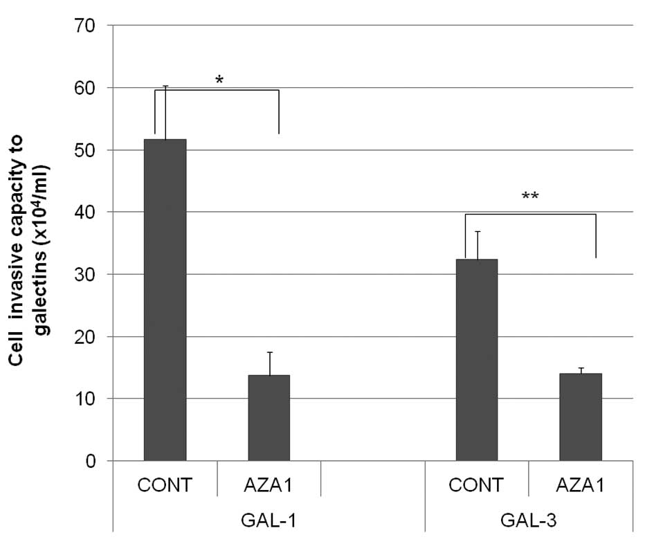
galectins (12). Pre-incubation of
H-ALCL cells with AZA1, an inhibitor of Rac 1 and Cdc42, also
significantly inhibited cell invasion of galectin-1 and galectin-3
(Fig. 8). The combined above data
suggested that the small GTPases Rho, Rac 1 and Cdc42 are involved
in H-ALCL invasion to galectin.
Discussion
The ability of cells to adhere to the ECM is known
to be closely associated with the metastatic capacity of malignant
tumor cells. Alteration of cell surface glycans can modulate the
metastatic rate to distant organs. In the present study, using
glycoengineering methods we showed a biological role of the surface
glycans of malignant lymphoma cells in cell adhesion and cell
invasion to extracellular matrix.
The presence of O-glycans and HPA reactive
oligosaccha-rides was demonstrated on HBL-8 cells using flow
cytometric analysis. BZ enhanced the expression of HPA reactive
oligosaccharides i.e., GalNAc residues. Adhesion of HBL-8 cells to
fibronectin was mediated by the VLA-4 adhesion molecule. This
fibronectin adhesion of HBL-8 was enhanced by treatment with BZ. It
was unclear whether the VLA-4 mediated cell adhesion was directly
modified by administration of BZ and we speculate that the effect
of BZ may be mediated by a GalNAc epitope that affects the
interactions between VLA-4 and fibronectin. Such a candidate GalNAc
epitope would be a mucin type epitope, or O-linked
oligosaccharides. It is known that O-linked oligosaccharides can
influence cell adhesive properties (4,5,13).
BZ treatment also enhanced the adhesion of H-ALCL cells to
fibronectin. BZ treatment is known to remove sialic acid which is
linked to the terminal residue of glycans. Therefore, desialylation
of O-glycans by BZ may also enhance cell adhesion to fibronectin.
Our present data obtained using H-ALCL cells indicated that this
effect is independent of VLA-5 and the RGD sequence of fibronectin.
Therefore, the sialic acid on O-glycans may regulate cell adhesion
to fibronectin independently of integrins. Previous reports have
suggested that desialylation of integrin upregulates cell adhesion
to fibronectin (14). On the other
hand α-2,6 sialic acid has been reported to be required for cell
adhesion to fibronectin (15). In
pancreatic carcinoma cells, α-2,6 sialic acid is required for tumor
cell adhesion to the ECM (16).
Based on these findings it appears that there is a difference in
the regulation of cell adhesion to the ECM by sialylation among
various cell types. Further study will be required to clarify the
detailed mechanisms by which sialic acid regulates cell adhesion to
the ECM.
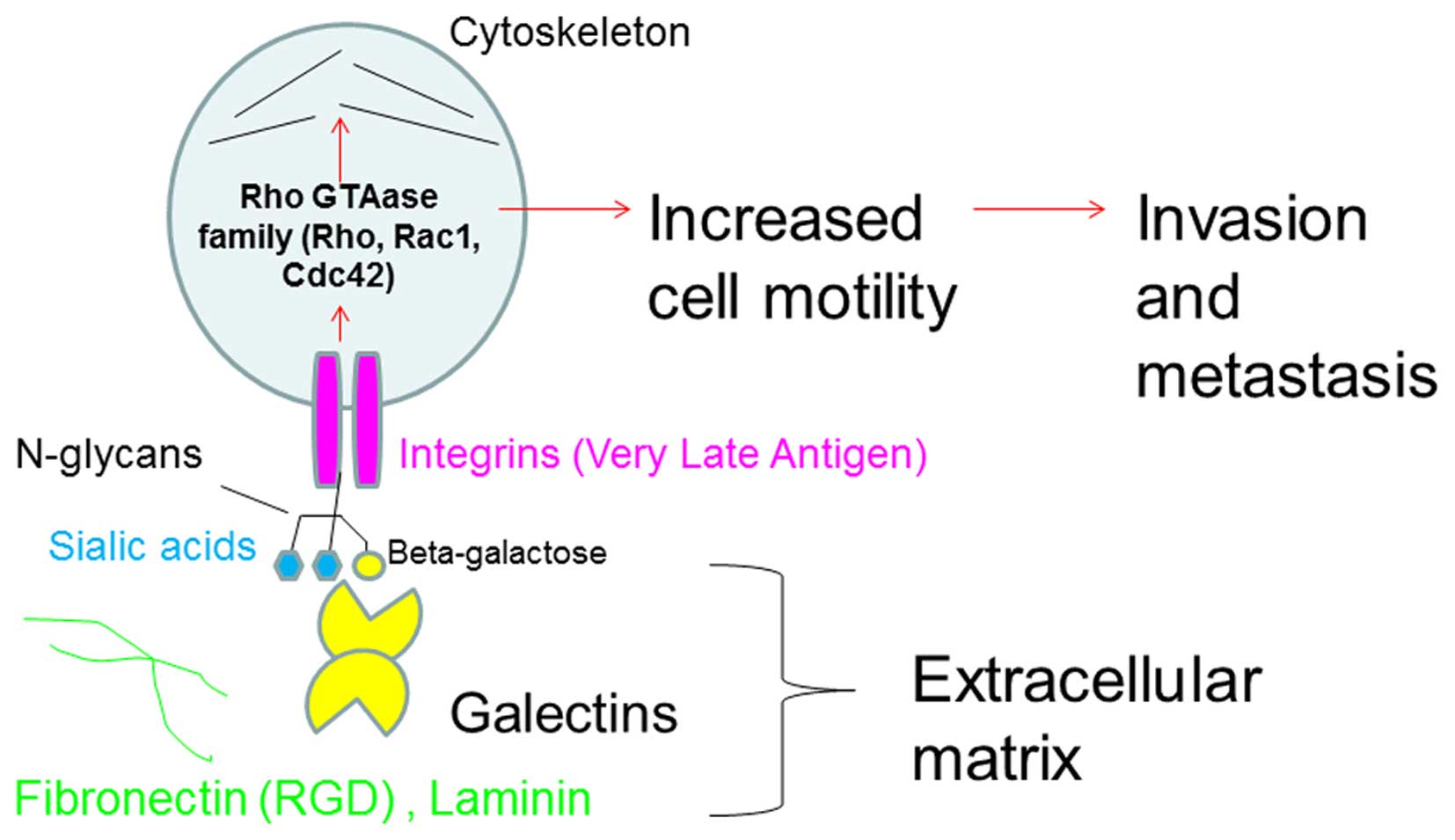
CD45 is a candidate receptor for galectin-1 and -3
(17). However, recent data
suggest that integrin is also a candidate galectin-3 receptor
(18). In our present data, H-ALCL
cell adhesion to galectin-3 was inhibited by pretreatment with the
RGD peptide suggesting that cell adhesion to galectin-3 was
mediated by integrin (VLA-5). Therefore, sialylation may regulate
cell adhesion to galectin-3 in an integrin-dependent manner and
integrin may act as a galectin receptor in cell adhesion to
galectins (Fig. 9). In this model
cell adhesion to galectin through integrins would activate
intrinsic signals leading to the activation of Rho, Rac 1 and
Ccd42, and resulting in rearrangement of the cytoskeleton. These
events act as a driving force of cell migration. Therefore, sialic
acid may regulate lymphoma cell invasion to galectin through
galectin-integrin interaction, subsequently inducing Rho family
activation and rearrangement of the cytoskeleton. These phenomena
may be associated with lymphoma cell invasion and metastasis.
N-linked oligosaccharides are known to be
transferred onto asparagine residues of proteins from a donor
substrate, dolichol-phosphate-oligosaccharide, in the endoplasmic
reticulum (19). Deglycosylation
of glycoproteins that is reflected by a decrease in L-PHA and ConA
lectin reactivity may be related to dysfunction of this
oligosaccharide transfer to proteins, which is catalyzed by the
enzyme oligosaccharyltransferase in the endoplasmic reticulum. The
N-glycan inhibitor, SW can inhibit complex type N-glycans,
resulting in the expression of hybrid type N-glycans. On the other
hand TM can inhibit both complex and hybrid type N-glycan
expression. Our data showed that SW cannot enhance cell adhesion to
fibronectin, whereas TM can. Therefore, differences in the detail
of the N-glycan structures resulting from SW and TM treatments can
influence cell adhesion to FN. Not only O-glycans, but also
N-glycans may regulate HBL-8 and H-ALCL cell adhesion to
fibronectin. Some previous studies have suggested that alteration
of the N-glycosylation of β-1 integrins may modulate the ability of
cells to adhere to the ECM (20).
In addition to this, it was suggested that VLA-4 expression may be
closely related to the ability of a tumor to metastasize to distant
organs in an animal model (21).
Therefore, cell surface N-glycans may modulate metastatic capacity
by modulation of VLA-4 function in lymphoma cells.
In a previous report we showed that L-PHA reactive
N-linked oligosaccharide on the lymphoma cell surface is closely
associated with clinical behavior of diffuse large B cell lymphoma
(DLBCL) cases (1,2). Loss of L-PHA reactive N-glycans on
the cell surface is correlated to a poor prognosis of DLBCL. In the
present study TM treatment resulted in enhancement of HBL-8 3G3
lymphoma cell adhesion to fibronectin or in the recent study,
inhibition of H-ALCL cell adhesion to fibro-nectin and laminin
(11). N-glycan bidirectionally
regulates cell adhesion to fibronectin suggesting that alteration
of N-glycans affects cell adhesion to the ECM resulting in
modulation of the biological behavior of lymphoma cells. Previous
reports showed that TM treatment modulates cell surface
glycosylation expression, resulting in alteration of cell adhesion
to the ECM (22), and that
alteration of cell surface glycosylation affects cell to cell
adhesion or cell-matrix interaction in several tumor cell lines
(22,23–26,27,28).
These findings and our present data may provide a new scientific
area of study of tumor cell biology that is focused on the
regulation of cell adhesion to the ECM by glycosylation.
GTPases are known to include Rac 1, Cdc42 and Rho.
Rac 1 forms lamellipodia, Cdc42 forms filopodia, and Rho forms
stress fibers, and these molecules are known to be associated with
cancer progression (29). The
function of these molecules is associated with cell motility of
lymphoma cells that is mediated through galectins in the stroma.
Furthermore, sialic acid on the cell surface may regulate cell
adhesion to galectins that is mediated by integrins. Sialic acid is
adequately regulated on cell adhesion to galectin resulting in
facilitation of cell motility, and subsequently enhances cell
motility resulting in more aggressive tumor behavior and
metastasis. In the present data, cell adhesion to galectin-3 was
inhibited by cell surface sialylation suggesting that cell surface
sialic acid may regulate cell adhesion to galectins. In terms of
cell surface glycosylation, especially of sialylation, we speculate
that sialylation enhances Rho GTPase family induced cell invasive
capacity and may result in a worse prognosis of the patients. Our
present data showed that desialylation enhanced cell adhesion to
galectin and inhibited cell invasion to galectin (12), suggesting that the effect of
desialylation on cell invasion of galectin is the opposite to the
effect of the Rho GTPase family on cell invasion.
In conclusion, cell surface sialylation appeared to
be a candidate molecule for the regulation of cell adhesion, and
the present data may provide a new scientific focus for lymphoma
glycobiology.
Acknowledgements
We are grateful to Ms. M. Satoh and Mrs. H. Kaneko
for their technical assistance and advice.
References
|
1
|
Suzuki O, Nozawa Y, Kawaguchi T and Abe M:
Phaseolus vulgaris leukoagglutinating lectin-binding reactivity in
human diffuse large B-cell lymphoma and its relevance to the
patient's clinical outcome: Lectin histochemistry and lectin blot
analysis. Pathol Int. 49:874–880. 1999. View Article : Google Scholar : PubMed/NCBI
|
|
2
|
Suzuki O, Nozawa Y, Kawaguchi T and Abe M:
Alpha-2,6-sialylation of L-PHA reactive oligosaccharides and
expression of N-acetylglucosaminyltransferase V in human diffuse
large B cell lymphoma. Oncol Rep. 10:1759–1764. 2003.PubMed/NCBI
|
|
3
|
Suzuki O, Nozawa Y and Abe M: Loss of
L-PHA-, PNA-, or ConA-reactive oligosaccharides is associated with
a poor prognosis in human Burkitt's lymphoma. Oncol Rep.
17:775–779. 2007.PubMed/NCBI
|
|
4
|
Porowska H, Paszkiewicz-Gadek A, Anchim T,
Wolczynski S and Gindzienski A: Inhibition of the O-glycan
elongation limits MUC1 incorporation to cell membrane of human
endometrial carcinoma cells. Int J Mol Med. 13:459–464.
2004.PubMed/NCBI
|
|
5
|
Kojima N, Saito M and Tsuji S: Role of
cell surface O-linked oligosaccharides in adhesion of HL60 cells to
fibronectin: Regulation of integrin-dependent cell adhesion by
O-linked oligosaccharide elongation. Exp Cell Res. 214:537–542.
1994. View Article : Google Scholar : PubMed/NCBI
|
|
6
|
Paszkiewicz-Gadek A, Porowska H,
Lemancewicz D, Wolczynski S and Gindzienski A: The influence of N-
and O-glycosylation inhibitors on the glycosylation profile of
cellular membrane proteins and adhesive properties of carcinoma
cell lines. Int J Mol Med. 17:669–674. 2006.PubMed/NCBI
|
|
7
|
Suzuki O, Nozawa Y and Abe M: Regulatory
roles of altered N- and O-glycosylation of CD45 in
galectin-1-induced cell death in human diffuse large B cell
lymphoma. Int J Oncol. 26:1063–1068. 2005.PubMed/NCBI
|
|
8
|
Guo HB, Lee I, Kamar M, Akiyama SK and
Pierce M: Aberrant N-glycosylation of beta1 integrin causes reduced
alpha5beta1 integrin clustering and stimulates cell migration.
Cancer Res. 62:6837–6845. 2002.PubMed/NCBI
|
|
9
|
Zhang Y, Zhao JH, Zhang XY, Guo HB, Liu F
and Chen HL: Relations of the type and branch of surface N-glycans
to cell adhesion, migration and integrin expressions. Mol Cell
Biochem. 260:137–146. 2004. View Article : Google Scholar : PubMed/NCBI
|
|
10
|
Suzuki O, Nozawa Y and Abe M: The
regulatory roles of cell surface sialylation and N-glycans in human
B cell lymphoma cell adhesion to galectin-1. Int J Oncol.
28:155–160. 2006.
|
|
11
|
Suzuki O, Abe M and Hashimoto Y:
Sialylation by β-galactoside α-2,6-sialyltransferase and N-glycans
regulate cell adhesion and invasion in human anaplastic large cell
lymphoma. Int J Oncol. 46:973–980. 2015.PubMed/NCBI
|
|
12
|
Suzuki O and Abe M: Galectin-1-mediated
cell adhesion, invasion and cell death in human anaplastic large
cell lymphoma: Regulatory roles of cell surface glycans. Int J
Oncol. 44:1433–1442. 2014.PubMed/NCBI
|
|
13
|
Zhang L and Ten Hagen KG: The cellular
microenvironment and cell adhesion: A role for O-glycosylation.
Biochem Soc Trans. 39:378–382. 2011. View Article : Google Scholar : PubMed/NCBI
|
|
14
|
Semel AC, Seales EC, Singhal A, Eklund EA,
Colley KJ and Bellis SL: Hyposialylation of integrins stimulates
the activity of myeloid fibronectin receptors. J Biol Chem.
277:32830–32836. 2002. View Article : Google Scholar : PubMed/NCBI
|
|
15
|
Yu S, Fan J, Liu L, Zhang L, Wang S and
Zhang J: Caveolin-1 up-regulates integrin α2,6-sialylation to
promote integrin α5β1-dependent hepatocarcinoma cell adhesion. FEBS
Lett. 587:782–787. 2013. View Article : Google Scholar : PubMed/NCBI
|
|
16
|
Bassagañas S, Pérez-Garay M and Peracaula
R: Cell surface sialic acid modulates extracellular matrix adhesion
and migration in pancreatic adenocarcinoma cells. Pancreas.
43:109–117. 2014. View Article : Google Scholar
|
|
17
|
Perillo NL, Pace KE, Seilhamer JJ and Baum
LG: Apoptosis of T cells mediated by galectin-1. Nature.
378:736–739. 1995. View
Article : Google Scholar : PubMed/NCBI
|
|
18
|
Zhuo Y, Chammas R and Bellis SL:
Sialylation of beta1 integrins blocks cell adhesion to galectin-3
and protects cells against galectin-3-induced apoptosis. J Biol
Chem. 283:22177–22185. 2008. View Article : Google Scholar : PubMed/NCBI
|
|
19
|
Kornfeld R and Kornfeld S: Assembly of
asparagine-linked oligosaccharides. Annu Rev Biochem. 54:631–664.
1985. View Article : Google Scholar : PubMed/NCBI
|
|
20
|
Janik ME, Lityńska A and Vereecken P: Cell
migration-the role of integrin glycosylation. Biochim Biophys Acta.
1800.545–555. 2010.
|
|
21
|
Matsuura N, Puzon-McLaughlin W, Irie A,
Morikawa Y, Kakudo K and Takada Y: Induction of experimental bone
metastasis in mice by transfection of integrin alpha 4 beta 1 into
tumor cells. Am J Pathol. 148:55–61. 1996.PubMed/NCBI
|
|
22
|
Kemmner W, Morgenthaler J and Brossmer R:
Alterations in cell surface carbohydrate composition of a human
colon carcinoma cell line affect adhesion to extracellular matrix
components. Biochimie. 74:117–122. 1992. View Article : Google Scholar : PubMed/NCBI
|
|
23
|
Przybyło M, Pocheć E, Link-Lenczowski P
and Lityńska A: Beta1-6 branching of cell surface glycoproteins may
contribute to uveal melanoma progression by up-regulating cell
motility. Mol Vis. 14:625–636. 2008.
|
|
24
|
Zhao H, Liang Y, Xu Z, Wang L, Zhou F, Li
Z, Jin J, Yang Y, Fang Z, Hu Y, et al: N-glycosylation affects the
adhesive function of E-Cadherin through modifying the composition
of adherens junctions (AJs) in human breast carcinoma cell line
MDA-MB-435. J Cell Biochem. 104:162–175. 2008. View Article : Google Scholar
|
|
25
|
Sato T, Takahashi M, Kawado T, Takayama E
and Furukawa K: Effect of staurosporine on N-glycosylation and cell
adhesion to fibronectin of SW480 human colorectal adenocarcinoma
cells. Eur J Pharm Sci. 25:221–227. 2005. View Article : Google Scholar : PubMed/NCBI
|
|
26
|
Schraen-Maschke S and Zanetta JP: Role of
oligomannosidic N-glycans in the proliferation, adhesion and
signalling of C6 glioblastoma cells. Biochimie. 85:219–229. 2003.
View Article : Google Scholar : PubMed/NCBI
|
|
27
|
Bironaite D, Nesland JM, Dalen H, Risberg
B and Bryne M: N-Glycans influence the in vitro adhesive and
invasive behaviour of three metastatic cell lines. Tumour Biol.
21:165–175. 2000. View Article : Google Scholar : PubMed/NCBI
|
|
28
|
de Freitas JC Junior, Silva BR, de Souza
WF, de Araújo WM, Abdelhay ES and Morgado-Díaz JA: Inhibition of
N-linked glycosylation by tunicamycin induces E-cadherin-mediated
cell-cell adhesion and inhibits cell proliferation in
undifferentiated human colon cancer cells. Cancer Chemother
Pharmacol. 68:227–238. 2011. View Article : Google Scholar
|
|
29
|
Rathinam R, Berrier A and Alahari SK: Role
of Rho GTPases and their regulators in cancer progression. Front
Biosci (Landmark Ed). 16:2561–2571. 2011. View Article : Google Scholar
|