Introduction
Infantile hemangioma (IH) is a common benign tumor
in childhood, and more frequent in prematures and low birth weight
infants (1). IH has three
distinctive clinical and histological phases: an initial
proliferative phase with rapid growth and undeveloped blood
channels, a plateau and the final involuted phase with scattered
capillaries and large draining vessels (2). IH is characterized by the spontaneous
regression of blood vessels. IHs are disfiguring in some cases,
which is also the leading cause of morbidity in affected children
(2,3). Recently, advances have been made in
the treatment of IH with clear curative effect, such as propranolol
(4), nadolol (5) and pulsed dye laser (6). However, the main clinical problem is
still the lack of reliable parameters to treat IH lesions in
patients. Therefore, the identification of genetic and epigenetic
alterations in IH lesions will help to establish molecular
mechanisms of this desease.
In recent years, miRNAs are considered as ideal
biomarkers for cancer identification, for the properties that
miRNAs are highly conserved in the genome and have a relatively
high degree of tissue specificity (7). Abnormal expression of miRNAs is
deeply associated with numerous pathological processes, such as
angiogenesis (8), carcinogenesis
(9) and inflammation (10). The microRNA (miR)-130 family is
composed of miR-130b, miR-301a and miR-301b, which share a common
seed sequence (9). miR-130a is
located at chromosome 11q12, close to the 11q13 area (11). miR-130a has been found to be
involved in multiple types of human diseases, including gastric
(12), cervical cancer (13), chronic myeloid leukemia (14) and metabolism-related inflammation
(15). Besides, reports indicate
that miR-130a promote the proliferation, migration and tube
formation of vascular endothelial cells (16). However, the comprehensive effect of
the miR-130a on tumor progression of hemangioma has not been
analyzed.
Tissue factor pathway inhibitor 2 (TFPI2) is
secreted as a glycosylated protein with a short acidic
amino-terminal region, three tandem Kunitztype domains and a
carboxy-terminal tail (17). TFPI2
belongs to a Kunitz family of serine protease inhibitors that
contain one or more Kunitz-type domains. The family has more than
20 members including pancreatic trypsin inhibitor (aprotinin) and
the homolog of TFPI2 (18). A
report indicated that human TFPI2 was first identified in the
placenta to inhibit the amidolytic activity of plasmin (19). Recent reports suggested that the
downregulation were associated with the development of multiple
deseases, including atherosclerotic plaque (20), pancreatic carcinoma (21), hepatocellular carcinoma (22) and cervical cancer (23). Considering TFPI2 is secreted into
the extracellular matrix (ECM) to help regulate ECM remodeling, the
role of TFPI2 involved in hemangioma is worthy of
investigation.
The present study explored the expression of
miR-130a in hemangioma tissues and cell lines. We found that the
inhibition of miR-130a effectively suppressed the growth of
hemangioma by targeting TFPI2 and inactivating focal adhesion
kinase (FAK)/phosphoinositide 3-kinase (PI3K)/Rac1/anti-mouse
double minute (mdm2) pathway. This study also revealed an
inhibitory effect of angiogenesis via downregulating miR-130a.
Materials and methods
Hemangioma samples
Twenty pairs of human hemangioma and adjacent normal
tissues were obtained from Shandong Provincial Hospital Affiliated
to Shandong University (Shandong, China). The tissues were frozen
in liquid nitrogen and stored at −80°C until use. Written informed
consent for tissue donation (for research purposes) was obtained
from the patients, and the protocol was approved by the
Institutional Review Board of the Provincial Affiliated Hospital of
the Shandong University.
Cell lines
The human vascular endothelial HUVEC-12 cells, human
IH cell lines XPTS-1 and mouse hemangioendothelioma cells (EOMA)
were obtained from the American Type Culture Collection (ATCC;
Manassas, VA, USA). These cell lines were cultured in Dulbecco's
modified eagle's medium (DMEM) plus 10% fetal bovine serum (FBS;
Life Technologies, Inc., Grand Island, NY, USA) and grown at 37°C
in a 5% CO2 atmosphere.
Quantitative real-time polymerase chain
reaction (qRT-PCR)
The total RNA was extracted from TRIzol reagent
(Invitrogen, Carlsbad, CA, USA) following the manufacturer's
instructions. The RT-PCR primers for miR-130a and U6 were purchased
from GeneCopoeia, Inc. (San Diego, CA, USA). The PCR primers for
TFPI2 were: forward, 5′-GTCGATTCTGCTGCTTTTCC-3′ and reverse,
5′-ATGGAATTTTCTTTGGTGCG-3′ (440 bp) (24). GAPDH and U6 SnRNA were used as the
internal control of the mRNA or miRNA, respectively. Fold-change of
miR-130a was calculated by the 2−ΔΔCt method.
Northern blot analysis
The expression levels of miR-130a in hemangioma
samples, adjacent normal tissues, HUVEC-12, XPTS-1 and EOMA cells
were further determined by northern blot assay according to
previously described procedures (25).
The transfection of miR-130a
mimic/inhibitor
The miR-130a mimic, negative control of miRNA mimic,
miR-130a inhibitor and negative control of miRNA inhibitor were
purchased from Shanghai GenePharma, Co., Ltd. (Shanghai, China),
and transfected into XPTS-1 and EOMA cells at a final concentration
of 100 nM with Lipofectamine 2000 (Invitrogen) according to the
manufacturer's instructions. After 6 h, the cells were returned to
normal medium and cultured for an additional 48 (for RNA isolation)
or 72 h (for protein extraction).
Colony formation assay
XPTS-1 and EOMA cells were transfected with miR-130a
inhibitor or control. After 48 h of infection, the cells were
plated in 6-well plates at 200/well and grown for 2 weeks. Then,
the cells were washed twice with phosphate-buffered saline (PBS),
fixed with methanol/acetic acid (3:1, vol/vol) and stained with
0.5% crystal violet. The number of colonies was counted under the
microscope.
Cell apoptosis assay and cell cycle
analysis
XPTS-1 (5×10³) and EOMA cells were plated
into each well of 96-well plates, respectively. Six hours later,
the cells were treated with miR-control or miR-130a inhibitor. The
procedures of cell cycle analysis were previously described
(21). Cell numbers were
determined by Scepter™ 2.0 Handheld Automated Cell Counter.
Western blotting
Cell samples were lysed in lysis buffer (Beyotime
Institute of Biotechnology, Haimen, China). The samples mixed with
loading buffer were incubated in boiling water for 10 min. Protein
(20–30 µg) was separated through SDS-PAGE and then
transferred onto polyvinylidene fluoride (PVDF) membranes
(Millipore, Billerica, MA, USA). The following primary antibodies
were used: anti-TFPI2, anti-Ki-67, anti-proliferating cell nuclear
antigen (PCNA), anti-FAK, anti-PI3K, anti-Rac1, anti-mdm2,
anti-p-FAK, anti-p-PI3K, anti-p-Rac1, anti-p-mdm2, anti-vascular
endothelial growth factor (VEGF), anti-matrix metalloproteinase
(MMP)-2, anti-MMP-9 and anti-GAPDH (Abcam, Hangzhou, China), which
was used as the internal reference. After incubation with the
appropriate horseradish peroxidase (HRP)-conjugated secondary
antibody, proteins were detected using a ChemiDoc XRS imaging
system and quantity One analysis software (Bio-Rad Laboratories,
San francisco, CA, USA).
Luciferase reporter assay
The cDNA of TFPI2 was amplified by PCR, and general
method of recombinant DNA was used to clone the wild-type (WT)
3′-UTR and mutant (MUT) 3′-UTR of TFPI2. The WT and MUT sequences
of TFPI2 were then cloned into a pMIR-Report luciferase reporter
vector (Ambion, Austin, TX, USA) to generate constructs of
Luc-TFPI2 and Luc-TFPI2-mut, followed by DNA sequencing
verification. The pMIR-Report control vector (Luc-control) was also
cloned. All three vectors were transfected into XPTS-1 cells/EOMA
cells in 6-well plates with a Lipofectamine 2000 reagent kit
(Sigma-Aldrich, St. Louis, MO, USA). After 1 day, the luciferase
activity was measured using a luciferase reporter assay system
(Promega, Madison, WI, USA). All relative luciferase activities
were normalized to the condition of Luc-control with
β-galactosidase transfection.
siRNA transfection
The small interfering RNA (siRNA), to genetically
downregulate TFPI2 (TFPI2-siRNA), as well as its non-specific
scramble siRNA (NC-siRNA), were purchased from Guangzhou RiboBio,
Co., Ltd. (Guangzhou, China). Transfection of siRNAs into XPTS-1
cells was conducted using Lipofectamine 2000 reagent kit. In XPTS-1
cells, 100 nM siRNAs (TFPI2-siRNA and NC-siRNA) were used. The
efficiency of siRNA interference was confirmed by western blot
analysis 48 h after transfection.
Overexpression of TFPI2 and FAK
TFPI2 and FAK overexpression was achieved by PCR
amplification using their cDNA as templates, separately, and the
TFPI2 and FAK expressing vectors were constructed by inserting
their cDNA into pcDNA 3.1 vector (Sigma-Aldrich). The recombinant
plasmids and other agents were co-transfected into 3×10⁶
XPTS-1 cells using a Nucleofector instrument. Forty-eight hours
later, subsequent experiments were performed on the cells. The
experiment was replicated thrice for data calculations.
Immunofluorescence staining
Fluorescent cells were cultured on an 8-well chamber
culture slides (Becton-Dickinson, Bedford, MA, USA). After 8 h,
cells were fixed in 3% paraformaldehyde in PBS at room temperature
for 8 min, then permeabilized with 0.2% Triton X-100 for 15 min at
room temperature. After washing in PBS, the cells were incubated
with primary mouse anti-FAK monoclonal antibody (1 mg/ml;
Transduction Laboratories, Lexington, KY, USA) at 4°C overnight.
After washing, cells were incubated with biotinylated goat
anti-mouse IgG (Pierce, Rockford, IL, USA) at room temperature for
1 h. The immunoreactivity was revealed using Alexa568-conjugated
streptavidin (Molecular Probes, Eugene, OR, USA) and cells were
counterstained with 10 mg/ml DAPI. The cells were examined under a
Nikon fluorescence microscope (Image Systems, Columbia, MD,
USA).
Hemangioma xenografts
Male athymic nude mice were housed and manipulated
according to the protocols approved by the Experimental Animal
Center of the Shandong Provincial Hospital Affiliated to Shandong
University. For each mouse, 5×10⁶ miR-130a-inhibiting
XPTS-1 cells were injected subcutaneously into the right scapula in
100 serum-free medium. After the development of a palpable tumor,
the tumor volume was monitored every 5 days and assessed by
measuring the 2 perpendicular dimensions using a caliper and the
formula (a × b2)/2, where a is the larger and b is the
smaller dimension of the tumor. At 25 days after inoculation, the
mice were sacrificed and tumor weights were assessed. A portion of
each tumor was selected for western blotting for TFPI2 and key
components of the FAK pathway and qRT-PCR analysis for
miR-130a.
Immunohistochemistry
Tumor tissues were fixed in formalin, and then were
embedded with paraffin for hemangioma C analysis. Briefly, 5
µm-thick paraffin sections were deparaffinized in xylene and
rehydrated in a 100, 95 and 75% ethanol gradually. In order to
quench the activity of endogenous peroxidase, the tissue sections
were incubated in 30% H2O2 for 30 min. After
antigen retrieval in heated 10 mM citrate buffer for 10 min, the
tissue sections were immunostained with mouse anti-human CD31 and
CD34 primary antibody overnight at 4°C. Corresponding mouse
horseradish peroxidase (HRP)-conjugated secondary antibody was
added for 1 h at room temperature. Images were obtained using an
Olympus IX71 inverted microscope.
Statistical analysis
All results are presented as mean ± SD from a
minimum of 3 replicates. Differences between groups were evaluated
by SPSS version 15.0 statistical software with Student's t-test
when comparing only 2 groups or assessed by one-way ANOVA when
>2 groups were compared. For the comparison of paired tissues, a
paired Student's t-test was used to determine statistical
significance. The relationship between TFPI2 and miR-130a
expression was explored by Spearman's correlation. Differences were
considered statistically significant at P<0.05.
Results
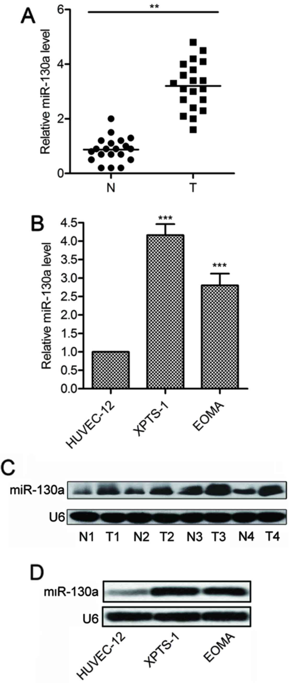
miR-130a is overexpressed in hemangioma
tissues and cell lines
The expression of miR-130a was measured in 21
fresh-frozen hemangioma specimens and matched tumor-adjacent normal
tissues. The results of qPCR showed that the average expression
levels of miR-130a were significantly higher in hemangioma tissues
than that in adjacent normal tissues (P<0.01; Fig. 1A). Consistently, miR-130a
expression was also increased in hemangioma cell lines (XPTS-1 and
EOMA), compared with normal vascular endothelial cells (HUVEC-12)
(P<0.001; Fig. 1B). Northern
blot analysis further confirmed the increased expression of
miR-130a in hemangioma specimens (Fig.
1C) and cell lines (Fig. 1D).
Taken together, these results reveal that miR-130a is upregulated
in human hemangioma tissues and cell lines.
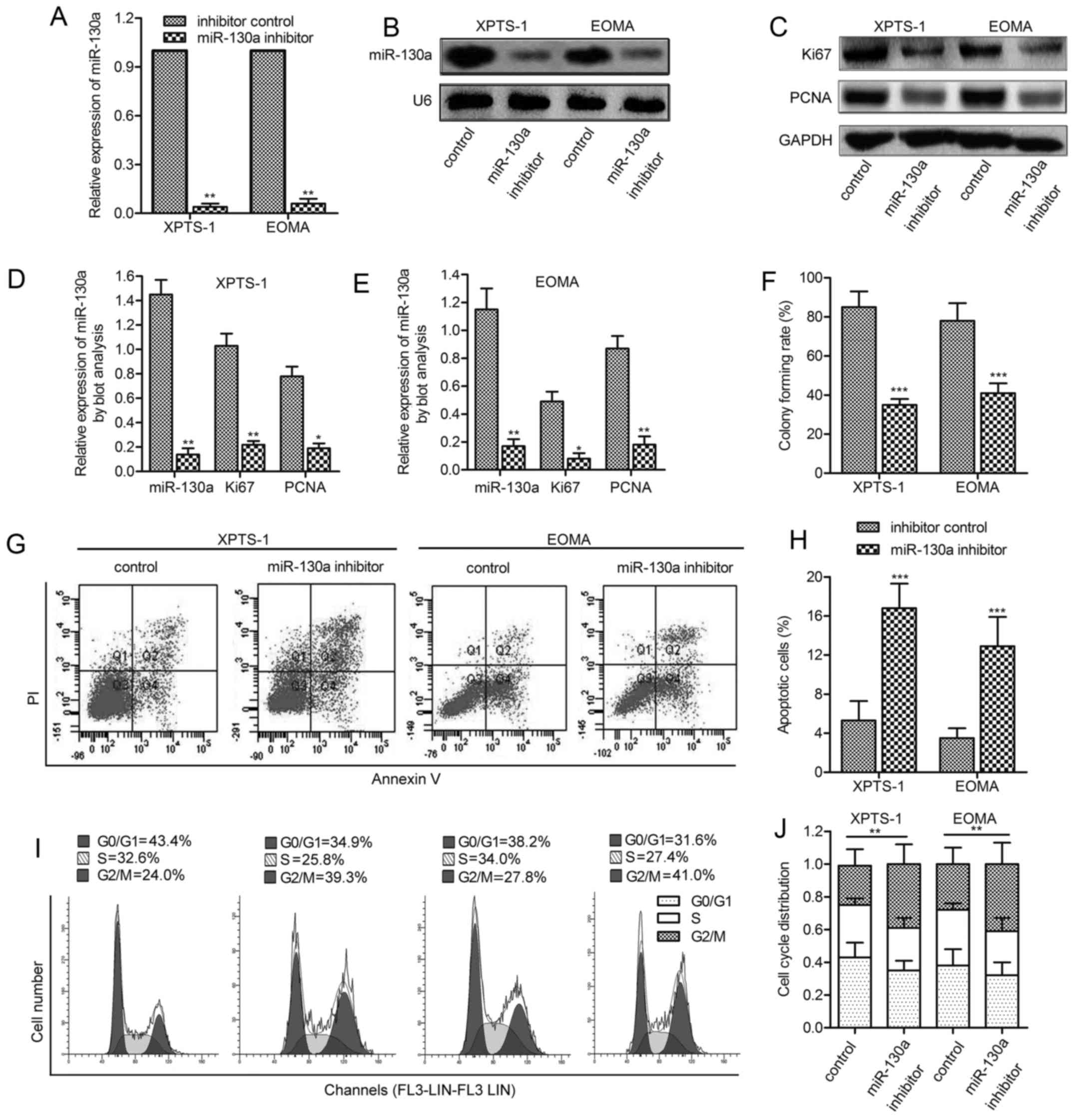
The inhibition of miR-130a restrains
hemangioma cell viability
To investigate whether miR-130a was involved in
regulating hemangioma cell growth, a synthetic inhibitor specific
for miR-130a was employed to reduce the level of endogenous
miR-130a. The efficiency of this miR-130a inhibitor was confirmed
by qPCR assay and northern blot analysis (Fig. 2A and B). Two cell proliferating
markers Ki-67 and PCNA were consistently found to be underexpressed
in XPTS-1 and EOMA cells under miR-130a inhibitor treatment
(P<0.05, P<0.01; Fig. 2C–E).
Consistently, inhibition of miR-130a significantly decreased colony
formation in XPTS-1 and EOMA cells compared with their
corresponding control (P<0.001; Fig. 2F). FACS analysis indicated that
miR-130a inhibition induced elevated level of cell apoptosis
(P<0.001; Fig. 2G and H) and an
accumulation in G2/M phase (P<0.01; Fig. 2I and J), implying the cell cycle
arrest of hemangioma cells. The above results indicate that
miR-130a inhibition reduces the viability of hemangioma in
vitro.
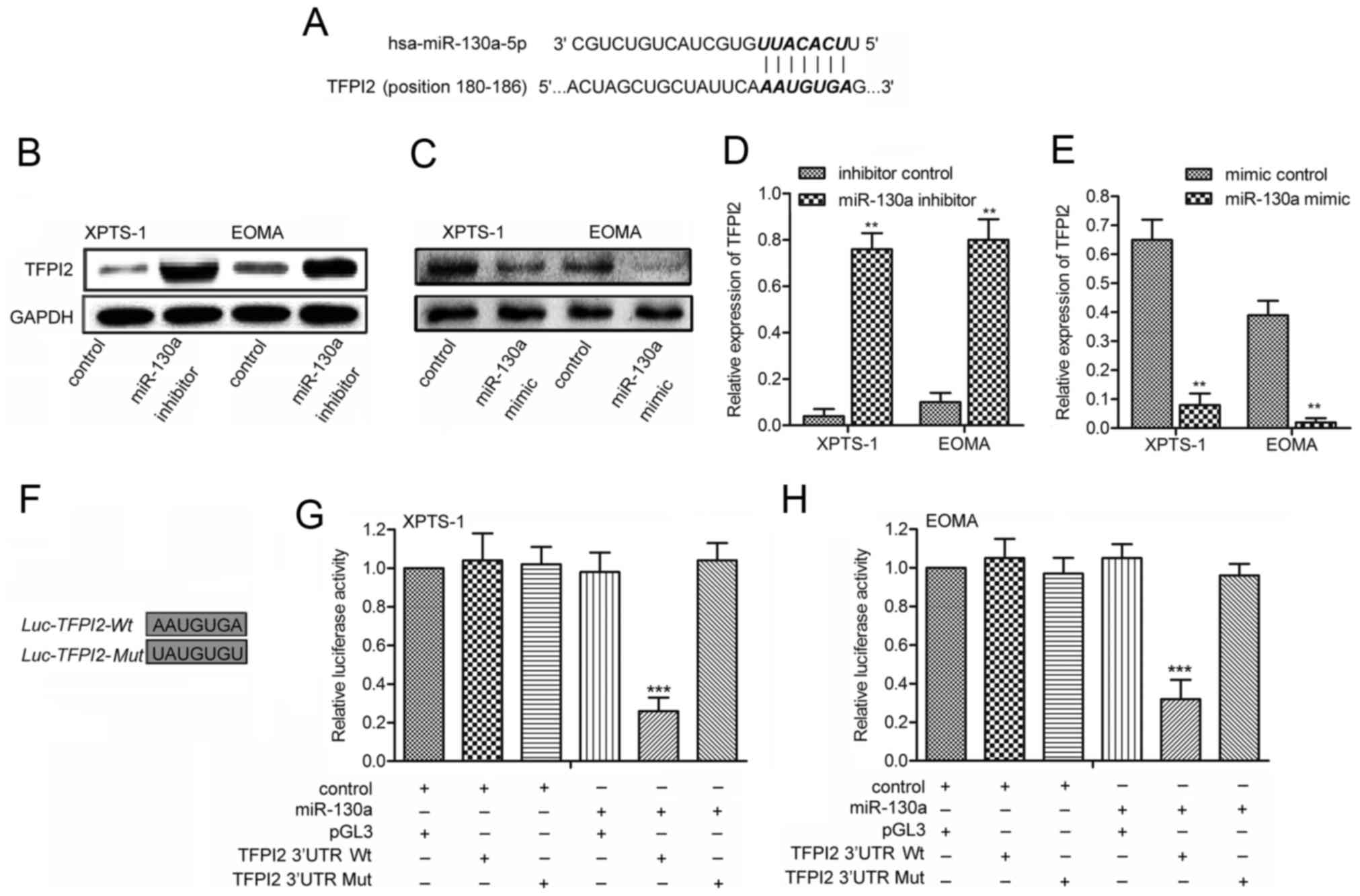
TFPI2 is a direct target of miR-130a
To explore the molecular mechanisms of miR-130a in
hemangioma cell viability, putative miR-130a targets were predicted
by TargetScan (http://www.targetscan.org/vert_71/). The results
revealed that TFPI2 was a potential target of miR-130a. The 3′-UTR
of TFPI2 mRNA contains a complementary site for the seed region of
miR-130a (Fig. 3A). To
experimentally confirm TFPI2 as an authentic target of miR-130a, a
synthetic mimic specific for miR-130a was employed to increased the
level of endogenous miR-130a. TFPI2 levels were immunoblot analyzed
adding miR-130a inhibitor or mimic in XPTS-1 and EOMA cells. The
data indicated that miR-130a inhibition induced the restoration of
TFPI2 (P<0.01; Fig. 3B and D),
whereas miR-130a overexpression caused the decline of TFPI2
expression (Fig. 3C and E). To
further verify the regulatory effect of miR-130a on TFPI2, the
plasmid pMIR-Report-TFPI2wt or pMIR-Report-TFPI2mut was transfected
into hemangioma cells together with miR-130a mimic (Fig. 3F). Luciferase expression levels of
pMIR-Report-TFPI2wt were suppressed by miR-130a overexpressing.
However, mutation of the predicted binding site of miR-130a on the
TFPI2 3′-UTR rescued the luciferase activity in XPTS-1 and EOMA
cells (P<0.001; Fig. 3G and H).
Taken together, these results support the bioinformatics
predictions indicating the TFPI2 3′-UTR to be a direct target of
miR-130a.
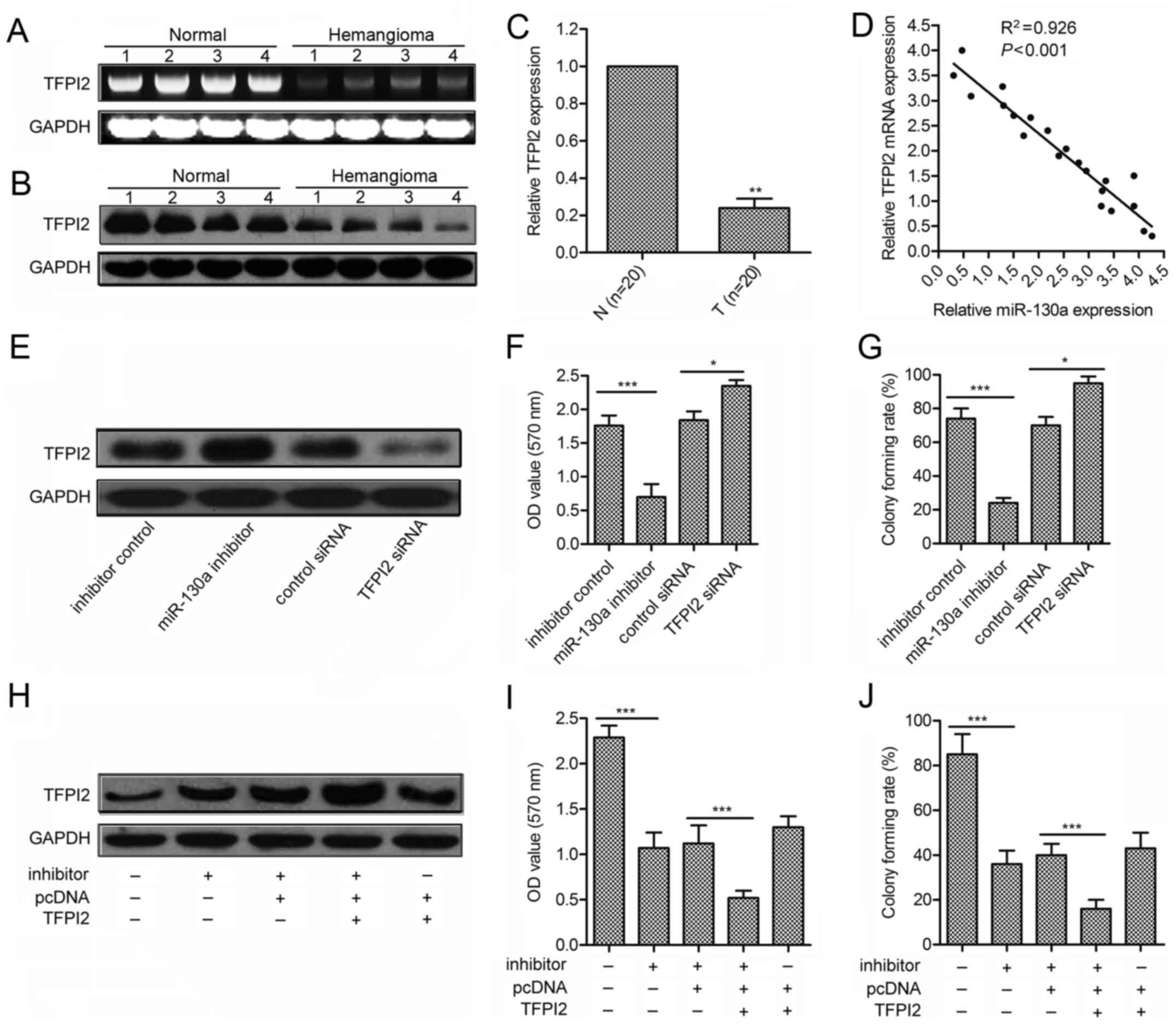
TFPI2 is involved in miR-130a-induced
hemangioma cell growth promotion
To investigate whether TFPI2 serves as a critical
mediator of miR-130a, we measured the expression of TFPI2 in 21
pairs of clinical hemangioma samples using western blotting and
qRT-PCR (Fig. 4A and B). Compared
with paired normal tissues, hemangioma showed significantly lower
TFPI2 expression (Fig. 4C). We
also analyzed the correlation between TFPI2 level and miR-130a
expression in the same patients. As shown in Fig. 4D, a significant inverse correlation
was observed (P<0.001, R=0.926). We further performed
loss-of-function and gain-of-function studies by transfecting TFPI2
siRNA or pcDNA-TFPI2 plasmids in XPTS-1 cells. As shown in Fig. 4E–G, siRNA against TFPI2 enhanced
cell growth and colony formation, which is contrary to those
induced by miR-130a inhibitor (P<0.001). Moreover,
overexpression of TFPI2 could strongly contribute to the inhibitive
effect of miR-130a inhibitor (P<0.001; Fig. 4H–J). These data suggest that TFPI2
is associated with miR-130a-induced increased hemangioma cell
viability.
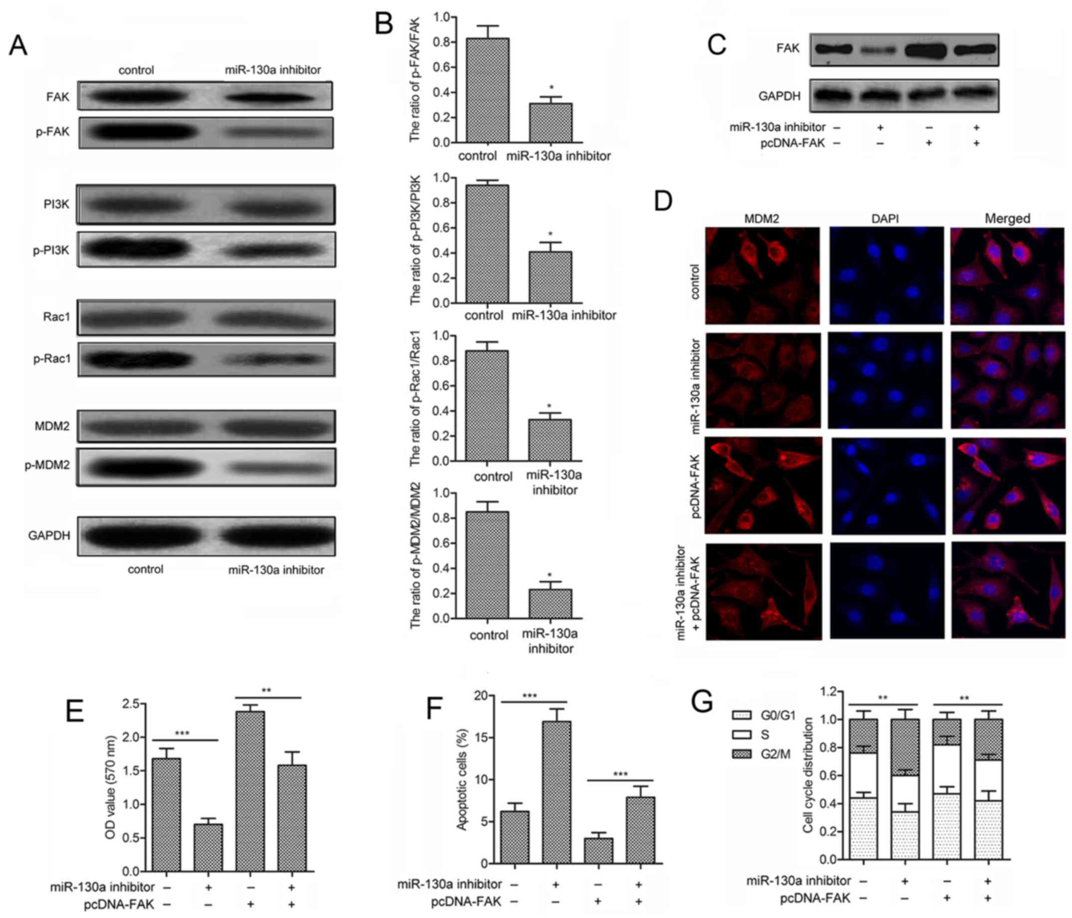
Inhibition of miR-130a suppresses the
activation of FAK/PI3K/Rac1/mdm2 signaling
To further investigate the mechanism of miR-130a in
hemangioma cell viability, the expression level of FAK signaling
(FAK, PI3K, Rac1 and mdm2) and their phosphorylated forms were
measured. No significant difference was observed in the expression
of unphosphorylated signaling kinases (FAK, PI3K, Rac1 and mdm2) in
XPTS-1 cells transfected with miR-130a inhibitor. However, the
expression of phosphorylated kinases (p-FAK, p-PI3K, p-Rac1 and
p-mdm2) was strongly suppressed in XPTS-1 cells treated with
miR-130a inhibitor transfection (Fig.
5A). The decreased ratios of p-FAK/FAK, p-PI3K/PI3K,
p-Rac1/Rac1 and p-mdm2/mdm2 further confirmed the inhibitory effect
of miR-130a inhibitor in the activation of FAK signaling
(P<0.05; Fig. 5B). To show
convincingly that miR-130a could regulate FAK signaling,
gain-of-function analyses were performed by overexpressing FAK in
XPTS-1 cells transfected with miR-130a inhibitor (Fig. 5C). Cells in miR-130a inhibitor
group showed minimal mdm2 nuclear staining, whereas cells
transfected with pcDNA-FAK exhibited maximum cytoplasmic and
nuclear staining of mdm2. Besides, cells with pcDNA-FAK
transfection displayed reduced nuclear staining of mdm2 under
miR-130a inhibitor treatment (Fig.
5D). Shown by MTT assays, miR-130a inhibitor counteracted the
promoted proliferation of XPTS-1 cells induced by pcDNA-FAK
transfection (P<0.01; Fig. 5E).
Similarly, flow cytometric analysis demonstrated that the reduced
cell apoptosis (P<0.001; Fig.
5F) and G2/M arrest (P<0.01; Fig. 5G) by pcDNA-FAK transfection was
prevented adding miR-130a inhibitor in XPTS-1 cells. These data
clearly suggest that the inhibition of miR-130a inactivates the
FAK/PI3K/Rac1/mdm2 pathway to suppress XPTS-1 cell viability.
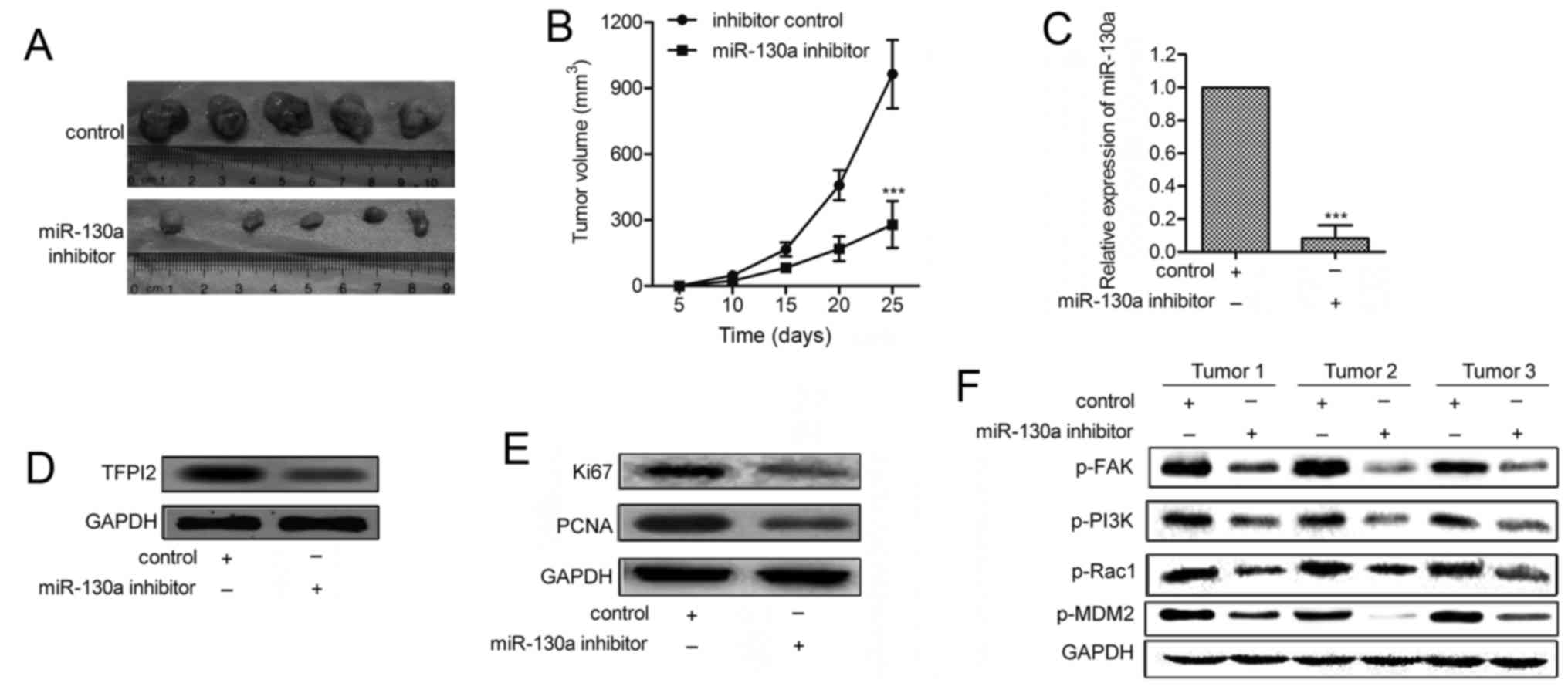
miR-130a inhibitor reduces tumor growth
in hemangioma xenograft models
To further investigate the effect of miR-130a on
tumor growth in vivo, miR-130a-downregulated and control
XPTS-1 cells were injected subcutaneously into the scapula of each
mouse (n=20). The tumor growth of the miR-130a-downregulated tumors
was significantly suppressed (Fig.
6A). The average tumor volume of miR-130a-downregulated tumors
was lower compared with control tumors (fig. 6B). The expressions of Ki-67 and
PCNA were significantly decreased in miR-130a inhibitor group
compared with control group (Fig.
6C). qRT-PCR and western blot analyses convincingly decreased
miR-130a with elevated TFPI2 levels in miR-130a-downregulated
tumors (P<0.001; Fig. 6D and
E). Finally, we examined the effect of miR-130a inhibitor on
the expression of p-FAK and its downstream genes. As shown in
Fig. 6F, the levels of p-FAK,
p-PI3K, p-Rac1and p-mdm2 were all decreased in
miR-130a-downregulated tumors. These findings indicate that
miR-130a inhibitor restrains tumor growth and the activation of
FAK/PI3K/Rac1/mdm2 pathway in vivo.
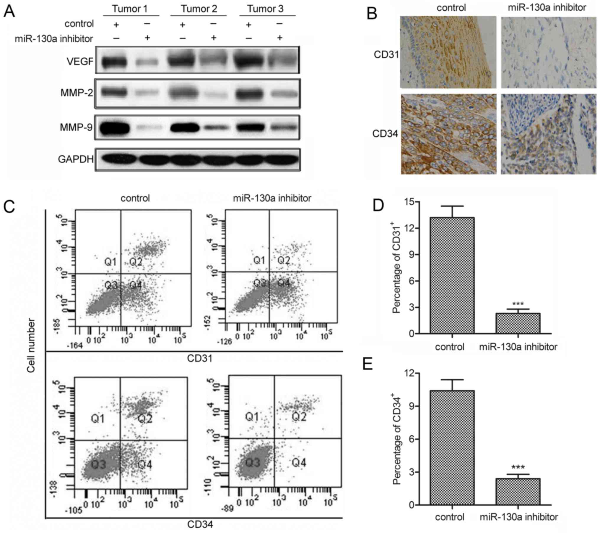
miR-130a inhibitor suppresses
angiogenesis
As the angiogenic process contribute to the
pathogenesis of hemangioma (26),
we measured the effect of miR-130a inhibitor on angiogenesis in
hemangioma xenograft models. The results revealed that miR-130a
inhibitor effectively decreased the level of angiogenesis-related
markers (VEGF, MMP-2 and MMP-9) (fig.
7A). Besides, immunohistochemistry for CD31+ and
CD34+ showed that the percentages of CD31+
and CD34+ tumor cells were significantly decreased
compared with control tumors (fig.
7B). Similarly, flow cytometric analysis convincingly showed
that, compared with control group, miR-130a inhibitor strongly
reduced the percentages of CD31+ and CD34+
cells in hemangioma xenograft models (P<0.001; Fig. 7C–E). These results indicate that
miR-130a inhibitor alleviates hemangioma via suppressing
angiogenesis.
Discussion
Infantile hemangiomas (IHs) are vascular tumors,
usually absent at birth or present as a premonitory mark with rapid
post-natal growth followed by slow involution (27). Topical antibiotics, occlusive
dressings, pulsed dye laser and triamcinolone acetonide have been
reported to be sometimes useful to limit pain and accelerate
healing (28,29). Despite the various clues that have
emerged regarding pathogenesis, many questions still remain. The
upregulation of miR-130a has been observed frequently in a variety
of human malignancies, including gastric (7,12),
bladder cancer (9), hepatocellular
carcinoma (30) and chronic
myeloid leukemia (14). Similarly,
the elevated level of miR-130a was observed in human hemangioma
tissues and cell lines in this study, and we revealed a
miR-130a/TFPI2/FAK/PI3K/Rac1/mdm2 axis that is involved in
promoting proliferation and angiogenesis in hemangioma.
The increased expression of miR-130a was reported to
be associated with tumor growth (7,9,12,31).
Duan et al (7) indicated
that overexpressed miR-130 could promote proliferation of SGC7901
cells, and miR-130 was considered as an oncogene in gastric cancer.
The miR-130/301 family has been reported to be a regulator of
multiple pro-proliferative pathways in pulmonary hypertension
(31). The research indicated that
the inhibition of miR-130a may prevent the proliferation of tumor
cells. In the present study, the designed miR-130a inhibitor was
transfected in XPTS-1 and EOMA cells. The results exhibited that
inhibition of miR-130a significantly decreased colony formation,
induced increased level of cell apoptosis and caused an
accumulation in G2/M phase. These findings suggest that miR-130a
inhibition reduces the viability of hemangioma in vitro.
Numerous reports confirmed that miRNA exerts its
effect in pathological process via regulating target genes. For
example, miR-130a exerted promoting effect on the metastasis and
epithelial-mesenchymal transition of osteosarcoma via targeting
PTEN (32). miR-130a regulated
cell migration and invasion via inhibition of Smad4 in
gemcitabine-resistant hepatoma cells (33). According to the report of Brock
et al (34) miR-130
enhanced hypoxia-induced smooth muscle proliferation and might be
involved in the development of right ventricular hypertrophy and
vascular remodeling in pulmonary hypertension, by directly
targeting tumor suppressor p21 (CDKN1A). On the basis of
bioinformatics analysis, we further predicted another miR-130a
target, TFPI2, which has been reported to regulate matrix
metalloproteinase activation and extracellular matrix degradation.
The deficiency of TFPI-2 may accelerate initiation of
atherosclerotic lesion in mice (20). Besides, methylation of TFPI2 has
been indicated to predict high risk of advanced tumor stage, early
tumor recurrence, and poor prognosis of hepatocellular carcinoma
(22). From the aspect mentioned
above, increasing the level of TFPI2 via miR-130a inhibitor may
offer a novel strategy to prevent the oncogenic effect of miR-130a
in hemangioma. In the present study, the experimental results
confirmed that TFPI2 is a functional target of miR-130a in
hemangioma cells by luciferase-reporter gene assays. The expression
of TFPI2 was significantly decreased in miR-130a overexpressing
cells. Besides, a significant inverse correlation was observed
between TFPI2 and miR-130a expression in patients. Moreover, the
knockdown of TFPI2 promoted cell growth contrary to the effect
induced by miR-130a inhibitor, whereas TFPI2 overexpression was
able to strengthen the growth inhibitive effect of miR-130a
inhibitor. These results strongly suggest that miR-130a-increased
cell growth is partly mediated by the repression of TFPI2
expression.
FAK acts as an adaptor for protein-protein
interaction or functions at sites of cell attachment to the ECM,
contributing to focal-adhesion 'scaffolding', and also transmits
adhesion-dependent signals into the cell interior (35). Rapid activation of FAK/PI3K has
been indicated to control actin reorganization in colon cancer
cells (36). Rac1, a small
GTP-binding protein, plays a pivotal role in regulating actin
dynamics. Substantial research has indicated that the loss of Rac1
activation contributed to cell migration and apoptosis (37,38).
Kallergi et al (39)
reported that activation of FAK/PI3K/Rac1 signaling regulated actin
reorganization and inhibits cell motility in malignant human breast
and prostate epithelial cells. Inhibition of the FAK/PI3K pathway
also caused less mesenchymal-like characteristics and reduced the
mobility, migration and invasion of Hep2 cells (40). Considering the effect of Rac1 in
controlling actin cytoskeletal organization in the cell, the
decreased p-Rac1/Rac1 ratio in this study suggests that miR-130a
inhibitor treatment inhibits hemangioma viability may involve actin
depolymerization. Reports also revealed that inhibiting the
phosphorylation of Akt and mdm2 could lead to the accumulation of
p53 and induced apoptosis of cancer cells (41). In accordance with these reports,
the present study demonstrated that XPTS-1 cells with pcDNA-FAK
transfection displayed reduced nuclear staining of mdm2 and cell
proliferation, increased cell apoptosis and G2/M cell cycle arrest
under miR-130a inhibitor treatment. These findings together with
in vivo experiments in hemangioma xenograft models convince
that the inhibition of miR-130a inactivates the FAK/PI3K/Rac1/mdm2
pathway to suppress hemangioma viability.
Angiogenesis is a complex process that depends on
the balance of pro- and anti-angiogenic factors that influence the
quiescence or proliferative state of the endothelium in various
diseases (8). A report revealed
that the formation of vascular tumors including hemangioma is
partly related to elevated level of angiogenic growth factors, such
as VEGF and MMPs, which lead to the development of a disorganized
blood vessel mass (42,43). According to the research of Zhao
et al (44), TFPI-2
overexpression may strongly inhibit the proliferation and migration
of VSMCs and suppress MMP-2/9 activity, making TFPI-2 a promising
candidate for treatment of atherosclerotic process. Similarly, this
study found that miR-130a inhibitor effectively decreased the level
of angiogenesis related markers and the percentages of
CD31+ and CD34+ tumor cells in hemangioma
xenograft models. These results indicate that miR-130a inhibitor
may alleviate hemangioma via targeting TFPI2 to suppress
angiogenesis.
Taken together, this study demonstrated that
miR-130a was generally upregulated in both hemangioma tissues and
cell lines. Inhibition of miR-130a reduced proliferation of
hemangioma cells and reduced tumor growth of hemangioma cells in
vivo. We also showed that TFPI2 was directly involved in the
regulation of miR-130a during hemangioma development, possibly
through inactivating FAK/PI3K/Rac1/mdm2 pathway. The final
experiments indicated that miR-130a inhibitor suppressed
angiogenesis in hemangioma xenograft models. Thus, this study
suggests that miR-130a may be a potential target for future
prevention and treatment of human hemangioma.
Abbreviations:
|
IH
|
infantile hemangioma
|
|
miR
|
microRNA
|
|
TFPI2
|
tissue factor pathway inhibitor 2
|
|
PCNA
|
proliferating cell nuclear antigen
|
|
FAK
|
focal adhesion kinase
|
|
PI3K
|
phosphoinositide 3-kinase
|
|
mdm2
|
mouse double minute
|
|
VEGF
|
vascular endothelial growth factor
|
|
MMP
|
matrix metalloproteinase
|
Acknowledgments
The present study was funded by The Natural Science
Foundation of Shandong Province, China (no. BS2014YY056).
References
|
1
|
Itinteang T, Tan ST, Brasch HD, Steel R,
Best HA, Vishvanath A, Jia J and Day DJ: Infantile haemangioma
expresses embryonic stem cell markers. J Clin Pathol. 65:394–398.
2012. View Article : Google Scholar : PubMed/NCBI
|
|
2
|
Greenberger S, Adini I, Boscolo E,
Mulliken JB and Bischoff J: Targeting NF-κB in infantile
hemangioma-derived stem cells reduces VEGF-A expression.
Angiogenesis. 13:327–335. 2010. View Article : Google Scholar : PubMed/NCBI
|
|
3
|
Waner M, North PE, Scherer KA, Frieden IJ,
Waner A and Mihm MC Jr: The nonrandom distribution of facial
hemangiomas. Arch Dermatol. 139:869–875. 2003. View Article : Google Scholar : PubMed/NCBI
|
|
4
|
Shao RZ, Zhao DH and Li J: Treatment of
infantile hemangioma by intralesional injection of propranolol
combined with compound betamethasone. Eur Rev Med Pharmacol Sci.
20:751–755. 2016.PubMed/NCBI
|
|
5
|
Villalba-Moreno AM, Cotrina-Luque J, Del
Vayo-Benito CA, Flores-Moreno S and Bautista-Paloma FJ: Nadolol for
the treatment of infantile hemangioma. Am J Health Syst Pharm.
72:44–46. 2015. View Article : Google Scholar
|
|
6
|
Furuta S, Sato H, Tsuji S, Murakami F and
Kitagawa H: Effective treatment for infantile hemangioma with
long-pulsed dye laser with oral propranolol medication: A
preliminary report. Pediatr Surg Int. 32:857–862. 2016. View Article : Google Scholar : PubMed/NCBI
|
|
7
|
Duan J, Zhang H, Qu Y, Deng T, Huang D,
Liu R, Zhang L, Bai M, Zhou L, Ying G, et al: Onco-miR-130 promotes
cell proliferation and migration by targeting TGFβR2 in gastric
cancer. Oncotarget. 7:44522–44533. 2016.PubMed/NCBI
|
|
8
|
Anand S, Majeti BK, Acevedo LM, Murphy EA,
Mukthavaram R, Scheppke L, Huang M, Shields DJ, Lindquist JN,
Lapinski PE, et al: MicroRNA-132-mediated loss of p120RasGAP
activates the endothelium to facilitate pathological angiogenesis.
Nat Med. 16:909–914. 2010. View
Article : Google Scholar : PubMed/NCBI
|
|
9
|
Egawa H, Jingushi K, Hirono T, Ueda Y,
Kitae K, Nakata W, Fujita K, Uemura M, Nonomura N and Tsujikawa K:
The miR-130 family promotes cell migration and invasion in bladder
cancer through FAK and Akt phosphorylation by regulating PTEN. Sci
Rep. 6:205742016. View Article : Google Scholar : PubMed/NCBI
|
|
10
|
Yang Y, Yang L, Liang X and Zhu G:
MicroRNA-155 promotes atherosclerosis inflammation via targeting
SOCS1. Cell Physiol Biochem. 36:1371–1381. 2015. View Article : Google Scholar : PubMed/NCBI
|
|
11
|
Zheng SL, Stevens VL, Wiklund F, Isaacs
SD, Sun J, Smith S, Pruett K, Wiley KE, Kim ST, Zhu Y, et al: Two
independent prostate cancer risk-associated loci at 11q13. Cancer
Epidemiol Biomarkers Prev. 18:1815–1820. 2009. View Article : Google Scholar : PubMed/NCBI
|
|
12
|
Jiang H, Yu WW, Wang LL and Peng Y:
miR-130a acts as a potential diagnostic biomarker and promotes
gastric cancer migration, invasion and proliferation by targeting
RUNX3. Oncol Rep. 34:1153–1161. 2015.PubMed/NCBI
|
|
13
|
Feng Y, Zhou S, Li G, Hu C, Zou W, Zhang H
and Sun L: Nuclear factor-κB-dependent microRNA-130a upregulation
promotes cervical cancer cell growth by targeting phosphatase and
tensin homolog. Arch Biochem Biophys. 598:57–65. 2016. View Article : Google Scholar : PubMed/NCBI
|
|
14
|
Li Q, Wu Y, Zhang J, Yi T and Li W:
MicroRNA-130a regulates cell malignancy by targeting RECK in
chronic myeloid leukemia. Am J Transl Res. 8:955–967.
2016.PubMed/NCBI
|
|
15
|
Zheng H, Dong X, Liu N, Xia W, Zhou L and
Chen X, Yang Z and Chen X: Regulation and mechanism of mouse
miR-130a/b in metabolism-related inflammation. Int J Biochem Cell
Biol. 74:72–83. 2016. View Article : Google Scholar : PubMed/NCBI
|
|
16
|
Chen Y and Gorski DH: Regulation of
angiogenesis through a microRNA (miR-130a) that down-regulates
antiangiogenic homeobox genes GAX and HOXA5. Blood. 111:1217–1226.
2008. View Article : Google Scholar
|
|
17
|
Herman MP, Sukhova GK, Kisiel W, Foster D,
Kehry MR, Libby P and Schönbeck U: Tissue factor pathway
inhibitor-2 is a novel inhibitor of matrix metalloproteinases with
implications for atherosclerosis. J Clin Invest. 107:1117–1126.
2001. View
Article : Google Scholar : PubMed/NCBI
|
|
18
|
Chand HS, Foster DC and Kisiel W:
Structure, function and biology of tissue factor pathway
inhibitor-2. Thromb Haemost. 94:1122–1130. 2005.
|
|
19
|
Seppälä M, Wahlström T and Bohn H:
Circulating levels and tissue localization of placental protein
five (PP5) in pregnancy and trophoblastic disease: Absence of PP5
expression in the malignant trophoblast. Int J Cancer. 24:6–10.
1979. View Article : Google Scholar : PubMed/NCBI
|
|
20
|
Hong J, Liu R, Chen L, Wu B, Yu J, Gao W,
Pan J, Luo X and Shi H: Conditional knockout of tissue factor
pathway inhibitor 2 in vascular endothelial cells accelerates
atherosclerotic plaque development in mice. Thromb Res.
137:148–156. 2016. View Article : Google Scholar
|
|
21
|
Zhai LL, Wu Y, Cai CY and Tang ZG:
Upregulated matrix metalloproteinase-2 and downregulated tissue
factor pathway inhibitor-2 are risk factors for lymph node
metastasis and perineural invasion in pancreatic carcinoma. Onco
Targets Ther. 8:2827–2834. 2015. View Article : Google Scholar : PubMed/NCBI
|
|
22
|
Sun FK, Sun Q, Fan YC, Gao S, Zhao J, Li
F, Jia YB, Liu C, Wang LY, Li XY, et al: Methylation of tissue
factor pathway inhibitor 2 as a prognostic biomarker for
hepatocellular carcinoma after hepatectomy. J Gastroenterol
Hepatol. 31:484–492. 2016. View Article : Google Scholar
|
|
23
|
Dong Y, Tan Q, Tao L, Pan X, Pang L, Liang
W, Liu W, Zhang W, Li F and Jia W: Hypermethylation of TFPI2
correlates with cervical cancer incidence in the Uygur and Han
populations of Xinjiang, China. Int J Clin Exp Pathol. 8:1844–1854.
2015.PubMed/NCBI
|
|
24
|
Crawley JT, Goulding DA, Ferreira V,
Severs NJ and Lupu F: Expression and localization of tissue factor
pathway inhibitor-2 in normal and atherosclerotic human vessels.
Arterioscler Thromb Vasc Biol. 22:218–224. 2002. View Article : Google Scholar : PubMed/NCBI
|
|
25
|
Liu J, Ma L, Li C, Zhang Z, Yang G and
Zhang W: Tumor-targeting TRAIL expression mediated by miRNA
response elements suppressed growth of uveal melanoma cells. Mol
Oncol. 7:1043–1055. 2013. View Article : Google Scholar : PubMed/NCBI
|
|
26
|
Wang FQ, Chen G, Zhu JY, Zhang W, Ren JG,
Liu H, Sun ZJ, Jia J and Zhao YF: M2-polarised macrophages in
infantile haemangiomas: Correlation with promoted angiogenesis. J
Clin Pathol. 66:1058–1064. 2013. View Article : Google Scholar : PubMed/NCBI
|
|
27
|
Liang MG and Frieden IJ: Infantile and
congenital hemangiomas. Semin Pediatr Surg. 23:162–167. 2014.
View Article : Google Scholar : PubMed/NCBI
|
|
28
|
Witman PM, Wagner AM, Scherer K, Waner M
and Frieden IJ: Complications following pulsed dye Laser treatment
of superficial hemangiomas. Lasers Surg Med. 38:116–123. 2006.
View Article : Google Scholar : PubMed/NCBI
|
|
29
|
Pandey A, Gangopadhyay AN, Sharma SP,
Kumar V, Gupta DK and Gopal SC: Evaluation of topical steroids in
the treatment of superficial hemangioma. Skinmed. 8:9–11.
2010.PubMed/NCBI
|
|
30
|
Xu N, Shen C, Luo Y, Xia L, Xue F, Xia Q
and Zhang J: Upregulated miR-130a increases drug resistance by
regulating RUNX3 and Wnt signaling in cisplatin-treated HCC cell.
Biochem Biophys Res Commun. 425:468–472. 2012. View Article : Google Scholar : PubMed/NCBI
|
|
31
|
Bertero T, Cottrill K, Krauszman A, Lu Y,
Annis S, Hale A, Bhat B, Waxman AB, Chau BN, Kuebler WM, et al: The
microRNA-130/301 family controls vasoconstriction in pulmonary
hypertension. J Biol Chem. 290:2069–2085. 2015. View Article : Google Scholar :
|
|
32
|
Chen J, Yan D, Wu W, Zhu J, Ye W and Shu
Q: MicroRNA-130a promotes the metastasis and epithelial-mesenchymal
transition of osteosarcoma by targeting PTEN. Oncol Rep.
35:3285–3292. 2016.PubMed/NCBI
|
|
33
|
Liu Y, Li Y, Wang R, Qin S, Liu J, Su F,
Yang Y, Zhao F, Wang Z and Wu Q: MiR-130a-3p regulates cell
migration and invasion via inhibition of Smad4 in gemcitabine
resistant hepatoma cells. J Exp Clin Cancer Res. 35:192016.
View Article : Google Scholar : PubMed/NCBI
|
|
34
|
Brock M, Haider TJ, Vogel J, Gassmann M,
Speich R, Trenkmann M, Ulrich S, Kohler M and Huber LC: The
hypoxia-induced microRNA-130a controls pulmonary smooth muscle cell
proliferation by directly targeting CDKN1A. Int J Biochem Cell
Biol. 61:129–137. 2015. View Article : Google Scholar : PubMed/NCBI
|
|
35
|
Mclean GW, Carragher NO, Avizienyte E,
Evans J, Brunton VG and Frame MC: The role of focal-adhesion kinase
in cancer - a new therapeutic opportunity. Nat Rev Cancer.
5:505–515. 2005. View Article : Google Scholar : PubMed/NCBI
|
|
36
|
Gu S, Kounenidakis M, Schmidt EM,
Deshpande D, Alkahtani S, Alarifi S, Föller M, Alevizopoulos K,
Lang F and Stournaras C: Rapid activation of
FAK/mTOR/p70S6K/PAK1-signaling controls the early
testosterone-induced actin reorganization in colon cancer cells.
Cell Signal. 25:66–73. 2013. View Article : Google Scholar : PubMed/NCBI
|
|
37
|
Bhat SS, Parray AA, Mushtaq U, Fazili KM
and Khanday FA: Actin depolymerization mediated loss of SNTA1
phosphorylation and Rac1 activity has implications on ROS
production, cell migration and apoptosis. Apoptosis. 21:737–748.
2016. View Article : Google Scholar : PubMed/NCBI
|
|
38
|
Tejada-Simon MV: Modulation of actin
dynamics by Rac1 to target cognitive function. J Neurochem.
133:767–779. 2015. View Article : Google Scholar : PubMed/NCBI
|
|
39
|
Kallergi G, Agelaki S, Markomanolaki H,
Georgoulias V and Stournaras C: Activation of FAK/PI3K/Rac1
signaling controls actin reorganization and inhibits cell motility
in human cancer cells. Cell Physiol Biochem. 20:977–986. 2007.
View Article : Google Scholar : PubMed/NCBI
|
|
40
|
Yang L, Zhou Q, Chen X, Su L, Liu B and
Zhang H: Activation of the FAK/PI3K pathway is crucial for
AURKA-induced epithelial-mesenchymal transition in laryngeal
cancer. Oncol Rep. 36:819–826. 2016.PubMed/NCBI
|
|
41
|
Wang SQ, Wang C, Chang LM, Zhou KR, Wang
JW, Ke Y, Yang DX, Shi HG, Wang R, Shi XL, et al: Geridonin and
paclitaxel act synergistically to inhibit the proliferation of
gastric cancer cells through ROS-mediated regulation of the
PTEN/PI3K/Akt pathway. Oncotarget. 7:72990–73002. 2016.PubMed/NCBI
|
|
42
|
Greenberger S, Boscolo E, Adini I,
Mulliken JB and Bischoff J: Corticosteroid suppression of VEGF-A in
infantile hemangioma-derived stem cells. N Engl J Med.
362:1005–1013. 2010. View Article : Google Scholar : PubMed/NCBI
|
|
43
|
Thaivalappil S, Bauman N, Saieg A, Movius
E, Brown KJ and Preciado D: Propranolol-mediated attenuation of
MMP-9 excretion in infants with hemangiomas. JAMA Otolaryngol Head
Neck Surg. 139:1026–1031. 2013. View Article : Google Scholar : PubMed/NCBI
|
|
44
|
Zhao B, Luo X, Shi H and Ma D: Tissue
factor pathway inhibitor-2 is downregulated by ox-LDL and inhibits
ox-LDL induced vascular smooth muscle cells proliferation and
migration. Thromb Res. 128:179–185. 2011. View Article : Google Scholar : PubMed/NCBI
|