Introduction
The International Society for the Study of Vascular
Anomalies (ISSVA) classifies vascular anomalies into vascular
tumors and vascular malformations, according to the biological
classification proposed by Mulliken and Glowacki (1,2).
Arteriovenous malformations (AVMs) are high-flow vascular
malformations in which well-formed arterial and venous elements
communicate directly, rather than through a normal capillary
network. Although the majority of AVMs occur in soft tissues,
osseous involvement is occasionally encountered, with symptoms such
as pain, mass effects with pulsation, swelling of the extremity,
skin ulceration, bone overgrowth, bone destruction, and heart
failure (3,4).
Intraosseous AVMs, which are relatively rare,
predominantly occur in craniofacial bones, such as the mandible and
maxilla (5,6). Pure intraosseous AVMs that do not
involve soft tissue are extremely rare (3,4).
Although several intraosseous AVMs have been described in the spine
or long bones (7–14), little is known on the diagnostic and
therapeutic strategies for pure intraosseous AVMs. It is crucial
for physicians, particularly orthopedic oncologists, to understand
the clinical characteristics of pure intraosseous AVMs in order to
distinguish them from primary or metastatic bone tumors. We herein
present a case of pure intraosseous AVM of the proximal femur in a
patient with polyostotic fibrous dysplasia, which was treated by
embolization and denosumab, and discuss the diagnostic
characteristics and therapeutic approach.
Case report
A 37-year-old woman was referred to the Hirosaki
University Hospital (Hirosaki, Japan) on December, 2014 with a
1-year history of pain in the left thigh while walking. The
patient's medical history included a diagnosis of polyostotic
fibrous dysplasia (poly-FD) when she was in her 20s at another
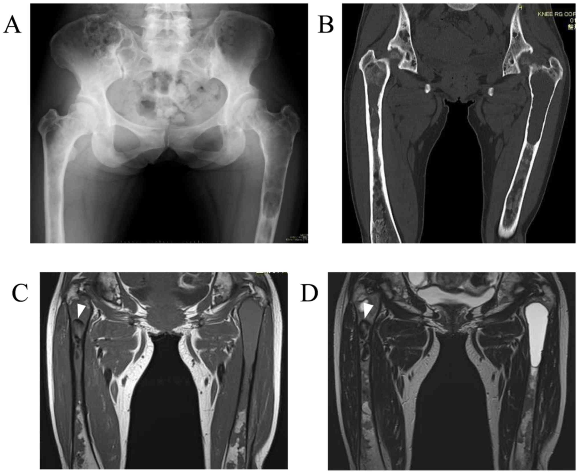
hospital, without endocrinopathy or skin pigmentation. Plain
radiographs and computed tomography (CT) examination revealed a
central, longitudinal, medullary lytic lesion of the left proximal
femur, with expansion and thinning of the bone cortex (Fig. 1A and B). An intramedullary lytic
lesion and endosteal scalloping were also observed in the right
proximal femur. There were multiple intramedullary lesions with a
ground-glass appearance and lytic lesions throughout the bilateral
femora and pelvis. Magnetic resonance imaging (MRI) revealed a
lesion in the left proximal femur that exhibited a homogeneous
intermediate signal on T1-weighted images and hyperintensity on the
T2-weighted images (Fig. 1C and D).
In the right proximal femur, an intramedullary hypointense lesion
was identified on both T1- and T2-weighted images. An impending
fracture of the left femur was diagnosed, resulting from a
secondary cyst formation of the FD, and intramedullary nailing of
the left femur was performed. The patient had an uneventful
recovery after surgery. At the 1-year follow-up on December, 2015,
she remained asymptomatic, without progressive changes in the right
lower extremity on plain radiographs, and was scheduled for routine
orthopedic follow-up at another hospital.
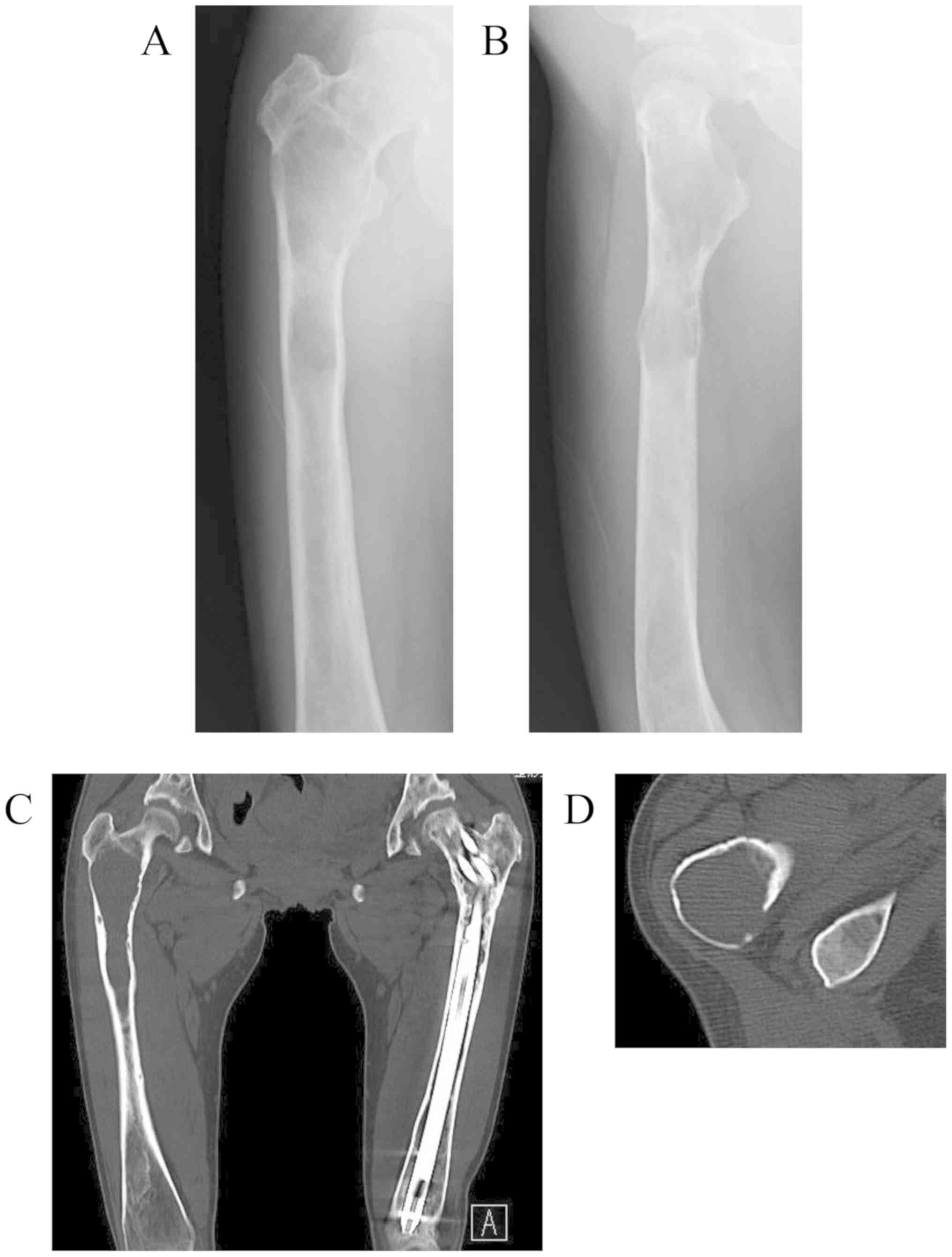
The patient visited our hospital again at the age of
39 years on November, 2016, with a 5-month history of right thigh
pain while walking. Plain radiographs and CT scans revealed
worsening of the findings compared with those 2 years earlier,
consisting of a central, longitudinal medullary lytic lesion with
expansion and thinning of the bone cortex (Fig. 2A-C). There was a mild disruption of
the intertrochanteric posterior bone cortex (Fig. 2D), but there were no soft tissue
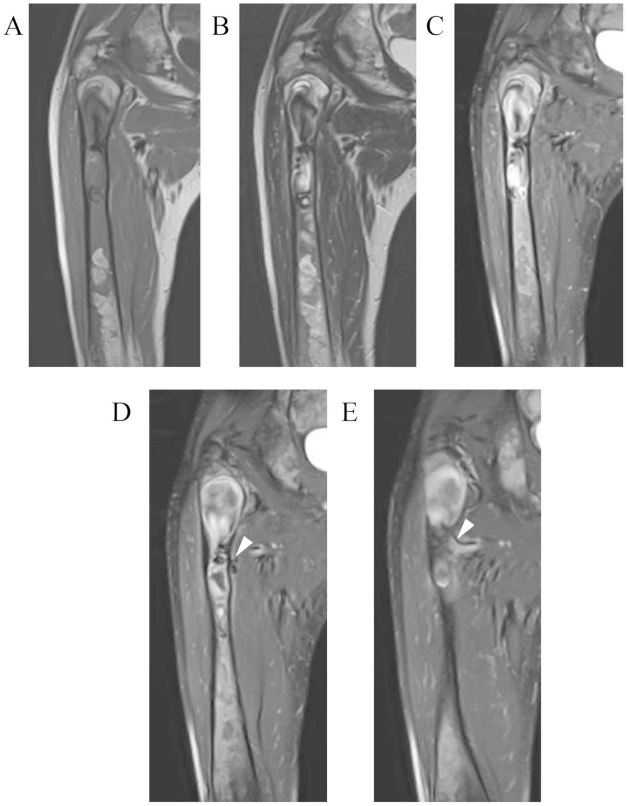
masses or periosteal reaction. MRI revealed an extended
intramedullary hypointense lesion on both T1- and T2-weighted
images (Fig. 3A and B). The
fat-suppressed post-contrast T1-weighted MRI revealed heterogeneous
enhancement around the intramedullary hypointense lesion (Fig. 3C). There were no soft tissue masses
near the right proximal femur. An impending fracture of the right
femur was diagnosed, resulting from progression of the FD.
Intramedullary nailing of the right femur was attempted, but the
operation was discontinued due to rapid profuse bleeding after
drilling the greater trochanter. The hemorrhage was controlled by
filling the drill hole with bone wax. A review of the preoperative
MRI revealed several feeding arteries arising from the deep femoral
artery that were responsible for the intramedullary hypointense
signal (signal void; Fig. 3D and E).
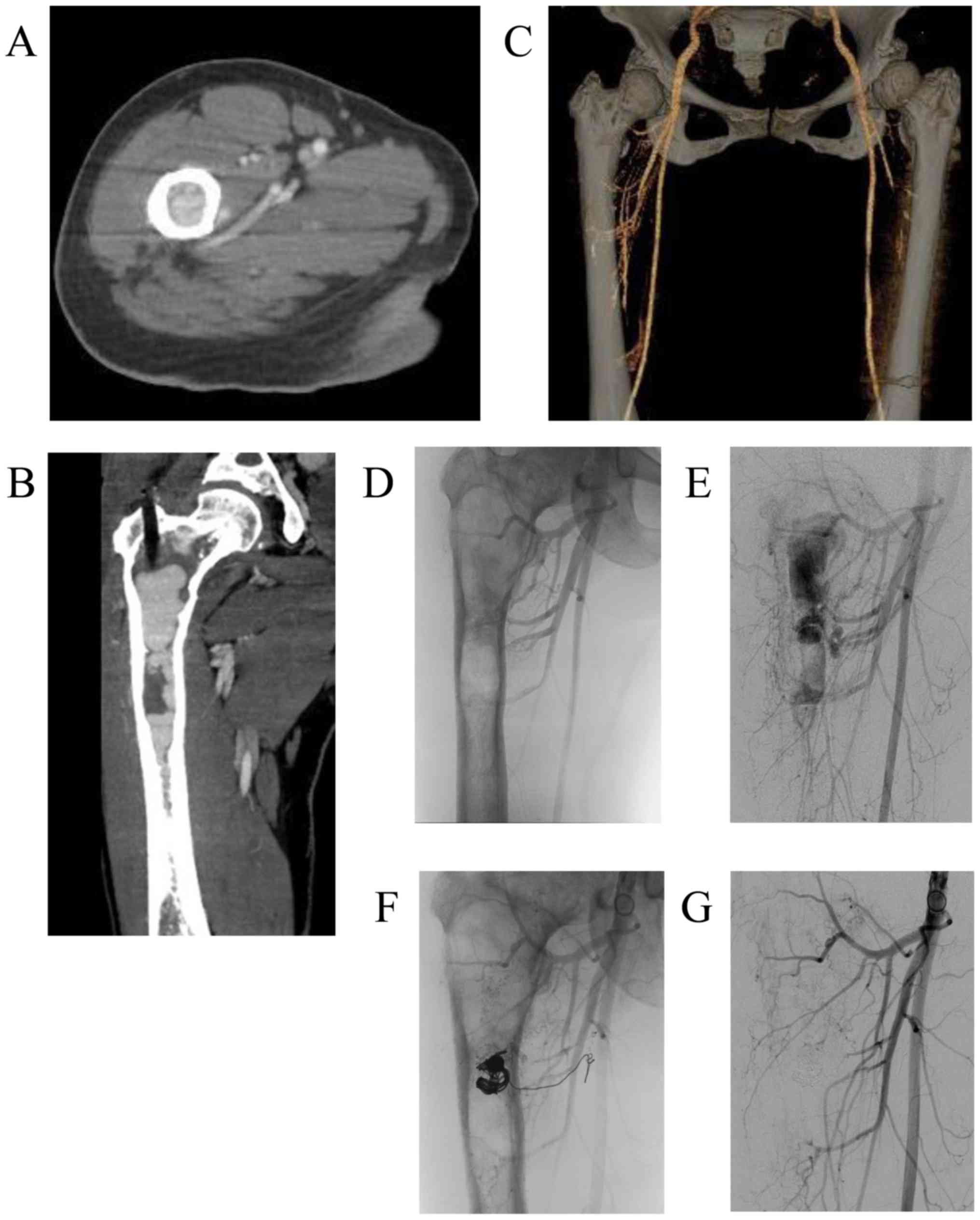
Contrast-enhanced CT and 3-dimensional CT images obtained on the
day after the discontinued surgery revealed marked intramedullary
enhancement of the proximal femoral lesion and multiple
hypertrophied perforating arteries arising from the deep femoral
artery (Fig. 4A-C). Selective
angiography of the deep femoral artery demonstrated multiple
hypertrophied perforating arteries penetrating the femoral cortex,
as well as early formation of the nidus and venous sac within the
medullary cavity of the femur (Fig. 4D
and E). These findings indicated that the proximal femoral
lesion was an intraosseous AVM.
Therapeutic embolization was then performed for a
total of 3 times (on days 2, 9 and 21 after the initial surgery).
In the first and second embolizations, transarterial n-butyl-2-
cyanoacrylate (NBCA) injections were performed. In the third
embolization, a small venous sac remaining within the medullary
cavity was completely obliterated by transvenous coil embolization
(Fig. 4F and G). The patient was
able to stand and walk with full weight-bearing without pain on the
day after the final embolization, but she was advised to walk with
crutches with load restriction on the right lower extremity and to
avoid activities that would increase the risk of pathological
fracture for 3 months after the embolization. In addition,
biochemical measurements, including serum total alkaline
phosphatase (ALP), bone alkaline phosphatase (BAP), calcium,
phosphate and urine N-terminal cross-linked telopeptide of type I
collagen (NTX), indicated high bone turnover rate (Table I). Therefore, subcutaneous injections
of 60 mg denosumab (Pralia®; once every 6 months) with
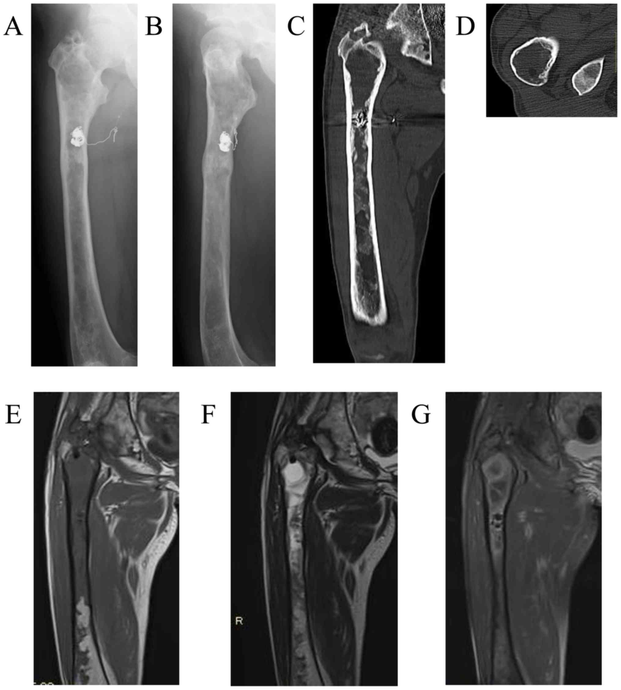
oral activated vitamin D and calcium were initiated. One year after
the embolization, the patient remained asymptomatic, and the right
proximal femur exhibited increased cortical thickness on plain
radiographs and CT images (Fig.
5A-D). No intramedullary signal void was detected on MRI
(Fig. 5E-G). Biochemical tests
before and after treatment revealed attenuation of the
high-turnover bone metabolism (Table
I). The bone mineral density (BMD) of the lumbar spine (L2-L4)
on dual-energy-X-ray absorptiometry was also increased on December,
2017 after 1 year treatment.
 | Table I.Changes in biochemical measurements
and lumbar spine (L2-L4) BMD before and after denosumab
administration. |
Table I.
Changes in biochemical measurements
and lumbar spine (L2-L4) BMD before and after denosumab
administration.
| Measurement time
point | Serum ALP (normal
range, 115–359 U/l) | Serum BAP (normal
range, 2.9–14.5 U/l) | Serum calcium
corrected for albumin level (normal range, 8.7–10.3 mg/dl) | Serum phosphate
(normal range, 2.5–4.7 mg/dl) | Urine NTX/Cr
(reference range, 9.3–54.3 nMBCE/mMCr) | BMD
(g/cm2) |
|---|
| Pretreatment | 627 | 66.6 | 9.3 | 3.4 | 440.5 | 1.253 |
| Post-treatment (1
week) | 524 | 57.2 | 8.8 | 1.9 | 89.4 |
|
| Post-treatment (1
year) | 187 | 13.3 | 9.4 | 2.7 | 15.1 | 1.459 |
Discussion
The present case demonstrated that careful imaging
review is crucial for distinguishing bone tumors from intraosseous
AVMs, which are characterized by signal void and the presence of
feeding arteries on MRI. Denosumab treatment also achieved a
satisfactory result, including increased cortical thickness of the
right proximal femur and attenuation of high-turnover bone
metabolism. To the best of our knowledge, this is the first report
of pure intraosseous AVM occurring in combination with poly-FD.
MRI examination in this case clearly demonstrated
intramedullary signal voids and feeding arteries in a pure
intraosseous AVM of a limb bone. MRI is an extremely important
modality for assessing vascular tumors and malformations, and the
presence of signal void is a hallmark MRI finding in AVMs (15–17). In
previous reports (7–14), intramedullary lesions exclusively
displayed a hypointense or isointense signal on T1-weighted images
and a hyperintense signal on T2-weighted images, without signal
voids. However, in the present case, MRI demonstrated
intramedullary signal voids and feeding arteries, indicating a
high-flow malformation. Although the signal voids were
misinterpreted as solid fibrous tissues on initial diagnosis,
contrast-enhanced CT pointed towards the correct diagnosis, as it
revealed marked intramedullary enhancement and hypertrophied
surrounding arteries. Therefore, careful imaging review is
warranted to avoid unnecessary bone biopsies and surgeries that may
cause excessive bleeding.
In this case, complete obliteration of the
intraosseous AVM was achieved by transarterial NBCA injections and
transvenous coil embolization. The management of patients with AVM
includes conservative treatment, transcatheter embolization,
sclerotherapy, surgical resection, or a combination of these
methods. Conservative treatment is recommended for asymptomatic or
mildly impaired patients, as incomplete treatment may stimulate
more aggressive growth (17).
Transcatheter embolization is recommended for symptomatic, impaired
patients with extensive high-flow lesions, either preoperatively or
as a first-line therapeutic strategy, when surgical resection
carries a significant risk of postoperative morbidity (5,6). In
addition, sclerotherapy may be applied preoperatively or as a
definitive treatment in selected patients (18). Do et al reported good results
of ethanol embolotherapy for extremity AVM involving bone (3). Complete surgical resection is
recommended when the AVM is small and located in a surgically
accessible area.
In the present case, denosumab achieved increased
cortical thickness of the proximal femur and attenuated the
high-turnover bone metabolism. Pathological fracture was a concern
in this case, due to the low blood supply post-embolization and the
high-turnover bone metabolism. Although surgical treatment is
usually recommended for FD of the lower limbs causing pain and
deformity (19), good results were
achieved with denosumab, which rapidly decreased the serum ALP, BAP
and urine NTX levels (Table I).
Several previous studies reported that intravenous pamidoronate or
oral alendronate markedly relieved bone pain, improved radiological
findings, increased bone density and decreased bone turnover in
children or adults with poly-FD (20,21).
Ganda and Seibel reported that denosumab induced a rapid
biochemical response and alleviated bone pain in two poly-FD
patients in whom the efficacy of prior long-term bisphosphonate
therapy was unsatisfactory (22). In
the present case, we hypothesized that the increased cortical
thickness of the proximal femur 1 year after embolization was the
result of improved bone metabolism by denosumab, as well as of the
spontaneous bone remodeling after the complete obliteration of the
AVM.
In conclusion, we herein report a case of pure
intraosseous AVM of the proximal femur in a patient with poly-FD.
Non-surgical treatment by embolization and denosumab achieved a
satisfactory result, which included complete obliteration of the
AVM, increased cortical thickness of the proximal femur, and
improvement of the high-turnover bone metabolism 1 year after
embolization. Careful imaging review is crucial for distinguishing
between bone tumors and intraosseous AVMs, which is characterized
by the presence of signal voids and feeding arteries on MRI, in
order to avoid unnecessary bone biopsy or surgery.
Acknowledgements
Not applicable.
Funding
No funding was received.
Availability of data and materials
All the data analyzed in this case are available
from the corresponding author on reasonable request.
Authors' contributions
SO, MY, YI participated in the treatment of the
patient including pharmacotherapy and surgery. FT performed
arterial embolization on the patient. All the authors have read and
approved the final version of this manuscript.
Ethics approval and consent to
participate
Not applicable.
Patient consent for publication
We received patient consent for the publication of
the case details and any associated laboratory data and images.
Competing interests
The authors declare that they have no competing
interests.
Glossary
Abbreviations
Abbreviations:
|
AVM
|
arteriovenous malformation
|
|
poly-FD
|
polyostotic fibrous dysplasia
|
References
|
1
|
Mulliken JB and Glowacki J: Hemangiomas
and vascular malformations in infants and children: A
classification based on endothelial characteristics. Plast Reconstr
Surg. 69:412–422. 1982. View Article : Google Scholar : PubMed/NCBI
|
|
2
|
Enjolras O: Classification and management
of the various superficial vascular anomalies: Hemangiomas and
vascular malformations. J Dermatol. 24:701–710. 1997. View Article : Google Scholar : PubMed/NCBI
|
|
3
|
Do YS, Park KB, Park HS, Cho SK, Shin SW,
Moon JW, Kim DI, Kim YW, Chang IS, Lee SH, et al: Extremity
arteriovenous malformations involving the bone: Therapeutic
outcomes of ethanol embolotherapy. J Vasc Interv Radiol.
21:807–816. 2010. View Article : Google Scholar : PubMed/NCBI
|
|
4
|
Do YS and Park KB: Special consideration
for intraosseous arteriovenous malformation. Semin Intervent
Radiol. 34:272–279. 2017. View Article : Google Scholar : PubMed/NCBI
|
|
5
|
Colletti G, Frigerio A, Giovanditto F,
Biglioli F, Chiapasco M and Grimmer JF: Surgical treatment of
vascular malformations of the facial bones. J Oral Maxillofac Surg.
72:1326.e1–18. 2014. View Article : Google Scholar
|
|
6
|
Theologie-Lygidakis N, Schoinohoriti O,
Tzermpos F, Christopoulos P and Iatrou I: Management of
intraosseous vascular malformations of the jaws in children and
adolescents: Report of 6 cases and literature review. J Oral
Maxillofac Res. 6:e52015. View Article : Google Scholar : PubMed/NCBI
|
|
7
|
Katzen BT and Said S: Arteriovenous
malformation of bone: An experience with therapeutic embolization.
AJR Am J Roentgenol. 136:427–429. 1981. View Article : Google Scholar : PubMed/NCBI
|
|
8
|
Nancarrow PA, Lock JE and Fellows KE:
Embolization of an intraosseous arteriovenous malformation. AJR Am
J Roentgenol. 146:785–786. 1986. View Article : Google Scholar : PubMed/NCBI
|
|
9
|
Savader SJ, Savader BL and Otero RR:
Intraosseous arteriovenous malformations mimicking malignant
disease. Acta Radiol. 29:109–114. 1988. View Article : Google Scholar : PubMed/NCBI
|
|
10
|
Knych SA, Goldberg MJ and Wolfe HJ:
Intraosseous arteriovenous malformation in a pediatric patient.
Clin Orthop Relat Res. 276:307–312. 1992.
|
|
11
|
Molina A, Martin C, Muñoz I, Aguilar L,
Serrano S and Ballester J: Spinal intraosseous arteriovenous
malformation as a cause of juvenile scoliosis. A case report. Spine
(Phila Pa 1976). 22:221–224. 1997. View Article : Google Scholar : PubMed/NCBI
|
|
12
|
Matsuyama A, Aoki T, Hisaoka M, Yokoyama K
and Hashimoto H: A case of intraosseous arteriovenous malformation
with unusual radiological presentation of low blood flow. Pathol
Res Pract. 204:423–426. 2008. View Article : Google Scholar : PubMed/NCBI
|
|
13
|
Louis RG Jr, Yen CP, Mohila CA, Mandell JW
and Sheehan J: A rare intraosseous arteriovenous malformation of
the spine. J Neurosurg Spine. 15:336–339. 2011. View Article : Google Scholar : PubMed/NCBI
|
|
14
|
Wang HH, Yeh TT, Lin YC and Huang GS:
Imaging features of an intraosseous arteriovenous malformation in
the tibia. Singapore Med J. 56:e21–e25. 2015. View Article : Google Scholar : PubMed/NCBI
|
|
15
|
Fayad LM, Hazirolan T, Bluemke D and
Mitchell S: Vascular malformations in the extremities: Emphasis on
MR imaging features that guide treatment options. Skeletal Radiol.
35:127–137. 2006. View Article : Google Scholar : PubMed/NCBI
|
|
16
|
Dubois J and Alison M: Vascular anomalies:
What a radiologist needs to know. Pediatr Radiol. 40:895–905. 2010.
View Article : Google Scholar : PubMed/NCBI
|
|
17
|
Flors L, Leiva-Salinas C, Maged IM, Norton
PT, Matsumoto AH, Angle JF, Hugo Bonatti M, Park AW, Ahmad EA,
Bozlar U, et al: MR imaging of soft-tissue vascular malformations:
Diagnosis, classification, and therapy follow-up. Radiographics.
31:1321–1341. 2011. View Article : Google Scholar : PubMed/NCBI
|
|
18
|
Kaji N, Kurita M, Ozaki M, Takushima A,
Harii K, Narushima M and Wakita S: Experience of sclerotherapy and
embolosclerotherapy using ethanolamine oleate for vascular
malformations of the head and neck. Scand J Plast Reconstr Surg
Hand Surg. 43:126–136. 2009. View Article : Google Scholar : PubMed/NCBI
|
|
19
|
Zhang X, Shang X, Wang Y, He R and Shi G:
Intramedullary nailing for fibrous dysplasia of lower limbs. Oncol
Lett. 4:524–528. 2012. View Article : Google Scholar : PubMed/NCBI
|
|
20
|
Chapurlat RD, Hugueny P, Delmas PD and
Meunier PJ: Treatment of fibrous dysplasia of bone with intravenous
pamidronate: Long-term effectiveness and evaluation of predictors
of response to treatment. Bone. 35:235–242. 2004. View Article : Google Scholar : PubMed/NCBI
|
|
21
|
Li GD, Ogose A, Hotta T, Kawashima H,
Ariizumi T, Xu Y and Endo N: Long-term efficacy of oral alendronate
therapy in an elderly patient with polyostotic fibrous dysplasia: A
case report. Oncol Lett. 2:1239–1242. 2011. View Article : Google Scholar : PubMed/NCBI
|
|
22
|
Ganda K and Seibel MJ: Rapid biochemical
response to denosumab in fibrous dysplasia of bone: Report of two
cases. Osteoporos Int. 25:777–782. 2014. View Article : Google Scholar : PubMed/NCBI
|