Spandidos Publications Logo
  • About
    • About Spandidos
    • Aims and Scopes
    • Abstracting and Indexing
    • Editorial Policies
    • Reprints and Permissions
    • Job Opportunities
    • Terms and Conditions
    • Contact
  • Journals
    • All Journals
    • Oncology Letters
      • Oncology Letters
      • Information for Authors
      • Editorial Policies
      • Editorial Board
      • Aims and Scope
      • Abstracting and Indexing
      • Bibliographic Information
      • Archive
    • International Journal of Oncology
      • International Journal of Oncology
      • Information for Authors
      • Editorial Policies
      • Editorial Board
      • Aims and Scope
      • Abstracting and Indexing
      • Bibliographic Information
      • Archive
    • Molecular and Clinical Oncology
      • Molecular and Clinical Oncology
      • Information for Authors
      • Editorial Policies
      • Editorial Board
      • Aims and Scope
      • Abstracting and Indexing
      • Bibliographic Information
      • Archive
    • Experimental and Therapeutic Medicine
      • Experimental and Therapeutic Medicine
      • Information for Authors
      • Editorial Policies
      • Editorial Board
      • Aims and Scope
      • Abstracting and Indexing
      • Bibliographic Information
      • Archive
    • International Journal of Molecular Medicine
      • International Journal of Molecular Medicine
      • Information for Authors
      • Editorial Policies
      • Editorial Board
      • Aims and Scope
      • Abstracting and Indexing
      • Bibliographic Information
      • Archive
    • Biomedical Reports
      • Biomedical Reports
      • Information for Authors
      • Editorial Policies
      • Editorial Board
      • Aims and Scope
      • Abstracting and Indexing
      • Bibliographic Information
      • Archive
    • Oncology Reports
      • Oncology Reports
      • Information for Authors
      • Editorial Policies
      • Editorial Board
      • Aims and Scope
      • Abstracting and Indexing
      • Bibliographic Information
      • Archive
    • Molecular Medicine Reports
      • Molecular Medicine Reports
      • Information for Authors
      • Editorial Policies
      • Editorial Board
      • Aims and Scope
      • Abstracting and Indexing
      • Bibliographic Information
      • Archive
    • World Academy of Sciences Journal
      • World Academy of Sciences Journal
      • Information for Authors
      • Editorial Policies
      • Editorial Board
      • Aims and Scope
      • Abstracting and Indexing
      • Bibliographic Information
      • Archive
    • International Journal of Functional Nutrition
      • International Journal of Functional Nutrition
      • Information for Authors
      • Editorial Policies
      • Editorial Board
      • Aims and Scope
      • Abstracting and Indexing
      • Bibliographic Information
      • Archive
    • International Journal of Epigenetics
      • International Journal of Epigenetics
      • Information for Authors
      • Editorial Policies
      • Editorial Board
      • Aims and Scope
      • Abstracting and Indexing
      • Bibliographic Information
      • Archive
    • Medicine International
      • Medicine International
      • Information for Authors
      • Editorial Policies
      • Editorial Board
      • Aims and Scope
      • Abstracting and Indexing
      • Bibliographic Information
      • Archive
  • Articles
  • Information
    • Information for Authors
    • Information for Reviewers
    • Information for Librarians
    • Information for Advertisers
    • Conferences
  • Language Editing
Spandidos Publications Logo
  • About
    • About Spandidos
    • Aims and Scopes
    • Abstracting and Indexing
    • Editorial Policies
    • Reprints and Permissions
    • Job Opportunities
    • Terms and Conditions
    • Contact
  • Journals
    • All Journals
    • Biomedical Reports
      • Information for Authors
      • Editorial Policies
      • Editorial Board
      • Aims and Scope
      • Abstracting and Indexing
      • Bibliographic Information
      • Archive
    • Experimental and Therapeutic Medicine
      • Information for Authors
      • Editorial Policies
      • Editorial Board
      • Aims and Scope
      • Abstracting and Indexing
      • Bibliographic Information
      • Archive
    • International Journal of Epigenetics
      • Information for Authors
      • Editorial Policies
      • Editorial Board
      • Aims and Scope
      • Abstracting and Indexing
      • Bibliographic Information
      • Archive
    • International Journal of Functional Nutrition
      • Information for Authors
      • Editorial Policies
      • Editorial Board
      • Aims and Scope
      • Abstracting and Indexing
      • Bibliographic Information
      • Archive
    • International Journal of Molecular Medicine
      • Information for Authors
      • Editorial Policies
      • Editorial Board
      • Aims and Scope
      • Abstracting and Indexing
      • Bibliographic Information
      • Archive
    • International Journal of Oncology
      • Information for Authors
      • Editorial Policies
      • Editorial Board
      • Aims and Scope
      • Abstracting and Indexing
      • Bibliographic Information
      • Archive
    • Medicine International
      • Information for Authors
      • Editorial Policies
      • Editorial Board
      • Aims and Scope
      • Abstracting and Indexing
      • Bibliographic Information
      • Archive
    • Molecular and Clinical Oncology
      • Information for Authors
      • Editorial Policies
      • Editorial Board
      • Aims and Scope
      • Abstracting and Indexing
      • Bibliographic Information
      • Archive
    • Molecular Medicine Reports
      • Information for Authors
      • Editorial Policies
      • Editorial Board
      • Aims and Scope
      • Abstracting and Indexing
      • Bibliographic Information
      • Archive
    • Oncology Letters
      • Information for Authors
      • Editorial Policies
      • Editorial Board
      • Aims and Scope
      • Abstracting and Indexing
      • Bibliographic Information
      • Archive
    • Oncology Reports
      • Information for Authors
      • Editorial Policies
      • Editorial Board
      • Aims and Scope
      • Abstracting and Indexing
      • Bibliographic Information
      • Archive
    • World Academy of Sciences Journal
      • Information for Authors
      • Editorial Policies
      • Editorial Board
      • Aims and Scope
      • Abstracting and Indexing
      • Bibliographic Information
      • Archive
  • Articles
  • Information
    • For Authors
    • For Reviewers
    • For Librarians
    • For Advertisers
    • Conferences
  • Language Editing
Login Register Submit
  • This site uses cookies
  • You can change your cookie settings at any time by following the instructions in our Cookie Policy. To find out more, you may read our Privacy Policy.

    I agree
Search articles by DOI, keyword, author or affiliation
Search
Advanced Search
presentation
Molecular and Clinical Oncology
Join Editorial Board Propose a Special Issue
Print ISSN: 2049-9450 Online ISSN: 2049-9469
Journal Cover
March-2017 Volume 6 Issue 3

Full Size Image

Sign up for eToc alerts
Recommend to Library

Journals

International Journal of Molecular Medicine

International Journal of Molecular Medicine

International Journal of Molecular Medicine is an international journal devoted to molecular mechanisms of human disease.

International Journal of Oncology

International Journal of Oncology

International Journal of Oncology is an international journal devoted to oncology research and cancer treatment.

Molecular Medicine Reports

Molecular Medicine Reports

Covers molecular medicine topics such as pharmacology, pathology, genetics, neuroscience, infectious diseases, molecular cardiology, and molecular surgery.

Oncology Reports

Oncology Reports

Oncology Reports is an international journal devoted to fundamental and applied research in Oncology.

Experimental and Therapeutic Medicine

Experimental and Therapeutic Medicine

Experimental and Therapeutic Medicine is an international journal devoted to laboratory and clinical medicine.

Oncology Letters

Oncology Letters

Oncology Letters is an international journal devoted to Experimental and Clinical Oncology.

Biomedical Reports

Biomedical Reports

Explores a wide range of biological and medical fields, including pharmacology, genetics, microbiology, neuroscience, and molecular cardiology.

Molecular and Clinical Oncology

Molecular and Clinical Oncology

International journal addressing all aspects of oncology research, from tumorigenesis and oncogenes to chemotherapy and metastasis.

World Academy of Sciences Journal

World Academy of Sciences Journal

Multidisciplinary open-access journal spanning biochemistry, genetics, neuroscience, environmental health, and synthetic biology.

International Journal of Functional Nutrition

International Journal of Functional Nutrition

Open-access journal combining biochemistry, pharmacology, immunology, and genetics to advance health through functional nutrition.

International Journal of Epigenetics

International Journal of Epigenetics

Publishes open-access research on using epigenetics to advance understanding and treatment of human disease.

Medicine International

Medicine International

An International Open Access Journal Devoted to General Medicine.

Journal Cover
March-2017 Volume 6 Issue 3

Full Size Image

Sign up for eToc alerts
Recommend to Library

  • Article
  • Citations
    • Cite This Article
    • Download Citation
    • Create Citation Alert
    • Remove Citation Alert
    • Cited By
  • Similar Articles
    • Related Articles (in Spandidos Publications)
    • Similar Articles (Google Scholar)
    • Similar Articles (PubMed)
  • Download PDF
  • Download XML
  • View XML
Case Report Open Access

Rare giant primary ureteral polyp: A case report and literature review

  • Authors:
    • Yunlin Cai
    • Zongping Zhang
    • Xiaofeng Yue
  • View Affiliations / Copyright

    Affiliations: Department of Urology, The Second Clinical Hospital of North Sichuan Medical College (Nanchong Central Hospital), Nanchong, Sichuan 637000, P.R. China
    Copyright: © Cai et al. This is an open access article distributed under the terms of Creative Commons Attribution License.
  • Pages: 327-330
    |
    Published online on: February 2, 2017
       https://doi.org/10.3892/mco.2017.1146
  • Expand metrics +
Metrics: Total Views: 0 (Spandidos Publications: | PMC Statistics: )
Metrics: Total PDF Downloads: 0 (Spandidos Publications: | PMC Statistics: )
Cited By (CrossRef): 0 citations Loading Articles...

This article is mentioned in:


Abstract

Primary ureteral fibroepithelial polyps (UFPs) are rare benign tumors of mesodermal origin. The majority UFPs are observed in the proximal part of the ureter and most commonly on the left side. The mean diameter of UFP is reported to be <5 cm. We herein present a rare case of a long primary ureteral polyp originating from the lower ureter in a 53-year-old woman. Following computed tomography, magnetic resonance imaging, retrograde pyelography and ureteroscopic examination, a preliminary diagnosis of giant primary lower ureteral polyp protruding into the bladder was obtained. Polyp resection and resection of the narrow ureteral segment with a V-shaped end-to-end anastomosis were performed. The length of this polyp was 15.0 cm, and the final pathological diagnosis was UFP. No recurrence or ureteral stenosis were observed at the 12-month follow-up. A supplementary review of previously published cases and related literature is also included.

Introduction

Primary ureteral polyps are rare benign tumors, comprising <1% of all genitourinary neoplasms (1). The presenting symptoms are vague; thus, preoperative diagnosis may be difficult. According to a recent systematic literature review, the median size of these polyps is 4.0 cm (2). However, polyps >15 cm have also been reported, although they are usually found in the proximal ureter. Due to its rarity, there is no standard treatment for this disease; however, complete excision is the optimal method to avoid recurrence. We herein present the case of a patient with a giant primary ureteral polyp located in the lower part of the ureter in an older woman, and discuss the diagnostic and therapeutic measures and related literature.

Case report

A 53-year-old woman was admitted to the Second Clinical Hospital of North Sichuan Medical College (Nanchong, China) with gross painless hematuria for ~2 years and frequency of urination for ~1 month. The physical examination was unremarkable. 
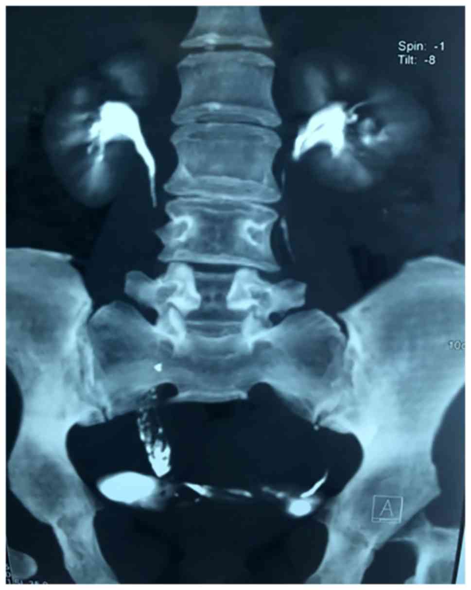
Multidetector computed tomography urography (CTU) revealed a 
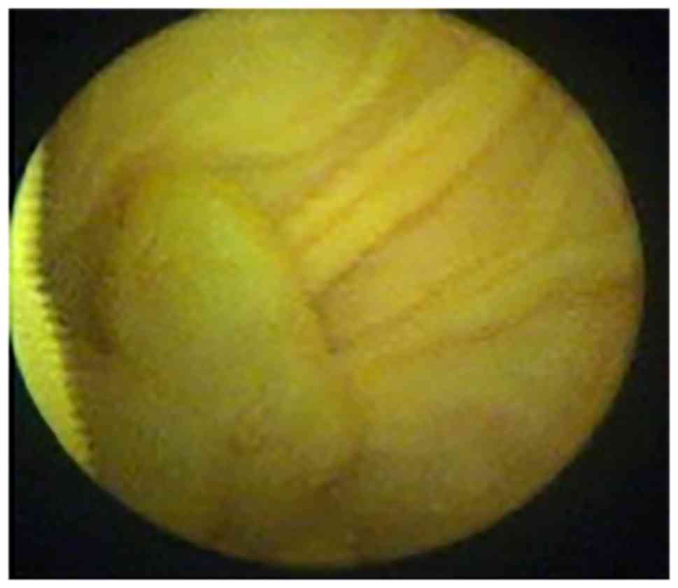
filling defect in the lower part of the right ureter (Fig. 1). Cystoscopy revealed a mass ~3 cm in length, with a smooth surface, protruding from the ureteral orifice into the bladder (Fig. 2). Examination of a bioptic specimen suggested that the mass was an inflammatory polyp. A 5F ureteral catheter was inserted from the right ureteral orifice; ~10 cm after the insertion point, resistance was encountered and the catheter could not be advanced 
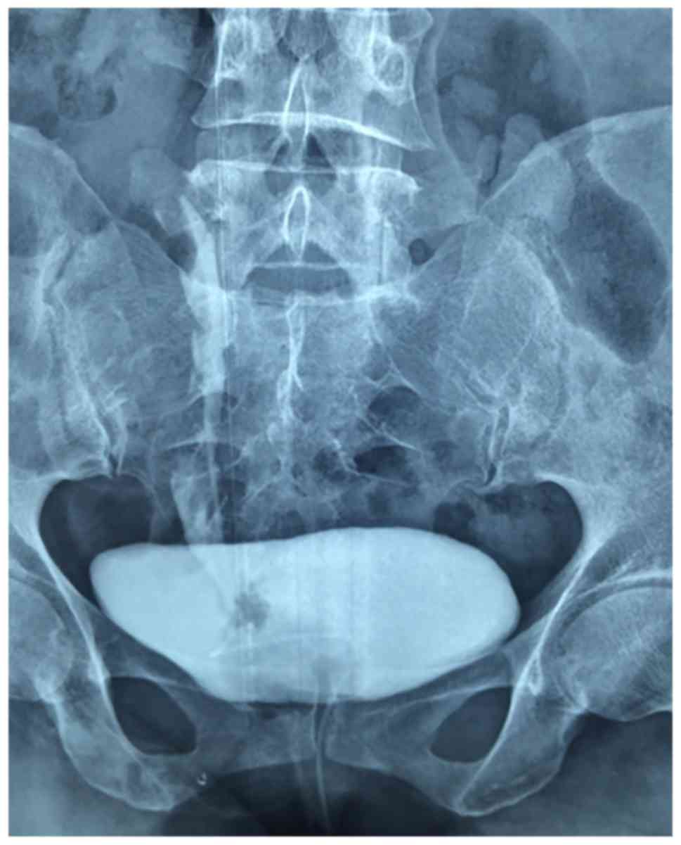
further. Retrograde urography was subsequently performed and the contrast media failed to reach the upper part of the ureter and the renal pelvis, whereas the lower part of the ureter exhibited a filling defect (Fig. 3).

Figure 1.

Computed tomography urography showing a filling defect at the lower part of the right ureter.

Figure 2.

Cystoscopy showing a neoplasm with a smooth surface, ~3 cm in length, arising from the right ureter.

Figure 3.

Retrograde urography showing a filling defect in the right lower ureter and no contrast medium in the upper ureter and renal pelvis.

Ureteroscopic assessment was performed under general anaesthesia and revealed a pedunculated tumor (~15 cm in length) arising from the lower segment of the right ureter, with a smooth surface, with the base of the tumor located ~13 cm from the ureteral orifice. The tumor macroscopically resembled a long grape. As we unable to find the upper ureteral orifice, the ureteroscope and guidewire could not pass through the base of the tumor. A small piece of tumor was resected by Holmium laser for histopathological examination and a Double-J stent was inserted, with the upper end of the stent located in the base of the tumor. Resectoscopy was then performed to incise the portion of the polyp protruding into the bladder. The histological examination of these two bioptic specimens suggested that the tumor was a fibroepithelial polyp. 
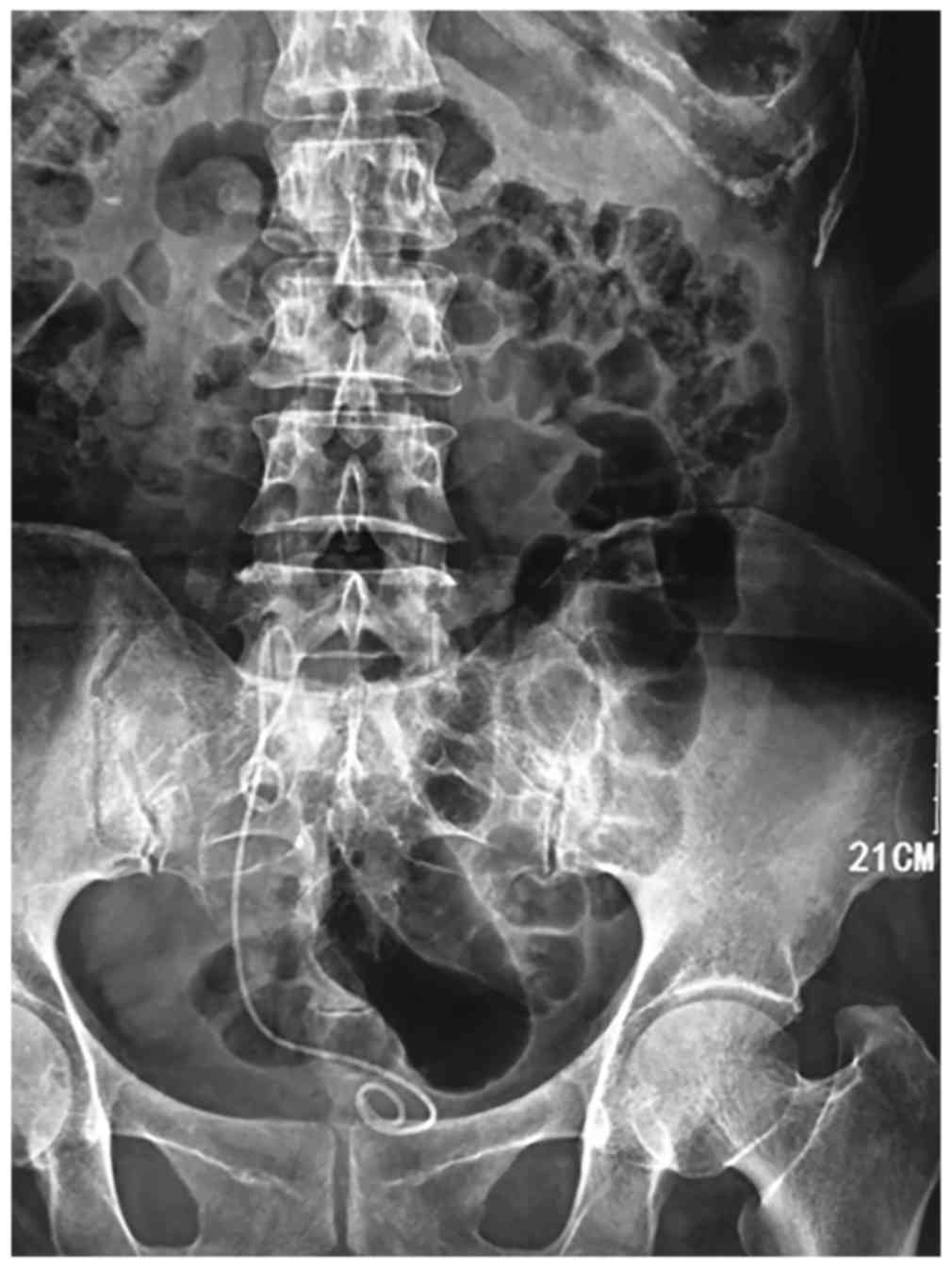
Kidney-ureter-bladder imaging was then performed, which revealed that the Double-J stent was folded and twisted in the fifth lumbar plane (Fig. 4).

Figure 4.

Kidney-ureter-bladder ultrasound, showing the Double-J stent folded and twisted in the fifth lumbar plane.

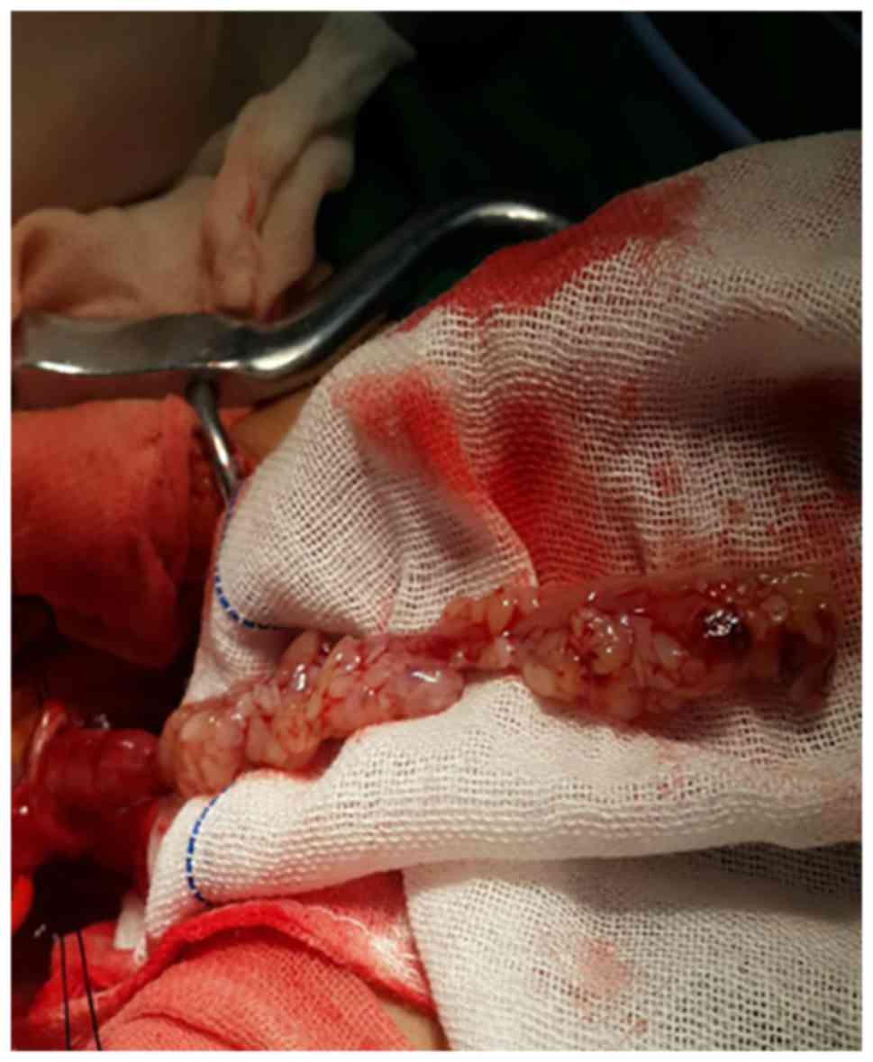
Open surgery was employed under general anaesthesia, Resection of the polyp and the narrow ureteral segment plus V-shaped end-to-end ureteral anastomosis were performed. An oblique incision on the right low abdomen was performed. Following exposure of the ureter, taking the apex of the Double J-tube as the longitudinal excision sign, the ureter was longitudinally opened for ~2 cm, the root of the polyp was immediately identified, and 
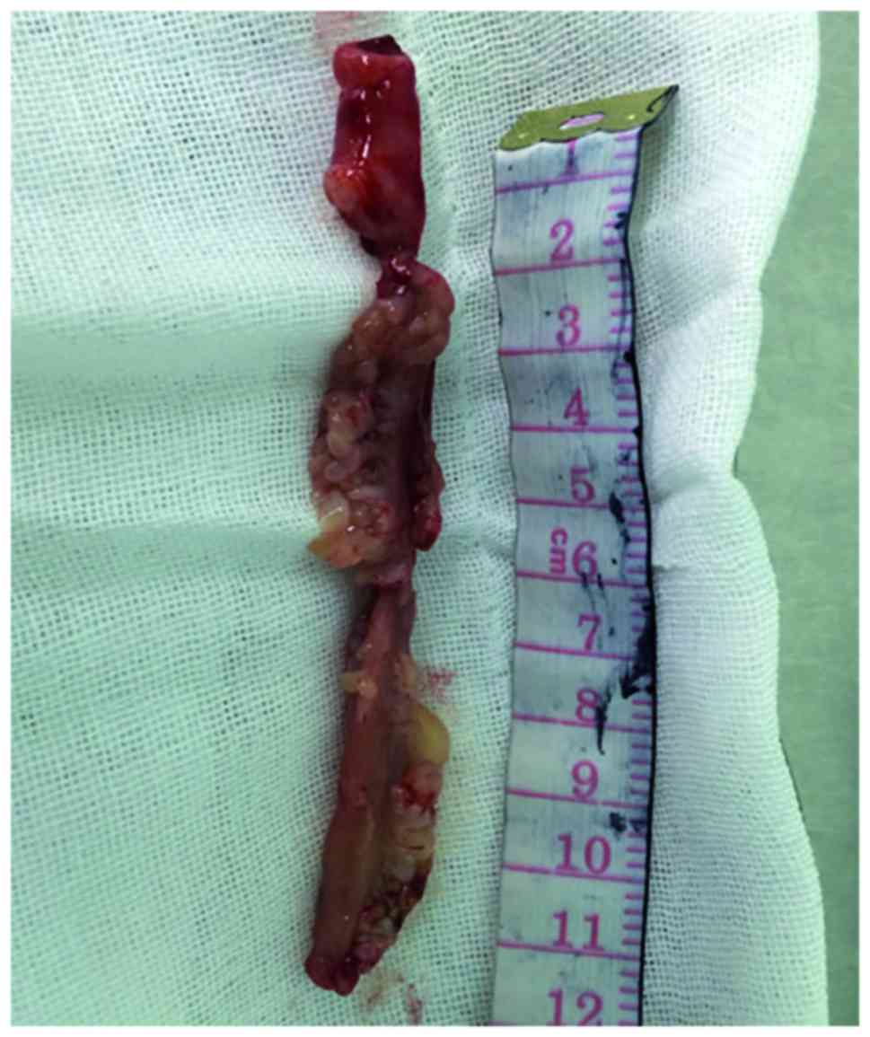
the ureter above the polyp was narrowed. The polyp was cut at the base and was completely removed from the ureter (Fig. 5); the length of the polyp was ~12 cm (Fig. 6). Approximately 0.5 cm of the narrow ureteral segment was resected and end-to-end anastomosis was performed. A Double-J stent was left in place.

Figure 5.

Gross appearance of the excised ureteral polyp, with a smooth surface and shaped similar to a long grape brunch.

Figure 6.

The 12.0-cm long fibroepithelial polyp removed by open surgery from the right ureter.

The postoperative histopathological examination findings revealed that the tumor was a ureteral fibroepithelial polyp (UFP). The Double-J stent was removed 2 months after the operation. The patient was asymptomatic, and no complications or recurrence have occurred during the 12 months of follow-up. gave their informed consent regarding the publication of the case details.

Discussion

Primary ureteral tumors are among the rarest in the spectrum of genitourinary tumors and are most commonly malignant. Only one-fifth of these tumors are benign and, among those, UFPs are considered to be the most common. The etiology of benign ureteral polyps is unclear. They are considered to be the result of various factors, including congenital (developmental anomaly), obstruction, trauma, irritation, infection, and specific endogenous hormonal imbalances (3). The clinical presentation of primary UFPs is non-specific. They most commonly present as a single, small polyp. Reports of multiple, bilateral polyps are extremely rare, whereas the mean diameter of UFPs was reported to be <5 cm; larger polyps may extend into the bladder cavity and may be difficult to distinguish from bladder tumors (4,5). In the majority of the cases, the upper ureter is the most common site of origin of these tumors, whereas polyps derived from the lower urinary tract are not as frequent (6). UFPs most commonly occur between the second and fourth decades, and they usually originate from the left ureter. The clinical symptoms are non-specific and the majority of the early primary UFPs are asymptomatic. Necrosis and bleeding on the polyp surface may be expressed as gross hematuria and, when obstruction occurs, hydronephrosis or renal colic may develop. In the present case, the polyp occurred in the lower part of the right ureter in a middle-aged woman; in addition, it was broad-based and had a total length of ~15 cm (portion removed by open surgery, ~12cm; and ureteroscopically removed bladder part, ~3 cm). Previous reports of such lower-segment ureteral giant primary UFPs are extremely rare.

Imaging examination (7) may be helpful and suggestive of a UFP diagnosis. Intravenous pyelography or CTU may show a filling defect, thereby hinting at the state of the renal function of the patient, and may also reveal whether hydronephrosis may be present. However, preoperative radiographic diagnosis may be challenging, as UFPs usually present as a filling defect, which may be attributed to blood clots, radiolucent calculi, neoplasms or a crossing vessel. Moreover, it may be difficult to differentiate UFPs from transitional cell carcinoma based only on imaging findings (8), as preoperative diagnosis confirmation is difficult and Li et al reported in 2014 that none of the UFPs in their study were detected prior to surgery (9). Mistaking these tumors for transitional cell carcinomas may result in unnecessary nephroureterectomy.

For patients with suspected UFPs who have been evaluated by intravenous pyelography, CT, or retrograde urography, a ureteroscopic examination is also required to confirm the diagnosis. Ureteroscopy may be attempted to obtain a diagnosis and determine the optimal treatment approach. Prior to ureteroscopy becoming widespread, differentiating malignant from benign lesions was difficult. The number of polyps, the diameter of the base and the location of the obstruction may be determined through ureteroscopy in the same session; ureteroscopic biopsy of the lesion may be performed for larger tumors, whereas smaller tumors may be completely resected during ureteroscopy, thus avoiding a second surgery. In the present case, preoperative ureteroscopic examination revealed that the polyp was extremely long, originated from the lower part of the ureter and its base was wide; the biopsy result revealed a benign fibroepithelial polyp, and surgical resection was selected. At the same time, a Double-J stent was also placed at the obstruction site, which was identified by searching the base of the polyp during open surgery.

The management of UFPs currently depends on the site, size and clinical expertise. Smaller lesions may simply be fulgurated endoscopically, while larger lesions require proper surgical excision. With the advent of new technology, minimally invasive techniques have become popular. Previous reports have described successful polypectomy through the use of ureteroscopy and endoscopic treatment (5,10). However, ureteroscopy and endoscopic resection may be difficult in patients with long or large polypoid lesions, due to poor visualization of the base of the stalk and limited working space, which make it difficult to differentiate the ureteral wall from the polyp, leading to incomplete resection or ureteral perforation. Laparoscopic surgery is another minimally invasive method (11,12). Successful laparoscopic treatment has been described in patients with large, long polyps, as well as in those with multiple polyps. Kijvikai et al (13) described the transperitoneal laparoscopic management of a 17-cm long fibroepithelial polyp in the proximal ureter associated with ureteral obstruction. However, this approach may not be suitable for polyps in the lower part of the ureter, particularly in cases without hydronephrosis, and laparoscopic surgery may cause difficulties in ureteral anastomosis following ureteral resection, particularly in patients with a lower ureteral polyp. Complete resection is considered the optimal method for avoiding recurrence of UFPs, as incomplete resection may result in recurrence. In our case, as the polyp was long, its stalk could not be fully visualized via ureterorenoscopy; furthermore, it occurred in an older woman and originated from the lower part of the ureter. Our main objective was to ensure complete resection, so open surgery was selected in this case. A small ureteral segment was resected, including the narrowed potion of the ureter and the entire stalk of the polyp, followed by end-to-end ureteral anastomosis.

In summary, an extremely long pedunculated UFP of the distal ureter, which protruded into the bladder, was excised via open surgery. Pedunculated urothelial polyps originating from the ureter should be taken into consideration in the differential diagnosis of a bladder mass on imaging. Primary ureteral polyps are benign and the prognosis is usually good. Ureteroscopy is not only a useful diagnostic method, but may also be an effective treatment for smaller, solitary ureteral polyps. For larger polyps, in patients with obstruction, ureteroscopy may help determine the polyp size, its location, the location of the ureteral stenosis, as well as the base of the polyp, which may help determine the optimal surgical approach.

Acknowledgements

The authors would like to thank Anguo Wang for assisting with the surgical procedure. This study was supported by the North Sichuan Medical College (grant no. 20131252).

References

1 

Piovesan AC, Torricelli FC, Borges LL, Chambô JL, Mesquita JL and Srougi M: Ureteral fibroepithelial polyps in a pregnant woman: Case report. Sao Paulo Med J. 127:238–240. 2009. View Article : Google Scholar : PubMed/NCBI

2 

Ludwig DJ, Buddingh KT, Kums JJM, Kropman RF, Roshani H and Hirdes WH: Treatment and outcome of fibroepithelial ureteral polyps: A systematic literature review. Can Urol Assoc J. 9:E631–E637. 2015. View Article : Google Scholar : PubMed/NCBI

3 

Ye L, Zhao LJ, Yue F, Song XS, Wei W, Jiang XJ and Yang JY: Large ureteral fibroepithelial polyp lacking epithelium due to ischemic infarction. Kaohsiung J Med Sci. 28:457–461. 2012. View Article : Google Scholar : PubMed/NCBI

4 

Kaba M, Kaba S, Kaya TY, Eren H and Pirincci N: A giant pedunculated urothelial polyp imicking bladder mass in a child: a rare case. Case Rep Pediatr 2014. 2014.1–3. View Article : Google Scholar

5 

Liu C, Liu XJ, Liu D and Yao DW: A giant ureteral polyp mimicking as a bladder mass resected ureteroscopically by diode laser: A case report and literature review. Int J Clin Exp Pathol. 8:14580–14583. 2015.PubMed/NCBI

6 

Childs MA, Umbreit EC, Krambeck AE, Sebo TJ, Patterson DE and Gettman MT: Fibroepithelial polyps of the ureter: A single-institutional experience. J Endourol. 23:1415–1419. 2009. View Article : Google Scholar : PubMed/NCBI

7 

Xu C, Zhang Z, Ye H, Wu C, Zhang C, Zhang Y, Wang Y, Cao Z, Wang H, Xu W, et al: Imaging diagnosis and endoscopic treatment for ureteral fibroepithelial polyp prolapsing into the bladder. J Xray Sci Technol. 21:393–399. 2013.PubMed/NCBI

8 

Patheyar V, Venkatesh SK, Siew EP, Consiglieri DT and Putti T: MR imaging features of fibroepithelial ureteral polyp in a patient with duplicated upper urinary tract. Singapore Med J. 52:e45–e47. 2011.PubMed/NCBI

9 

Li R, Lightfoot M, Alsyouf M, Nicolay L, Baldwin DD and Chamberlin DA: Diagnosis and management of ureteral fibroepithelial polyps in children: A new treatment algorithm. J Pediatr Urol. 11:22.e1–22.e6. 2015. View Article : Google Scholar

10 

Hubosky SG and Bagley DH: Laser Resection of Fibroepithelial Polyps with Digital Ureteroscopy. J Endourol Case Rep. 1:36–38. 2015. View Article : Google Scholar : PubMed/NCBI

11 

Dai LN, Chen CD, Lin XK, Wang YB, Xia LG, Liu P, Chen XM and Li ZR: Retroperitoneal laparoscopy management for ureteral fibroepithelial polyps causing hydronephrosis in children: A report of five cases. J Pediatr Urol. 11:257.e1–257.e5. 2015. View Article : Google Scholar

12 

Bian Z, Liu X, Hua Y, Liu F, Lin T and He D: Laparoscopic management of multiple ureteral polyps in children. J Urol. 186:1444–1449. 2011. View Article : Google Scholar : PubMed/NCBI

13 

Kijvikai K, Maynes LJ and Herrell SD: Laparoscopic management of large ureteral fibroepithelial polyp. Urology. 70:3732007. View Article : Google Scholar : PubMed/NCBI

Related Articles

  • Abstract
  • View
  • Download
  • Twitter
Copy and paste a formatted citation
Spandidos Publications style
Cai Y, Zhang Z and Yue X: Rare giant primary ureteral polyp: A case report and literature review. Mol Clin Oncol 6: 327-330, 2017.
APA
Cai, Y., Zhang, Z., & Yue, X. (2017). Rare giant primary ureteral polyp: A case report and literature review. Molecular and Clinical Oncology, 6, 327-330. https://doi.org/10.3892/mco.2017.1146
MLA
Cai, Y., Zhang, Z., Yue, X."Rare giant primary ureteral polyp: A case report and literature review". Molecular and Clinical Oncology 6.3 (2017): 327-330.
Chicago
Cai, Y., Zhang, Z., Yue, X."Rare giant primary ureteral polyp: A case report and literature review". Molecular and Clinical Oncology 6, no. 3 (2017): 327-330. https://doi.org/10.3892/mco.2017.1146
Copy and paste a formatted citation
x
Spandidos Publications style
Cai Y, Zhang Z and Yue X: Rare giant primary ureteral polyp: A case report and literature review. Mol Clin Oncol 6: 327-330, 2017.
APA
Cai, Y., Zhang, Z., & Yue, X. (2017). Rare giant primary ureteral polyp: A case report and literature review. Molecular and Clinical Oncology, 6, 327-330. https://doi.org/10.3892/mco.2017.1146
MLA
Cai, Y., Zhang, Z., Yue, X."Rare giant primary ureteral polyp: A case report and literature review". Molecular and Clinical Oncology 6.3 (2017): 327-330.
Chicago
Cai, Y., Zhang, Z., Yue, X."Rare giant primary ureteral polyp: A case report and literature review". Molecular and Clinical Oncology 6, no. 3 (2017): 327-330. https://doi.org/10.3892/mco.2017.1146
Follow us
  • Twitter
  • LinkedIn
  • Facebook
About
  • Spandidos Publications
  • Careers
  • Cookie Policy
  • Privacy Policy
How can we help?
  • Help
  • Live Chat
  • Contact
  • Email to our Support Team