Introduction
Follicular dendritic cells (FDCs) are an accessory
part of the lymphoid system and their function is to present
antigens to specialized lymphocytes (1). In the liver, FDCs are distributed
around portal spaces, and the probability of them forming tumors is
quite small. According to the World Health Organization
classification in 2001 and 2008, inflammatory pseudotumor
(IPT)-like FDC tumor was considered to be a distinct variant of
low-grade FDC tumor/sarcoma (2).
This tumor is characterized by a lymphoplasmacytic infiltrate mixed
with neoplastic spindle cells, forming an inflammatory pseudotumor
(3). Due to the low incidence and
our limited experience with the disease, IPT-like FDC tumor in the
liver may not be included in the differential diagnosis during the
initial evaluation and may be misdiagnosed; the differential
diagnosis includes hepatic focal nodular hyperplasia and
interdigitating dendritic cell sarcoma. We herein present a rare
case of a young female patient diagnosed with hepatic IPT-like FDC
tumor.
Case report
A 19-year-old woman, with no prior significant
disease history, initially presented to the The Second People's
Hospital of Changzhou with complaints of abdominal discomfort for 1
month. The α-fetoprotein, carcinoembryonic antigen and carbohydrate
antigen 19–9 levels were within the normal range. The laboratory
values obtained during the initial work-up were as follows: White
blood cell count 11.61×10⁹/l, lymphocytes 23.8%,
neutrophils 57.1%, hemoglobin level 133 g/l, total protein 85 g/l,
albumin 46.2 g/l, total bilirubin 7.7 µmol/l, direct bilirubin 2.3
µmol/l, aspartate aminotransferase 13 U/l, alanine aminotransferase
10 U/l and γ-glutamyltransferase 56 U/l. Ultrasonographic
examination revealed a solitary well-defined hypoechoic mass in the
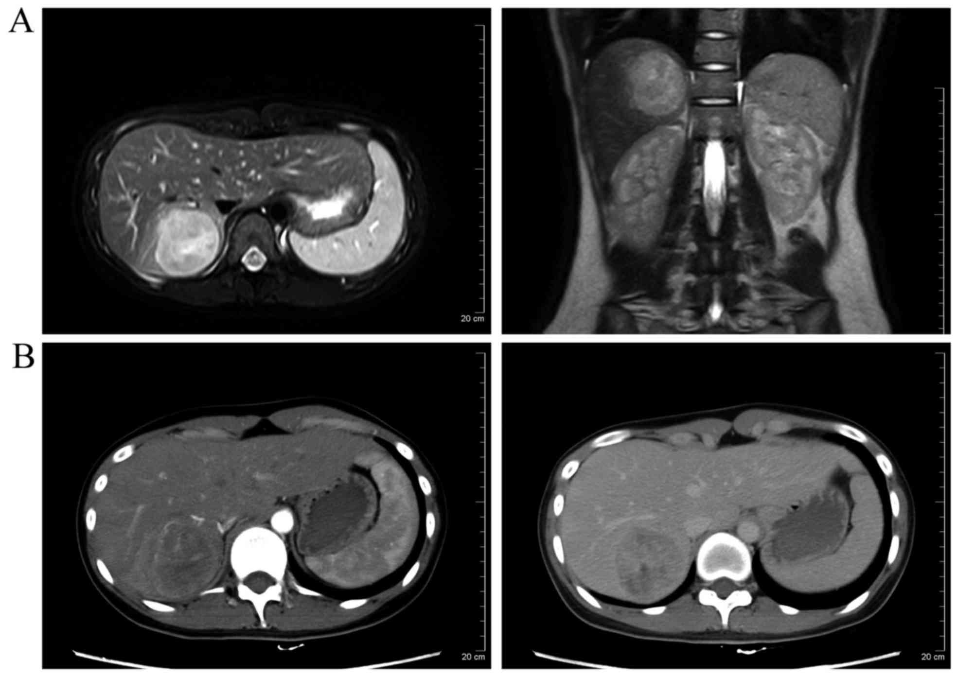
liver. Enhanced abdominal computed tomography and magnetic
resonance imaging demonstrated a heterogeneous mass in the right
hepatic lobe with several low attenuation areas, suggesting
necrosis (Fig. 1). No other mass or
evidence of lymphadenopathy was present in the abdomen, pelvis or
retroperitoneum. Intraoperatively, no lesions were found in the
spleen and small bowel, and frozen section pathological examination
revealed inflammatory cellular infiltration. The patient underwent
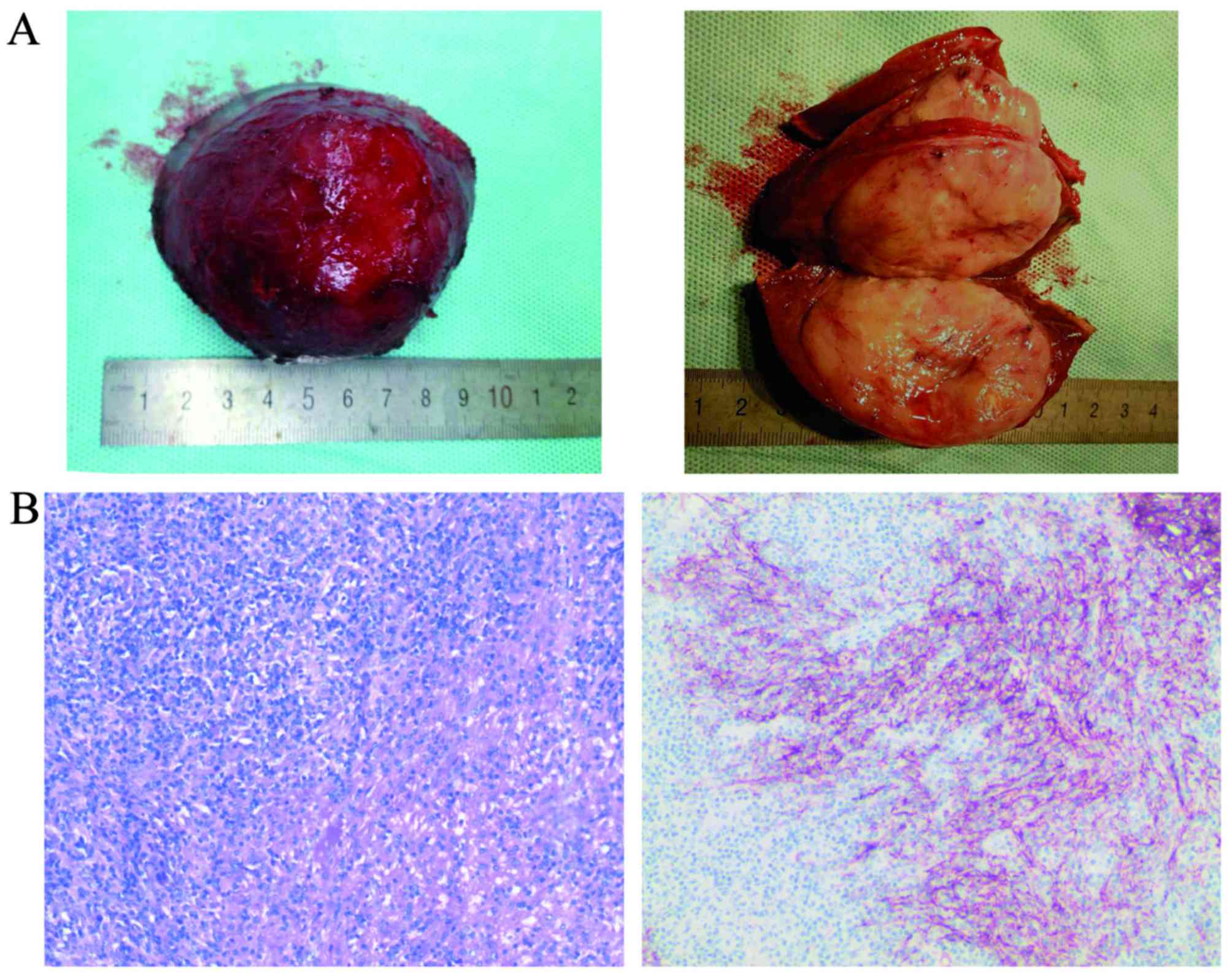
hepatic VII segmental resection and the solid nodule was sized
~6×5×4 cm (Fig. 2A). The
postoperative pathological diagnosis was IPT-like FDC tumor with
negative margins. Immunohistochemical analysis showed strong
reactivity of the tumor cells for CD21, CD35, CD45RO and CD79α. A
proportion of the lymphocytes were positive for CD20 (Fig. 2B) and negative for actin, anaplastic
lymphoma kinase (ALK), smooth muscle actin-α (α-SMA) and S-100. The
infiltrating spindle cells were positive for Epstein-Barr virus
(EBV)-encoded small RNAs as determined by in situ hybridization.
Finally, the patient was discharged on postoperative day 7 and no
metastases or recurrence had developed within a period of 1 year,
with normal laboratory values.
The patient provided a signed informed consent
regarding the publication of the case details and associated
images.
Discussion
Hepatic FDC tumor is a rare neoplasm with only 24
cases reported in the English literature to date (4–8).
IPT-like FDC tumors of liver are even more rare, and the
clinicopathological nature of this type of disease remains unknown.
First, the most common presentation of patients with FDC tumors is
abdominal pain or weight loss, but IPT-like FDC tumors often grow
without clinical symptoms or abdominal discomfort, as was the case
in our patient (9). Second, IPT-like
FDC tumors always exhibit a marked female predominance (10). Third, IPT-like FDC tumors are
strongly associated with the presence of EBV (11,12),
although with a lower positivity rate compared with conventional
FDC sarcomas (13). On pathological
examination, IPT-like FDC tumors cells have a more prominent
inflammatory component. In addition, on immunohistochemical
staining, the tumor cells are positive for CD21, CD23, CD35 and CD3
(in some cases), and negative for ALK, actin and α-SMA, which helps
with the differentiation from interdigitating dendritic cell tumors
and histiocytic neoplasms (14).
Finally, although both IPT-like FDC tumors and conventional FDC
tumors are treated by surgical resection with no adjuvant therapy,
such as radiotherapy or chemotherapy (15), IPT-like FDC tumors exhibit a less
aggressive pattern of growth, and fewer recur/metastasize or have a
fatal outcome (16).
In conclusion, compared with FDC sarcomas, hepatic
IPT-like FDC tumors exhibit a female predominance, association with
EBV infection, and generally have a good outcome, with only rare
cases of recurrence or metastasis. However, the pathophysiological
process underlying this type of disease remains unclear and
requires further investigation.
Acknowledgements
The present study was supported by a grant from the
Foundation for Changzhou's High-level Health Talent Cultivation
(no. 2016CZBJ007).
References
|
1
|
van Nierop K and de Groot C: Human
follicular dendritic cells: Function, origin and development. Semin
Immunol. 14:251–257. 2002. View Article : Google Scholar : PubMed/NCBI
|
|
2
|
Chan J, Pileri SA and Desol G: Follicular
dendritic cell sarcoma. IARC Press; Lyon: 2008
|
|
3
|
Cheuk W, Chan JK, Shek TW, Chang JH, Tsou
MH, Yuen NW, Ng WF, Chan AC and Prat J: Inflammatory
pseudotumor-like follicular dendritic cell tumor: A distinctive
low-grade malignant intra-abdominal neoplasm with consistent
Epstein-Barr virus association. Am J Surg Pathol. 25:721–731. 2001.
View Article : Google Scholar : PubMed/NCBI
|
|
4
|
Martins PN, Reddy S, Martins AB and
Facciuto M: Follicular dendritic cell sarcoma of the liver: Unusual
presentation of a rare tumor and literature review. Hepatobiliary
Pancreat Dis Int. 10:443–445. 2011. View Article : Google Scholar : PubMed/NCBI
|
|
5
|
Bai LY, Kwang WK, Chiang IP and Chen PM:
Follicular dendritic cell tumor of the liver associated with
Epstein-Barr virus. Jpn J Clin Oncol. 36:249–253. 2006. View Article : Google Scholar : PubMed/NCBI
|
|
6
|
Granados R, Aramburu JA, Rodríguez JM and
Nieto MA: Cytopathology of a primary follicular dendritic cell
sarcoma of the liver of the inflammatory pseudotumor-like type.
Diagn Cytopathol. 36:42–46. 2008. View
Article : Google Scholar : PubMed/NCBI
|
|
7
|
Nguyen BD, Roarke MC and Yang M:
Synchronous hepatic and splenic inflammatory pseudotumour-like
follicular dendritic cell sarcomas. Liver Int. 35:19172015.
View Article : Google Scholar : PubMed/NCBI
|
|
8
|
Ma Y, Sun J, Yang C, Yuan D and Liu J:
Follicular dendritic cell sarcoma: Two rare cases and a brief
review of the literature. Onco Targets Ther. 8:1823–1830. 2015.
View Article : Google Scholar : PubMed/NCBI
|
|
9
|
Chen Y, Shi H, Li H, Zhen T and Han A:
Clinicopathological features of inflammatory pseudotumor-like
follicular dendritic cell tumor of the abdomen with 10 cases
report. Histopathology. 2015.
|
|
10
|
Zhang ZX, Cheng J, Shi QL, et al:
[Follicular dendritic cell sarcoma: a clinicopathologic study of 8
cases]. Zhonghua bing li xue za zhi Chinese journal of pathology.
37:395–399. 2008.PubMed/NCBI
|
|
11
|
Selves J, Meggetto F, Brousset P, Voigt
JJ, Pradère B, Grasset D, Icart J, Mariamé B, Knecht H and Delsol
G: Inflammatory pseudotumor of the liver. Evidence for follicular
dendritic reticulum cell proliferation associated with clonal
Epstein-Barr virus. Am J Surg Pathol. 20:747–753. 1996. View Article : Google Scholar : PubMed/NCBI
|
|
12
|
Chen TC, Kuo TT and Ng KF: Follicular
dendritic cell tumor of the liver: A clinicopathologic and
Epstein-Barr virus study of two cases. Mod Pathol. 14:354–360.
2001. View Article : Google Scholar : PubMed/NCBI
|
|
13
|
Torres U, Hawkins WG, Antonescu CR and
DeMatteo RP: Hepatic follicular dendritic cell sarcoma without
Epstein-Barr virus expression. Arch Pathol Lab Med. 129:1480–1483.
2005.PubMed/NCBI
|
|
14
|
Ge R, Liu C, Yin X, Chen J, Zhou X, Huang
C, Yu W and Shen X: Clinicopathologic characteristics of
inflammatory pseudotumor-like follicular dendritic cell sarcoma.
Int J Clin Exp Pathol. 7:2421–2429. 2014.PubMed/NCBI
|
|
15
|
Hu J, Chen LL, Ding BW, Jin DY and Xu XF:
Resection is an effective treatment for recurrent follicular
dendritic cell sarcoma from retroperitoneum: Unusual presentation
of a rare tumor. Int J Clin Exp Med. 8:8218–8221. 2015.PubMed/NCBI
|
|
16
|
Pang J, Mydlarz WK, Gooi Z, et al:
Follicular dendritic cell sarcoma of the head and neck: Case
report, literature review, and pooled analysis of 97 cases. Head
Neck. 2015.PubMed/NCBI
|