Introduction
Colorectal cancer (CRC) is the third most common
cancer in men and the second in women worldwide (1). According to the World Health
Organization GLOBOCAN database, in 2012, ~1.4 million new cases of
CRC were diagnosed and 694,000 patients succumbed to this disease
(2). CRC is one of the major causes
of cancer-related mortality, with a 5-year survival of ~50%
(3). Metastasis to the liver and/or
lungs is the main cause of death and is already present in ≤25% of
patients at presentation (4). Early
diagnosis and timely intervention are crucial for therapeutic
effectiveness and prolongation of survival. Adequate preoperative
staging is important for the management of CRC patients so that the
appropriate treatment plan may be devised. Following treatment
completion, patients must be monitored for the development of
recurrence or distant metastasis. Tumour markers are widely used
for the diagnosis, staging and monitoring of CRC patients in
clinical practice. These markers are biological or biochemical
substances that are usually produced by tumour cells and then
secreted into the circulation in detectable amounts and used for
screening and staging (5).
Carcinoembryonic antigen (CEA), an oncofetal glycoprotein that is
overexpressed in adenocarcinoma of the colorectum, is the most
frequently used biomarker in CRC (6). CEA has low sensitivity and specificity
for diagnosis; therefore, it should not be used for screening.
However, in patients with newly diagnosed CRC, the absolute level
of the serum CEA is correlated with disease burden increase in 75%
of patients with distant metastasis and has prognostic value
(7). High levels of CEA may also be
observed in association with benign conditions, such as smoking,
peptic ulcer, inflammatory bowel disease, pancreatitis,
hypothyroidism, biliary obstruction and cirrhosis (8). Another tumour marker, carbohydrate
antigen 19-9 (CA19-9), is produced by adenocarcinomas of the
pancreas, stomach, gallbladder, colon, ovary and lung. The
incidence of high serum CA19-9 levels in CRC ranges from 20 to 40%
(9).
At present, the sensitivity and specificity of the
clinically used markers of CRC are low for early diagnosis and
screening; thus, a number of studies have been conducted to
identify more effective markers. Several studies have recently
investigated the overexpression of human epididymis protein 4 (HE4)
in certain cancer types. HE4 was first identified in 1991 by
Kirckhoff et al (10) and its
overexpression has been found in carcinomas of the ovary,
endometrium, lung, breast, gastrointestinal system and
uroepithelium (11,12). HE4 was originally isolated from the
epithelium of the distal epididymis and was predicted to be a
protease inhibitor essential for sperm maturation (2,13). The
HE4 gene product is also referred to as whey acidic protein and
four-disulphide core domain protein 2 (14). This protein acts in a manner similar
to antiproteinases, secretory leukocyte protease 1 and elafin; they
all exhibit antibacterial properties and exert anti-inflammatory
effects (15). HE4 is generally
considered to play a role in natural immunity; however, its true
role in carcinogenesis has not been fully elucidated (10). Multiple studies have reported its
specifity and sensitivity as an ovarian cancer biomarker (16). In 2009, the United States Food and
Drug Administration (FDA) approved HE4 as a useful marker for
monitoring patients with epithelial ovarian cancer (17); and in September, 2011, the FDA also
approved marketing of HE4 for the detection of ovarian masses
(18). There is accumulating
evidence that HE4 may also be a useful marker for endometrial
cancer. Recently, HE4 expression was also investigated in breast,
lung, gastric and pancreatic cancer (19). To the best of our knowledge, this is
the first clinical study to evaluate the diagnostic and
preoperative predictive value of serum HE4 levels in newly
diagnosed CRC patients.
Patients and methods
Patients
The study comprised 46 newly diagnosed CRC patients
who were diagnosed and treated at the Ondokuz Mayis University
Hospital (Samsun, Turkey) between January, 2014 and July, 2014, and
36 age- and gender-matched healthy subjects as the control group.
The control subjects were individually selected from patients
attending the outpatient clinic for a routine check-up. Approval
for the study was granted by the Ethics Committee of the Ondokuz
Mayıs University Hospital and all the patients and controls
provided written informed consent.
The pathological staging of the CRC patients was
performed according to the 7th edition of the
tumour-node-metastasis (TNM) classification (http://www.uicc.org/sites/main/files/private/TNM_Classification_of_Malignant_Tumours_Website_15%20MAy2011.pdf).
Patients with renal failure were excluded, as they have high HE4
levels. All the subjects were treatment-naive (no operation,
chemotherapy or chemoradiotherapy) prior to blood sample
collection.
Blood sampling and serum marker
levels
Blood samples were obtained by venous puncture,
centrifuged, and stored at −50°C until the analyses were performed.
An ELISA kit (Cusabio Biotech Co., Ltd., Wuhan, China) was used for
HE4 evaluation. All measurements were performed according to the
manufacturers' instructions. The plates were read at the wavelength
of 450 nm 10 min after the administration of the stop solution. The
minimum detectable dose of HE4 was <0.30 pmol/ml according to
this measurement. The HE4 cut-off level of ≤0.30 pmol/ml was
considered to be HE4-negative and all cases with HE4 levels
>0.30 pmol/ml were considered as HE4-positive.
The serum CEA and CA19-9 levels were measured using
the enzyme immunoassay method, using a HITACHI automatic analyzer
(Hitachi Corporation, Tokyo, Japan). According to the
manufacturer's instructions, the normal range for serum CEA was
0–3.4 ng/ml and for CA19-9 0–37 U/ml.
Statistical analysis
The statistical analysis was performed with SPSS
software, version 17.0 (SPSS Inc., Chicago, IL, USA). The results
were expressed as mean ± standard deviation and frequency (%). The
normality of distribution was checked initially and Student's
t-test was then used to compare the mean values. Yates's corrected
Chi-squared and Fisher's exact tests were also used for frequency
(data obtained by counting) comparisons. Receiver operator
characteristics (ROC) curves were applied and the area under the
curve (AUC) was calculated to determine the sensitivity and
specificity of HE4. P<0.05 was considered to indicate
statistically significant differences.
Results
Subject characteristics
A total of 46 newly diagnosed CRC patients and 36
healthy age- and gender-matched control subjects were included in
the study. The demographic characteristics of the patients and
controls are shown in Table I. There
were no significant demographic differences between the two groups.
HE4 positivity was determined in 13 of the 46 cases (28.3%) in the
CRC group; however, no HE4-positive subjects were identified in the
control group (0%; P=0.002).
 | Table I.Characteristics of colorectal cancer
patients and controls. |
Table I.
Characteristics of colorectal cancer
patients and controls.
| Variables | Patients, n (%)
(n=46) | Controls, n (%)
(n=36) | P-value |
|---|
| Age, years (mean ±
SD) | 60.5±11.8 | 58±12.5 | 0.360 |
| Gender |
|
| 0.901 |
|
Female | 16 (34.8) | 13 (36.1) |
|
| Male | 30 (65.2) | 23 (63.9) |
|
| HE4 |
|
| 0.002 |
|
Positive | 13 (28.3) | 0 (0.0) |
|
|
Negative | 33 (71.7) | 36 (100.0) |
|
Patient characteristics
The detailed clinicopathological characteristics of
the CRC patients are shown in Table
II. CEA positivity was determined in 28 (66.7%) and CA19-9
positivity in 17 (40.5%) CRC cases. Of the 46 patients, 33 were
lymph node-positive and 22 of those had stage IV disease.
| Table II.Characteristics of colorectal cancer
patients. |
Table II.
Characteristics of colorectal cancer
patients.
| Variables | n (%) |
|---|
| HE4 |
|
|
Positive | 13 (28.3) |
|
Negative | 33 (71.7) |
| Lymph node
status |
|
|
Positive | 33 (71.7) |
|
Negative | 13 (28.3) |
| CEAa |
|
|
Positive | 28 (66.7) |
|
Negative | 14 (33.3) |
| CA199b |
|
|
Positive | 17 (40.5) |
|
Negative | 25 (59.5) |
| Grade |
|
| 1 | 13 (35.1) |
| 2 | 21 (56.8) |
| 3 | 3 (8.1) |
| Stage |
|
| I | 2 (4.3) |
| II | 11 (23.9) |
| III | 11 (23.9) |
| IV | 22 (47.9) |
Comparison between HE4-positive and
-negative patients
As shown in Table
III, nodal involvement was significantly associated with HE4
positivity. None of the node-negative patients were HE4-positive,
whereas 13 of the 33 patients with nodal involvement were
HE4-positive (P=0.009). When comparing HE4 positivity between
cancer stages I–II and III–IV, the difference was statistically
significant (P=0.009). HE4 was also statistically significantly
positive in patients with high CA19-9 levels (P<0.001). No
statistically significant associations were observed between tumour
grade, CEA levels and HE4.
| Table III.Comparison of HE4-positive and
-negative patients. |
Table III.
Comparison of HE4-positive and
-negative patients.
| Variables | HE4-positive, n
(%) | HE4-negative, n
(%) | P-value |
|---|
| Age, years (mean ±
SD) | 63.3±11.7 | 59.4±11.8 | 0.315 |
| Grade |
|
| 0.162 |
| I | 2 (16.7) | 11 (44.0) |
|
| II | 8 (66.6) | 13 (52.0) |
|
| III | 2 (16.7) | 1 (4.0) |
|
| Lymph node
status |
|
| 0.009 |
|
Positive | 13 (100.0) | 20 (60.6) |
|
|
Negative | 0 (0.0) | 13 (39.4) |
|
| Stage |
|
| 0.009 |
|
I–II | 0 (0.0) | 20 (60.6) |
|
|
III–IV | 13 (39.4) | 13 (100.0) |
|
| CEA, ng/ml |
|
| 0.159 |
|
≥3.7 | 11 (84.6) | 17 (58.6) |
|
|
<3.7 | 2 (15.4) | 12 (41.4) |
|
| CA199, U/ml |
|
|
<0.001 |
|
≥37 | 11 (84.6) | 6 (20.7) |
|
|
<37 | 2 (15.4) | 23 (79.3) |
|
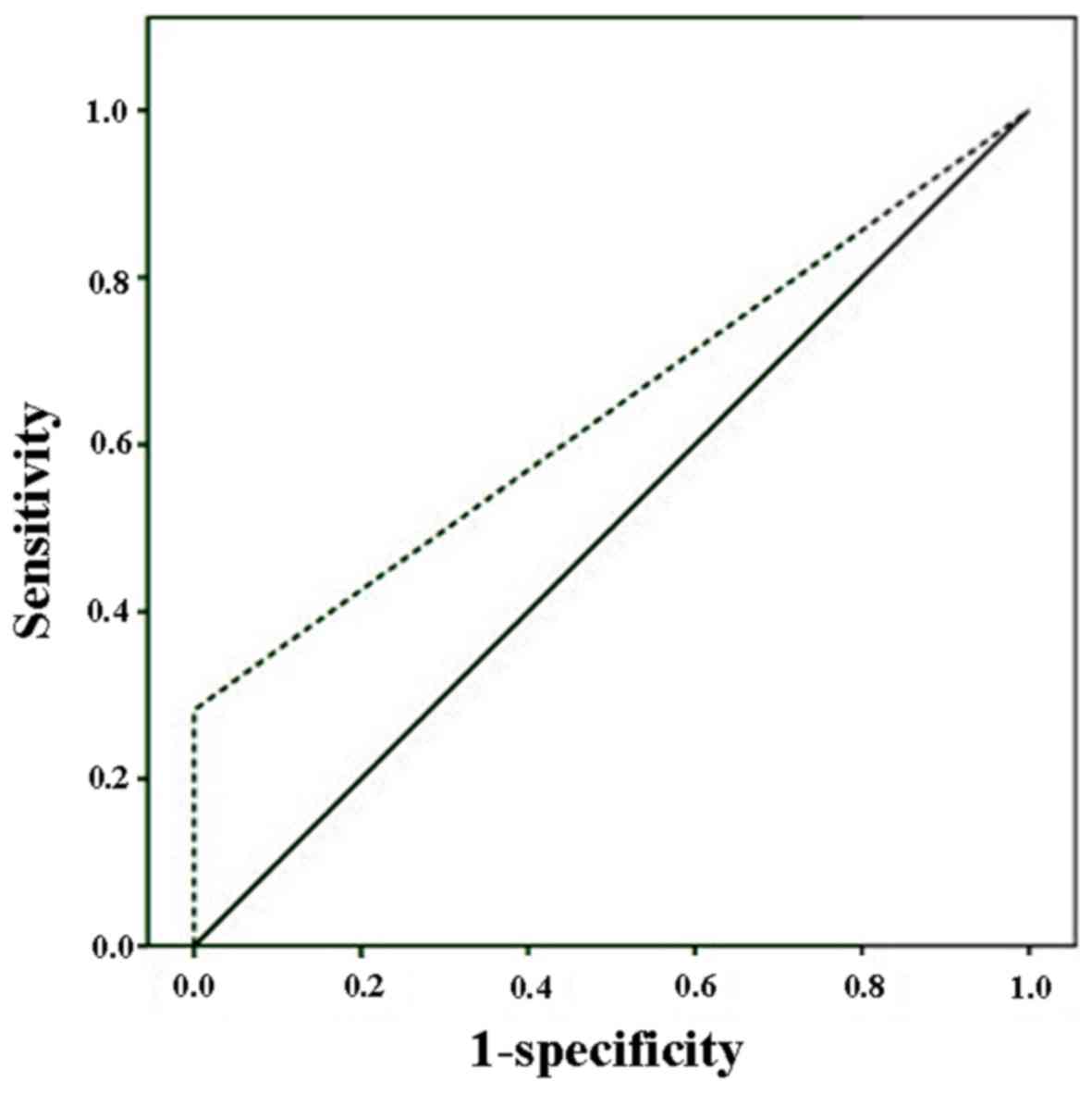
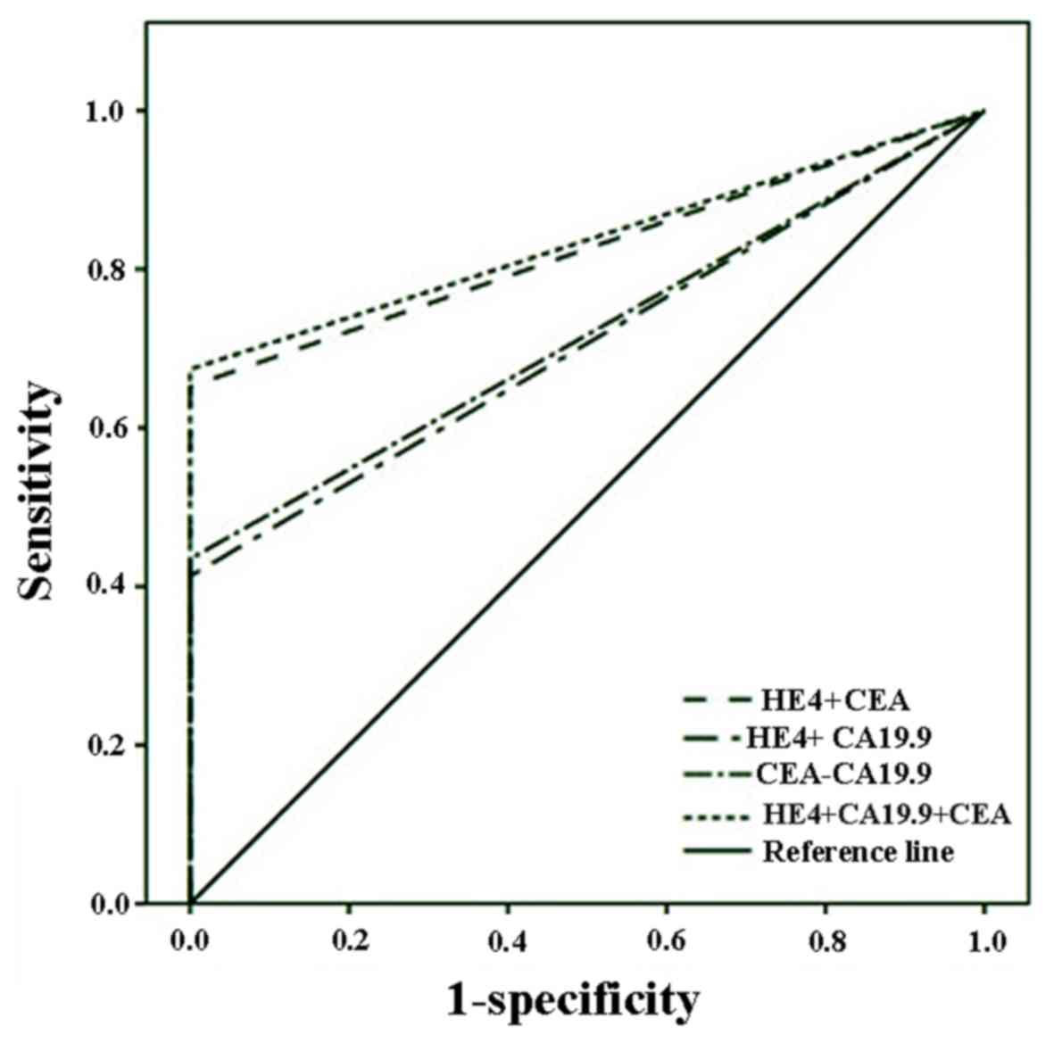
Diagnostic performance of HE4
The ROC curve analyses of the diagnostic performance
of HE4 in healthy controls and CRC patients revealed AUC=0.641 [95%
confidence interval (CI): 0.523–0.760, P=0029; sensitivity=28.3%,
specificity=100.0%; Fig. 1]. When
ROC curve analyses were applied to stage III and IV, the AUC
increased to 0.697 (95% CI): 0.569–0.825, P=0.005;
sensitivity=39.4%, specificity=100.0%). In another ROC curve
analysis, HE4 was combined with CEA and CA19-9 and it was
demonstrated that the combinations may enhance diagnostic accuracy
(AUC of HE4+CEA+CA19-9=0.837, P<0.001; Fig. 2).
Discussion
The present study demonstrated that the rate of HE4
positivity in CRC patients is significantly higher compared with
that in healthy controls, and this positivity is strongly
associated with nodal involvement, advanced stage and CA19-9
positivity.
Although a number of tumour markers are widely used
for the diagnosis, staging and monitoring of CRC patients in
clinical practice, their role in the early detection of colorectal
tumours is limited. Therefore, novel biomarkers with better
clinical utility are needed. Serum HE4 was recently introduced in
the routine diagnostics of ovarian cancer (20). In September, 2011, the US FDA
approved marketing of HE4 for the detection of ovarian masses, as
HE4 has a higher sensitivity and specificity compared with CA125
for the diagnosis of ovarian cancer (21).
HE4 was first described by Kirchhoff et al in
1991 (10); it acts as a proteinase
inhibitor and belongs to the ‘four-disulphide core’ family of
proteins (10). HE4 is expressed in
normal as well as malignant tissues (10). Galgano et al reported that
breast epithelium, female genital tract, epididymis, vas deferens,
distal renal tubules, respiratory epithelium, colonic mucosa and
salivary glands all show HE4 immunoreactivity (12). HE4 expression has also been confirmed
in several cancers, including ovarian cancer, mesothelioma, lung,
endometrial, breast, gastrointestinal, renal and transitional cell
carcinomas (12). The highest
expression levels have been found in ovarian and endometrial
cancer, and moderate levels have been found in lung cancer
(12). In 2003, Hellstrom et
al reported the potential role of HE4 as a secreted biomarker
(22), after which time serum HE4
was investigated as a possible valuable biomarker for ovarian and
endometrial cancers. In a meta-analysis, Zhen et al reported
that the diagnostic accuracy of HE4 in distinguishing ovarian
cancer from other benign gynecological diseases was found to be
superior to that of CA125 (23).
More recently, Brennan et al reported in a large
population-based study that serum HE4 may be a useful prognostic
biomarker in endometrial cancer (17). Furthermore, Yang et al
determined its importance as a preoperative predictor for optimal
tumour cytoreductive surgery in epithelial ovarian cancer (18). In 2012, researchers found that serum
HE4 levels were significantly elevated in lung cancer patients
compared with healthy controls (24). In 2014, Nagy et al, confirmed
that HE4 levels were significantly elevated in lung cancer patients
compared with controls, and also demonstrated that HE4 levels were
significantly correlated with tumour size and the presence of lymph
node metastasis, similar to the findings of the present study
(25). In another more recent study,
again consistent with the present findings, Guo et al
confirmed that HE4 was more commonly observed in gastric carcinoma
tissues compared with normal tissues and was significantly
correlated with advanced stage and tumour size (26); moreover, they found that silencing
HE4 in vitro diminishes the phosphorylation level of Akt,
Erk1/2, Fak and Src, all of which are important for the
angiogenesis, migration and survival of cancer cells (26).
To the best of our knowledge, this is the first
report to examine plasma HE4 levels in CRC patients. In the
literature, only Galgano et al (12) have investigated HE4 expression in CRC
tissues compared with other human neoplasms, and reported weak
positivity in this group. The results of the present study not only
show the significance of HE4 positivity in CRC, but also
demonstrate the positive association with cancer stage and CA19-9
levels, which may be simply correlated with tumour load or be due
to complex tumour biology.
This study has certain limitations: First, the
sample size was relatively small; and second, information on
follow-up is lacking. However, it was designed as a preliminary
study and has reached all the end points; thus, it may be
considered a valuable basis for further research.
In conclusion, serum HE4 may be a useful biomarker
in CRC, particularly in patients with stage III–IV disease. Further
studies are required to confirm these findings and, in the future,
HE4 may be used as a new target for therapeutic interventions.
References
|
1
|
Labianca R, Nordlinger B, Beretta GD,
Mosconi S, Mandalà M, Cervantes A and Arnold D: ESMO Guidelines
Working Group: Early colon cancer: ESMO clinical practice
guidelines for diagnosis, treatment and follow-up. Ann Oncol. 24
Suppl 6:vi64–vi72. 2013. View Article : Google Scholar : PubMed/NCBI
|
|
2
|
World Cancer Report 2014. World Health
Organization. Chapter 1.1. 2014
|
|
3
|
Welch JP and Donaldson GA: The clinical
correlation of an autopsy study of recurrent colorectal cancer. Ann
Surg. 189:496–502. 1979.PubMed/NCBI
|
|
4
|
Borner MM: Neoadjuvant chemotherapy for
unresectable liver metastases of colorectal cancer-too good to be
true? Ann Oncol. 10:623–326. 1999. View Article : Google Scholar : PubMed/NCBI
|
|
5
|
Pamies RJ and Crawford DR: Tumor markers.
An update. Med Clin North Am. 80:185–199. 1996. View Article : Google Scholar : PubMed/NCBI
|
|
6
|
Perkins GL, Slater ED, Sanders GK and
Prichard JG: Serum tumor markers. Am Fam Physician. 68:1075–1082.
2003.PubMed/NCBI
|
|
7
|
Clinical practice guidelines for the use
of tumor markers in breast and colorectal cancer. Adopted on May
17, 1996 by the American Society of Clinical Oncology. J Clin
Oncol. 14:2843–2877. 1996. View Article : Google Scholar : PubMed/NCBI
|
|
8
|
Fletcher RH: Carcinoembryonic antigen. Ann
Intern Med. 104:66–73. 1986. View Article : Google Scholar : PubMed/NCBI
|
|
9
|
Gupta MK, Arciaga R, Bocci L, Tubbs R,
Bukowski R and Deodhar SD: Measurement of a
monoclonal-antibody-defined antigen (CA19-9) in the sera of
patients with malignant and nonmalignant diseases. Comparison with
carcinoembryonic antigen. Cancer. 56:277–283. 1985. View Article : Google Scholar : PubMed/NCBI
|
|
10
|
Kirchhoff C, Habben I, Ivell R and Krull
N: A major human epididymis-specific cDNA encodes a protein with
sequence homology to extracellular proteinase inhibitors. Biol
Reprod. 45:350–357. 1991. View Article : Google Scholar : PubMed/NCBI
|
|
11
|
Drapkin R, von Horsten HH, Lin Y, Mok SC,
Crum CP, Welch WR and Hecht JL: Human epididymis protein 4 (HE4) is
a secreted glycoprotein that is overexpressed by serous and
endometrioid ovarian carcinomas. Cancer Res. 65:2162–2169. 2005.
View Article : Google Scholar : PubMed/NCBI
|
|
12
|
Galgano MT, Hampton GM and Frierson HF Jr:
Comprehensive analysis of HE4 expression in normal and malignant
human tissues. Mod Pathol. 19:847–853. 2006.PubMed/NCBI
|
|
13
|
Kirchhoff C: Molecular characterization of
epididymal proteins. Rev Reprod. 3:86–95. 1998. View Article : Google Scholar : PubMed/NCBI
|
|
14
|
Clauss A, Lilja H and Lundwall A: A locus
on human chromosome 20 contains several genes expressing protease
inhibitor domains with homology to whey acidic protein. Biochem J.
368:233–242. 2002. View Article : Google Scholar : PubMed/NCBI
|
|
15
|
Thompson RC and Ohlsson K: Isolation,
properties, and complete amino acid sequence of human secretory
leukocyte protease inhibitor, a potent inhibitor of leukocyte
elastase. Proc Natl Acad Sci USA. 83:6692–6696. 1986. View Article : Google Scholar : PubMed/NCBI
|
|
16
|
Plebani M: HE4 Study Group: HE4 in
gynecological cancers: Report of a European investigators and
experts meeting. Clin Chem Lab Med. 50:2127–2136. 2012. View Article : Google Scholar : PubMed/NCBI
|
|
17
|
Brennan DJ, Hackethal A, Metcalf AM,
Coward J, Ferguson K, Oehler MK, Quinn MA, Janda M, Leung Y,
Freemantle M, et al: Serum HE4 as a prognostic marker in
endometrial cancer-a population based study. Gynecol Oncol.
132:159–165. 2014. View Article : Google Scholar : PubMed/NCBI
|
|
18
|
Yang Z, Luo Z, Zhao B, Zhang W, Zhang J,
Li Z and Li L: Diagnosis and preoperative predictive value of serum
HE4 concentrations for optimal debulking in epithelial ovarian
cancer. Oncol Lett. 6:28–34. 2013.PubMed/NCBI
|
|
19
|
Simmons AR, Baggerly K and Bast RC Jr: The
emerging role of HE4 in the evaluation of epithelial ovarian and
endometrial carcinomas. Oncology (Williston Park). 27:548–556.
2013.PubMed/NCBI
|
|
20
|
Yu S, Yang HJ, Xie SQ and Bao YX:
Diagnostic value of HE4 for ovarian cancer: A meta-analysis. Clin
Chem Lab Med. 50:1439–1446. 2012. View Article : Google Scholar : PubMed/NCBI
|
|
21
|
Lin J, Qin J and Sangvatanakul V: Human
epididymis protein 4 for differential diagnosis between benign
gynecologic disease and ovarian cancer: A systematic review and
meta-analysis. Eur J Obstet Gynecol Reprod Biol. 167:81–85. 2013.
View Article : Google Scholar : PubMed/NCBI
|
|
22
|
Hellstrom I, Raycraft J, Hayden-Ledbetter
M, Ledbetter JA, Schummer M, McIntosh M, Drescher C, Urban N and
Hellström KE: The HE4 (WFDC2) protein is a biomarker for ovarian
carcinoma. Cancer Res. 63:3695–3700. 2003.PubMed/NCBI
|
|
23
|
Zhen S, Bian LH, Chang LL and Gao X:
Comparison of serum human epididymis protein 4 and carbohydrate
antigen 125 as markers in ovarian cancer: A meta-analysis. Mol Clin
Oncol. 2:559–566. 2014.PubMed/NCBI
|
|
24
|
Iwahori K, Suzuki H, Kishi Y, Fujii Y,
Uehara R, Okamoto N, Kobayashi M, Hirashima T, Kawase I and Naka T:
Serum HE4 as a diagnostic and prognostic marker for lung cancer.
Tumour Biol. 33:1141–1149. 2012. View Article : Google Scholar : PubMed/NCBI
|
|
25
|
Nagy B Jr, Bhattoa HP, Steiber Z, Csobán
M, Szilasi M, Méhes G, Müller M, Lázár J, Kappelmayer J and
Antal-Szalmás P: Serum human epididymis protein 4 (HE4) as a tumor
marker in men with lung cancer. Clin Chem Lab Med. 52:1639–1648.
2014. View Article : Google Scholar : PubMed/NCBI
|
|
26
|
Guo YD, Wang JH, Lu H, Li XN, Song WW and
Zhang XD: The human epididymis protein 4 acts as a prognostic
factor and promotes progression of gastric cancer. Tumour Biol.
36:2457–2464. 2015. View Article : Google Scholar : PubMed/NCBI
|