Introduction
Medullary thyroid carcinoma (MTC) is a rare
neuroendocrine tumor, which arises from parafollicular
calcitonin-secreting cells (C-cells), and accounts for 3–10% of
thyroid malignancies, although MTC is responsible for up to 13.4%
of thyroid cancer-related deaths (1). MTC is of intermediate malignancy,
characterized by partly invasive growth, and is more prone to
generating lymph node metastasis (LNM) and distant metastasis (DM)
compared with differentiated thyroid cancer, with ~10–15% of MTC
patients having DM when first diagnosed. Unfortunately, the number
of available studies on the predictors of metastasis in patients
with MTC is limited.
Sex, age, tumor size, calcitonin (Ctn) and
carcinoembryonic antigen (CEA) concentrations, among other factors,
have been found to be associated with MTC (2–4). Ctn is
a 32-amino acid residue peptide, encoded by a cognate gene located
on the short arm of chromosome 11 in the C-cell. Ctn serves as a
very sensitive and specific tumor marker for the diagnosis of MTC
(5).
Neutrophils are the most abundant leukocyte
population in the circulation and are the first cells recruited to
the site of infection or inflammation (6). Neutrophils are common infiltrating
cells in acute inflammation and a component of chronic inflammatory
infiltrates. Research suggests that an inflammatory environment
contributes to tumor angiogenesis, mutations, cell migration and
metastatic progression through this particular environment that
promotes the growth of malignant cells (7–9).
Numerous studies have reported that neutrophils play
a critical role in tumor progression through the release of
cytokines and angiogenic factors (10). Lymphocytes, which are key factors
involved in the immune surveillance of tumor cells (11), kill cancer cells to suppress the
progression of cancer. Furthermore, the development of several
malignant tumors is closely correlated with an imbalance in the
size of the populations of functional lymphocytes. A growing body
of evidence highlights the role of lymphocytes as a central factor
associated with the prognosis of patients with locally advanced
cancer (12).
The systemic inflammatory response, a subject of
intensive research, suppresses the activity of host immune cells
through promoting microvascular regeneration, which facilitates the
proliferation and differentiation of tumor cells, thus enhancing
the invasion of tissues by tumor cells (13–15). The
neutrophil-to-lymphocyte ratio (NLR) serves as an accurate and
reliable index of systemic inflammation and, thus, serves as a
systemic marker of inflammation (16). Furthermore, strong evidence indicates
that a higher NLR is associated with the growth and migration of
malignant cells.
Numerous studies suggest that the initiation and
progression of thyroid cancer is closely associated with
inflammation (17). Moreover,
neutrophils indicate the presence of inflammation, and lymphocytes
reflect the function of the immune system. The NLR is a relevant
factor of several malignancies, such as urothelial carcinoma
(18), lung cancer (19), gastric cancer (20) and colorectal cancer (21). Unfortunately, the number of studies
assessing the clinical factors that may help predict those MTC
patients who have metastases is limited. The aim of the present
study was to investigate the clinical data of 61 patients with MTC
to identify predictors of metastasis.
Patients and methods
Patients
A retrospective analysis of 61 patients newly
diagnosed with MTC at the General Hospital of the Chinese People's
Liberation Army (PLAGH) between January 2001 and January 2016 was
performed. The patients were diagnosed according to the findings of
the histopathological examination. The inclusion criteria were as
follows: i) MTC was confirmed using thyroid tissue obtained by
ultrasound-guided aspiration biopsy or surgery; ii) the patients
were not administered chemotherapy, radiotherapy or hormone
therapy; iii) routine blood tests were conducted within 3 days
prior to surgery or within 1 week prior to biopsy; iv) serum Ctn
and CEA assays were conducted within 3 days prior to surgery or
within 1 week after diagnosis by biopsy; and v) the patients
underwent preoperative ultrasonography of the thyroid gland and
lymph nodes, as well as fluorodeoxyglucose (FDG)-positron emission
tomography PET/computed tomography (CT). Patients with suspicious
foci detected using FDG-PET/CT underwent CT or magnetic resonance
imaging (MRI) of the neck, chest and abdomen, as well as
emission-CT (ECT) of bone. The exclusion criteria were as follows:
i) Infectious disease, ii) other malignancies and iii) inflammatory
conditions within 1 month prior to the surgery or biopsy. The final
study population comprised 61 patients (27 men and 34 women, aged
25–73 years), 59 of whom had sporadic and 2 hereditary MTC. All the
patients in the present study signed written informed consent forms
prior to admission, and they acknowledged that their clinical data
would be used for clinical studies. The study protocol was granted
ethical approval by the Ethics Committee of the General Hospital of
the Chinese People's Liberation Army (Beijing, China).
Ultrasound-guided fine-needle
aspiration
The fine-needle aspiration procedure was performed
using a standard 21-gauge needle. Sampling typically targeted the
solid component of the lesion. If there was more than one nodule, a
sample was taken from the nodule with suspicious or atypical
ultrasound characteristics.
Surgery
When MTC is clinically apparent (thyroid nodule and
a positive fine-needle aspiration), measurements of serum Ctn, CEA
and calcium concentrations, as well as ultrasound imaging of the
neck should be performed. The surgeon should examine the results of
thoracic and superior mediastinal CT/MRI scans if enlarged lymph
nodes are identified, or if the preoperative serum Ctn
concentration is >400 pg/ml.
The surgical approaches used at our hospital to
treat patients with MTC were as follows: i) Total thyroidectomy
(TT) with central lymph node dissection for unilateral
intrathyroidal tumors sized ≤1 cm; preoperative serum Ctn ≤400
pg/ml; and absence of suspected central neck or lateral cervical
lymph nodes metastases on ultrasound. ii) TT with dissection of the
central lymph node compartment (level VI) and dissection of the
involved lateral neck compartments (levels II–V) when any of these
features are present: Tumor >1 cm, suspected lateral cervical
lymph node metastases on ultrasound, and preoperative serum Ctn
concentrations >400 pg/ml. iii) TT for patients with confined
bilateral nodularity, dissection of the central lymph node
compartment (level VI), and dissection of the bilateral lateral
neck compartments (levels II–V). Patients' sex, age at diagnosis,
histological characteristics of the tumors and neck lymph nodes
(such as tumor size, focality and extrathyroidal extension) were
recorded. All thyroid and lymph node specimens were evaluated by
pathologists at the PLAGH.
Blood tests
Routine blood tests were performed within 3 days
prior to surgery or within 1 week prior to biopsy. Samples were
analyzed using a Sysmex XE-2100 hematology automated analyzer
(Sysmex, Kobe, Japan) with the supplied reagents, red blood cell
controls and calibrators (Sysmex, Kobe, Japan). The concentrations
of serum Ctn and CEA were determined within 3 days prior to surgery
or within 1 week after diagnostic biopsy. The analyzer employs a
chemiluminescent immunoassay to measure the concentrations of Ctn
and CEA. Ctn assessment was performed using an IMMUNITE1000
chemiluminescence analyzer (Siemens AG, Munich, Germany). The
detection limits ranged from 2 to 2,000 pg/ml. The optimal Ctn
cut-off value was 8.4 and 5.0 pg/ml for men and women,
respectively. CEA measurement was performed using an ARCHITECT
i4000 SR chemiluminescence analyzer (Abbott, Chicago, IL, USA). The
detection limits of this method range from 0.5 to 1,500 ng/ml. The
optimal cut-off value of CEA was 5.0 ng/ml.
Statistical analysis
Statistical analyses were performed using SPSS 21.0
software (IBM, Armonk, NY, USA). The Kolmogorov-Smirnov test was
used to ensure that the data were normally distributed, and such
data are presented as mean ± standard deviation, which were
subjected to an independent samples t-test to evaluate the
significance of differences between groups. Data not normally
distributed are presented as median and quartiles, and they were
evaluated using the Mann-Whitney test. Data are presented as
frequency and rate. Chi-squared tests were used to analyze
categorical variables. Receiver operating characteristic (ROC)
curves and the area under the ROC curve (AUC) were used to
determine the optimal cut-off values for the NLR to predict LNM and
DM in MTC. Then, multivariate logistic regression analyses were
used to identify factors independently associated with metastasis.
P-values <0.05 were considered to indicate statistically
significant differences.
Results
Identification of independent
predictors of metastasis
Of the 61 patients with MTC, 41 were confirmed to
have metastases (group M), and 20 were confirmed to be
metastasis-free (group NM). Single-factor analysis revealed
statistically significant differences between groups regarding sex,
tumor size, Ctn and CEA concentrations and NLR (P<0.05). There
was no significant difference between the groups according to age
and solitary focal/multifocal thyroid carcinoma (P>0.05)
(Table I). Multivariate logistic
regression analysis revealed that Ctn concentrations >500 pg/ml
and the NLR were independent predictors of metastasis (Table II).
 | Table I.Single-factor analysis results of
lymphatic metastasis and/or distant metastasis of MTC. |
Table I.
Single-factor analysis results of
lymphatic metastasis and/or distant metastasis of MTC.
| Variables | N=61 (%) | Group M, n=41
(%) | Group NM, n=20
(%) | P-value |
|---|
| Sex (%) |
|
|
| 0.034 |
| Male | 27 (100) | 22 (81.5) | 5 (18.5) |
|
|
Female | 34 (100) | 19 (55.9) | 15 (44.1) |
|
| Age (years) |
|
|
| 0.689 |
|
<45 | 28 (100) | 20 (71.4) | 8 (28.6) |
|
| ≥45 | 33 (100) | 22 (66.7) | 11 (33.3) |
|
| Maximum nodal axis
(cm) |
|
|
|
|
| ≤2 | 32 (100) | 18 (56.2) | 14 (43.8) |
|
| 2–4 | 21 (100) | 15 (71.4) | 6 (28.6) | 0.055 |
|
>4 | 8 (100) | 8 (100) | 0 (0) | 0.034 |
| Focality |
|
|
| 0.098 |
|
Solitary | 40 (100) | 24 (60) | 16 (40) |
|
|
Multifocal | 21 (100) | 17 (81) | 4 (19) |
|
| Ctn (pg/ml) |
|
|
|
|
|
<20 | 3 (100) | 1 (33.3) | 2 (66.7) |
|
|
20–50 | 5 (100) | 1 (20) | 4 (80) | 0.2 |
|
50–200 | 7 (100) | 2 (28.6) | 5 (71.4) | 0.006 |
|
200–500 | 4 (100) | 2 (50) | 2 (50) | 0.000 |
|
>500 | 42 (100) | 35 (83.3) | 7 (16.7) | 0.000 |
| CEA (µg/l), median
(range) | 34.3
(0.6–731.4) | 46.9
(4.2–731.4) | 16.6
(0.66–136.3) | 0.007 |
| NLR, Median
(range) | 1.8
(0.69–5.53) | 1.92
(0.76–5.53) | 1.69
(0.69–2.76) | 0.019 |
| Table II.Multivariate logistic analysis. |
Table II.
Multivariate logistic analysis.
| Variables | B | SE | Wald
χ2 | OR | P-value | 95% CI |
|---|
| Ctn (>500
pg/ml) | 3.064 | 1.074 | 8.145 | 21.422 | 0.004 | 2.611–175.731 |
| NLR | 1.778 | 0.837 | 4.509 | 5.918 | 0.034 | 1.147–30.541 |
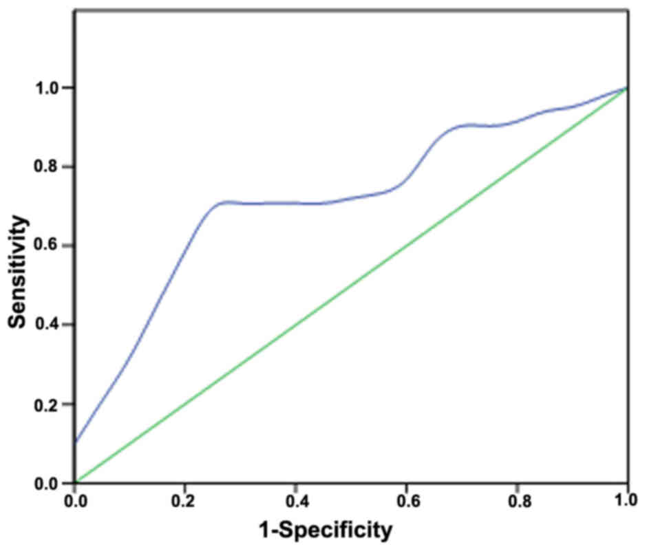
ROC curve analysis and cut-off value
of the NLR
ROC curve analysis indicated that the optimum NLR
cut-off for lymphatic metastasis and/or distant metastasis was
1.784 (the sensitivity and specificity were 68.3 and 80%,
respectively). The AUC value of the NLR was 0.717 (95% confidence
interval: 0.583–0.851) (Fig. 1).
Discussion
To the best of our knowledge, metastatic MTC is
incurable; therefore, the clinician must select patients that are
likely to benefit from therapy, balancing the often slow rate of
tumor progression associated with good quality of life against the
limited efficacy and potential toxicities of local and systemic
therapies (22). There are no known
predictive factors for LNM and DM of MTC. Ctn produced by thyroid
C-cells is the most sensitive diagnostic marker of MTC. The NLR is
a simple index of a systemic inflammatory response, and an elevated
NLR may reflect an imbalanced inflammatory state that facilitates
tumor growth (23).
In the present study, among the 41 patients with
metastasis, 40 (97.56%, 40/41) had Ctn concentrations >500 pg/ml
or an NLR >1.784. Therefore, Ctn concentration and NLR may help
predict metastasis. These findings provide compelling evidence that
the serum Ctn concentration and the NLR may be used as a primary
screen of patients with MTC to detect LNM and DM. Routine
implementation of these measurements may facilitate earlier
detection of LNM and DM, allowing earlier medical intervention.
MTC is an aggressive tumor, prone to hematogenous
and lymphatic metastasis (24). The
prognosis of patients with MTC differs between those with
differentiated thyroid carcinoma or anaplastic thyroid carcinoma.
The tumor metastasizes early to paratracheal and lateral cervical
lymph nodes, and distant metastases may develop in the liver, lungs
and bones. Given its propensity to spread to lymph nodes and
distant organs, MTC is often difficult to cure if not detected
early. When lymph node involvement or distant metastases are
present at diagnosis, the prognosis is usually poor, even when the
primary tumor is quite small (25–27).
Therefore, the treatment of such patients differs from that of
patients without metastasis, and accurate assessment of a patient's
metastatic status is crucial for prescribing optimal individualized
treatment that will increase the survival rate.
Ctn, which is produced by thyroid C-cells, is the
most sensitive marker available for the diagnosis of MTC. Moreover,
the concentration of serum Ctn represents an accurate and sensitive
marker for preoperative diagnosis and post-surgical follow-up. For
example, Ctn concentrations >100 pg/ml have a positive
predictive value (PPV) of 100% for MTC (27). The PPV associated with Ctn
concentrations ≥50 and <100 pg/ml vs. Ctn concentrations ≥20 and
<50 pg/ml is 25 and 8.3%, respectively. The preoperative basal
serum Ctn concentration helps assess the extent of lymph node
metastasis (22). By contrast, CEA
is not a specific biomarker of MTC, although serum CEA
concentrations are useful for evaluating disease progression in
patients with clinically evident MTC. Furthermore, the preoperative
serum CEA concentration is correlated with the number of metastatic
lymph nodes, and combining the Ctn and CEA data increases the rate
of successful diagnosis.
Kazaure et al (1) advocate that the size of the primary
tumor and extrathyroid extension is independently associated with
the prognosis of MTC, whereas Dequanter and Lothaire (3) consider age to be an important
prognostic factor. Furthermore, the basal mortality rate of MTC
reveals that survival is independent of age (28). Inflammation is implicated in the
initiation and progression of thyroid cancer. For example, the
molecular inflammatory process plays a central role in the
malignant progression of transformed thyroid cells (29,30). The
NLR is a simple index of the systemic inflammatory response and
serves as a prognostic indicator in certain cancers. Liu et
al (23) suggested that an
elevated NLR may serve as a marker of an imbalanced inflammatory
state that facilitates tumor growth. Moreover, the NLR serves an
independent prognostic predictor of patients with cancer (31–33).
The present study retrospectively analyzed 61
patients with newly diagnosed MTC. Univariate logistic regression
analysis revealed that sex, tumor size, preoperative peripheral
blood NLR and serum concentrations of CEA and Ctn were associated
with LNM and DM. Multivariate analysis revealed that Ctn
concentrations >500 pg/ml and NLR are independent predictors of
metastasis. The Ctn concentrations in men were markedly higher
compared with those in women, which may be explained by the
abundance of thyroid C-cells in men (34). Furthermore, Ctn concentrations are
inversely correlated with those of CEA and with tumor size
(35,36).
The optimal cut-off value of the NLR for predicting
metastasis obtained from ROC analysis was 1.784 (sensitivity 68.3%
and specificity 80%, AUC 0.717). Furthermore, patients with an NLR
>1.784 had a significantly higher rate of LNM and DM compared
with patients with an NLR ≤1.784. Among the 41 patients with
metastasis, 40 (97.56%) had Ctn concentrations >500 pg/ml or an
NLR >1.784. Previous studies demonstrated that Ctn
concentrations are associated with tumor load (37). Of the 42 patients with Ctn
concentrations >500 pg/ml in the present study, 35 had LNM
or/and DM. By contrast, other studies reported that high Ctn
concentrations are consistent with LNM and higher TNM stage
(38). In the present study, among
the 32 patients with NLR >1.784, 28 had LNM or/and DM,
supporting the conclusion that a high NLR is associated with
metastasis.
There is a strong association between inflammation
and MTC. For example, activated neutrophils may directly and
indirectly stimulate tumor growth. Lymphocytes participate in the
immune process, construct immune barriers, and kill tumor cells. An
elevated NLR may indicate an excessive but ineffective immune
response to the tumor load, or serve as a marker of an imbalanced
inflammatory state, which facilitates tumor growth.
Whole-body imaging of all patients with MTC is
costly and labor-intensive. Serum Ctn assays and NLR determination,
which are cost-effective and universally available, may be used as
a primary screen for MTC patients to detect LNM and DM.
Furthermore, high-risk patients should undergo imaging examinations
to facilitate early detection of metastasis, earlier diagnosis, and
earlier medical intervention.
There were certain limitations to this study. First,
this was a retrospective single-center study. Second, the limited
sample size of MTC patients did not permit us to perform more
intensive analyses. Finally, only the NLR in the prediction of LNM
and DM in MTC patients was analyzed, whereas no information was
reported on other immunological parameters. A multicenter
community-based prospective study, a study with a higher number of
cases, and a more sophisticated study including eosinophils,
basophils or IgG levels and other immunological parameters, are
required in the future in order to investigate a more useful and
precise method for predicting LNM and MD in patients with MTC.
In summary, the findings of the present study
demonstrated that the serum Ctn concentration and the NLR helped
predict the presence of LNM and DM in patients with MTC. Therefore,
physicians should pay more attention to patients with Ctn
concentrations >500 pg/ml or an NLR >1.784, and then perform
imaging examinations to detect metastases as early as possible.
These interventions are critically important for efforts to
implement individualized treatment that improves the quality of
life and prolongs survival.
Acknowledgements
Not applicable.
Funding
This study was funded by Center for Diagnosis and
Treatment of Thyroid Diseases in Chinese PLA General Hospital.
Availability of data and materials
All the data collected and analyzed in the study are
available from the corresponding author on reasonable request.
Authors' contributions
All the authors have read and approved the final
version of this manuscript. WT and NX designed the present study.
NX, YJ and YW performed the experiments. All authors participated
in the writing of the manuscript.
Ethics approval and consent to
participate
The study protocol was granted ethical approval by
the Ethics Committee of the General Hospital of the Chinese
People's Liberation Army (Beijing, China). All the patients in the
present study signed written informed consent forms prior to
admission, and they acknowledged that their clinical data would
been used for clinical studies.
Patient consent to publication
Not applicable.
Competing interests
The authors declare that they have no competing
interests to disclose.
References
|
1
|
Kazaure HS, Roman SA and Sosa JA:
Medullary thyroid microcarcinoma: A population-level analysis of
310 patients. Cancer. 118:620–627. 2012. View Article : Google Scholar : PubMed/NCBI
|
|
2
|
Chen H, Sippel RS, O'Dorisio MS, Vinik AI,
Lloyd RV and Pacak K; North American Neuroendocrine Tumor Society
(NANETS), : The North American Neuroendocrine Tumor Society
consensus guideline for the diagnosis and management of
neuroendocrine tumors: Pheochromocytoma, paraganglioma, and
medullary thyroid cancer. Pancreas. 39:775–783. 2010. View Article : Google Scholar : PubMed/NCBI
|
|
3
|
Dequanter D and Lothaire P: Medullary
thyroid cancer: Surgical results and prognostic factors. Rev Med
Liege. 65:450–452. 2010.(In French). PubMed/NCBI
|
|
4
|
Skinner MA, Moley JA, Dilley WG, Owzar K,
Debenedetti MK and Wells SA Jr: Prophylactic thyroidectomy in
multiple endocrine neoplasia type 2A. N Engl J Med. 353:1105–1113.
2005. View Article : Google Scholar : PubMed/NCBI
|
|
5
|
Lee CR, Lee S, Son H, Ban E, Kang SW, Lee
J, Jeong JJ, Nam KH, Chung WY and Park CS: Medullary thyroid
carcinoma: a 30-year experience at one institution in Korea. Ann
Surg Treat Res. 91:278–287. 2016. View Article : Google Scholar : PubMed/NCBI
|
|
6
|
Sionov RV, Assi S, Gershkovitz M, Sagiv
JY, Polyansky L, Mishalian I, Fridlender ZG and Granot Z: Isolation
and characterization of neutrophils with anti-tumor properties. J
Vis Exp: e52933. 2015. View
Article : Google Scholar
|
|
7
|
Sionov RV, Fridlender ZG and Granot Z: The
multifaceted roles neutrophils play in the tumor microenvironment.
Cancer Microenviron. 8:125–158. 2015. View Article : Google Scholar : PubMed/NCBI
|
|
8
|
Tecchio C and Cassatella MA:
Neutrophil-derived cytokines involved in physiological and
pathological angiogenesis. Chem Immunol Allergy. 99:123–137. 2014.
View Article : Google Scholar : PubMed/NCBI
|
|
9
|
Jiang K, Lei J, Chen W, Gong Y, Luo H, Li
Z, Gong R and Zhu J: Association of the preoperative
neutrophil-to-lymphocyte and platelet-to-lymphocyte ratios with
lymph node metastasis and recurrence in patients with medullary
thyroid carcinoma. Medicine (Baltimore). 95:e50792016. View Article : Google Scholar : PubMed/NCBI
|
|
10
|
Giraldo NA, Becht E, Vano Y,
Sautès-Fridman C and Fridman WH: The immune response in cancer:
From immunology to pathology to immunotherapy. Virchows Arch.
467:127–135. 2015. View Article : Google Scholar : PubMed/NCBI
|
|
11
|
Jiang K, Lei J, Li C, Shu K, Li W, Zhang
Y, Li Z, Gong R and Zhu J: Comparison of the prognostic values of
selected inflammation based scores in patients with medullary
thyroid carcinoma: A pilot study. J Surg Oncol. 116:281–287. 2017.
View Article : Google Scholar : PubMed/NCBI
|
|
12
|
Yu Y, Wang H, Yan A, Wang H, Li X, Liu J
and Li W: Pretreatment neutrophil to lymphocyte ratio in
determining the prognosis of head and neck cancer: A meta-analysis.
BMC Cancer. 18:3832018. View Article : Google Scholar : PubMed/NCBI
|
|
13
|
Brücher BL and Jamall IS: Epistemology of
the origin of cancer: A new paradigm. BMC Cancer. 14:3312014.
View Article : Google Scholar : PubMed/NCBI
|
|
14
|
Kim S, Miller BJ, Stefanek ME and Miller
AH: Inflammation-induced activation of the indoleamine
2,3-dioxygenase pathway: Relevance to cancer-related fatigue.
Cancer. 121:2129–2136. 2015. View Article : Google Scholar : PubMed/NCBI
|
|
15
|
Hainaut P and Plymoth A: Targeting the
hallmarks of cancer: Towards a rational approach to next-generation
cancer therapy. Curr Opin Oncol. 25:50–51. 2013. View Article : Google Scholar : PubMed/NCBI
|
|
16
|
Liu JF, Ba L, Lv H, Lv D, Du JT, Jing XM,
Yang NJ, Wang SX, Li C and Li XX: Association between
neutrophil-to-lymphocyte ratio and differentiated thyroid cancer: A
meta-analysis. Sci Rep. 6:385512016. View Article : Google Scholar : PubMed/NCBI
|
|
17
|
Kocer D, Karakukcu C, Karaman H, Gokay F
and Bayram F: May the neutrophil/lymphocyte ratio be a predictor in
the differentiation of different thyroid disorders? Asian Pac J
Cancer Prev. 16:3875–3879. 2015. View Article : Google Scholar : PubMed/NCBI
|
|
18
|
Taguchi S, Nakagawa T, Matsumoto A, Nagase
Y, Kawai T, Tanaka Y, Yoshida K, Yamamoto S, Enomoto Y, Nose Y, et
al: Pretreatment neutrophil-to-lymphocyte ratio as an independent
predictor of survival in patients with metastatic urothelial
carcinoma: A multi-institutional study. Int J Urol. 22:638–643.
2015. View Article : Google Scholar : PubMed/NCBI
|
|
19
|
Huang C, Yue J, Li Z, Li N, Zhao J and Qi
D: Usefulness of the neutrophil-to-lymphocyte ratio in predicting
lymph node metastasis in patients with non-small cell lung cancer.
Tumour Biol. 36:7581–7589. 2015. View Article : Google Scholar : PubMed/NCBI
|
|
20
|
Chen J, Hong D, Zhai Y and Shen P:
Meta-analysis of associations between neutrophil-to-lymphocyte
ratio and prognosis of gastric cancer. T World J Surg Oncol.
13:1222015. View Article : Google Scholar
|
|
21
|
Choi WJ, Cleghorn MC, Jiang H, Jackson TD,
Okrainec A and Quereshy FA: Preoperative neutrophil-to-lymphocyte
ratio is a better prognostic serum biomarker than
platelet-to-lymphocyte ratio in patients undergoing resection for
nonmetastatic colorectal cancer. Ann Surg Oncol. 22 Suppl
3:S603–S613. 2015. View Article : Google Scholar : PubMed/NCBI
|
|
22
|
Wells SA Jr, Asa SL, Dralle H, Elisei R,
Evans DB, Gagel RF, Lee N, Machens A, Moley JF, Pacini F, et al:
Revised American Thyroid Association guidelines for the management
of medullary thyroid carcinoma. Thyroid. 25:567–610. 2015.
View Article : Google Scholar : PubMed/NCBI
|
|
23
|
Liu CL, Lee JJ, Liu TP, Chang YC, Hsu YC
and Cheng SP: Blood neutrophil-to-lymphocyte ratio correlates with
tumor size in patients with differentiated thyroid cancer. J Surg
Oncol. 107:493–497. 2013. View Article : Google Scholar : PubMed/NCBI
|
|
24
|
Hazard JB, Hawk WA and Crile G Jr:
Medullary (solid) carcinoma of the thyroid; a clinicopathologic
entity. J Clin Endocrinol Metab. 19:152–161. 1959. View Article : Google Scholar : PubMed/NCBI
|
|
25
|
Yamazaki M, Straus FH, Messina M, Robinson
BG, Takeda T, Hashizume K and DeGroot LJ: Adenovirus-mediated
tumor-specific combined gene therapy using Herpes simplex virus
thymidine/ganciclovir system and murine interleukin-12 induces
effective antitumor activity against medullary thyroid carcinoma.
Cancer Gene Ther. 11:8–15. 2004. View Article : Google Scholar : PubMed/NCBI
|
|
26
|
Hyer SL, Vini L, A'Hern R and Harmer C:
Medullary thyroid cancer: Multivariate analysis of prognostic
factors influencing survival. Eur J Surg Oncol. 26:686–690. 2000.
View Article : Google Scholar : PubMed/NCBI
|
|
27
|
Costante G, Meringolo D, Durante C,
Bianchi D, Nocera M, Tumino S, Crocetti U, Attard M, Maranghi M,
Torlontano M and Filetti S: Predictive value of serum calcitonin
levels for preoperative diagnosis of medullary thyroid carcinoma in
a cohort of 5817 consecutive patients with thyroid nodules. J Clin
Endocrinol Metab. 92:450–455. 2007. View Article : Google Scholar : PubMed/NCBI
|
|
28
|
de Groot JW, Plukker JT, Wolffenbuttel BH,
Wiggers T, Sluiter WJ and Links TP: Determinants of life expectancy
in medullary thyroid cancer: Age does not matter. Clin Endocrinol
(Oxf). 65:729–736. 2006. View Article : Google Scholar : PubMed/NCBI
|
|
29
|
De Santis E, Di Vito M, Perrone GA, Mari
E, Osti M, De Antoni E, Coppola L, Tafani M, Carpi A and Russo MA:
Overexpression of pro-inflammatory genes and down-regulation of
SOCS-1 in human PTC and in hypoxic BCPAP cells. Biomed
Pharmacother. 67:7–16. 2013. View Article : Google Scholar : PubMed/NCBI
|
|
30
|
Guarino V, Castellone MD, Avilla E and
Melillo RM: Thyroid cancer and inflammation. Mol Cell Endocrinol.
321:94–102. 2010. View Article : Google Scholar : PubMed/NCBI
|
|
31
|
Malietzis G, Giacometti M, Askari A,
Nachiappan S, Kennedy RH, Faiz OD, Aziz O and Jenkins JT: A
preoperative neutrophil to lymphocyte ratio of 3 predicts
disease-free survival after curative elective colorectal cancer
surgery. Ann Surg. 260:287–292. 2014. View Article : Google Scholar : PubMed/NCBI
|
|
32
|
Feng JF, Huang Y, Zhao Q and Chen QX:
Clinical significance of preoperative neutrophil lymphocyte ratio
versus platelet lymphocyte ratio in patients with small cell
carcinoma of the esophagus. ScientificWorldJournal.
2013:5043652013. View Article : Google Scholar : PubMed/NCBI
|
|
33
|
Lee S, Oh SY, Kim SH, Lee JH, Kim MC, Kim
KH and Kim HJ: Prognostic significance of neutrophil lymphocyte
ratio and platelet lymphocyte ratio in advanced gastric cancer
patients treated with FOLFOX chemotherapy. BMC Cancer. 13:3502013.
View Article : Google Scholar : PubMed/NCBI
|
|
34
|
Basuyau JP, Mallet E, Leroy M and Brunelle
P: Reference intervals for serum calcitonin in men, women, and
children. Clin Chem. 50:1828–1830. 2004. View Article : Google Scholar : PubMed/NCBI
|
|
35
|
Boschin IM, Torresan F, Toniato A, Zane M,
Ide EC, Pennelli G, Rampin L, Colletti PM, Rubello D and Pelizzo
MR: Incidental medullary thyroid microcarcinoma revealed by mild
increase of preoperative serum calcitonin levels: Therapeutic
implications. Endocrine. 45:448–453. 2014. View Article : Google Scholar : PubMed/NCBI
|
|
36
|
Saltiki K, Rentziou G, Stamatelopoulos K,
Georgiopoulos G, Stavrianos C, Lambrinoudaki E and Alevizaki M:
Small medullary thyroid carcinoma: Post-operative calcitonin rather
than tumour size predicts disease persistence and progression. Eur
J Endocrinol. 171:117–126. 2014. View Article : Google Scholar : PubMed/NCBI
|
|
37
|
Kihara M, Miyauchi A, Kudo T, Hirokawa M
and Miya A: Reference values of serum calcitonin with calcium
stimulation tests by electrochemiluminescence immunoassay
before/after total thyroidectomy in Japanese patients with thyroid
diseases other than medullary thyroid carcinoma. Endocr J.
63:627–632. 2016. View Article : Google Scholar : PubMed/NCBI
|
|
38
|
Romero-Lluch AR, Cuenca-Cuenca JI,
Guerrero-Vázquez R, Martínez-Ortega AJ, Tirado-Hospital JL,
Borrego-Dorado I and Navarro-González E: Diagnostic utility of
PET/CT with 18F-DOPA and 18F-FDG in persistent or recurrent
medullary thyroid carcinoma: The importance of calcitonin and
carcinoembryonic antigen cutoff. Eur J Nucl Med Mol Imaging.
44:2004–2013. 2017. View Article : Google Scholar : PubMed/NCBI
|