Introduction
Tongue analysis, one of the essential methods of
traditional Chinese medicine diagnosis, helps understand
pathological changes of internal organs and meridians (1). Tongue coating is the main content
used for tongue analysis, and serves a role in reflecting the
occurrence, development and prognosis of the disease (2). Tongue coating is a visible layer
adherent to the tongue dorsum, mainly comprised of desquamated
epithelial cells and four different types of papillae (3).
These features have been examined via electron
microscopy studies, with the formation of the tongue coating found
to be dependent on the physiological state of filiform papillae
(4). For example, the filiform
papillae within the cuticle of a thick coating are prominent and
dense, while the cuticle of a thin coating is obviously atrophied
(5). The epidermis of the tongue
dorsum contains a highly specialized epithelium consisting of
several stratified cell layers called squamous epithelium, with
these epithelial cells continuously renewed by the mitotic activity
of stem cells within the basal layer (6). Thus, the area and thickness of the
tongue coating are closely associated with the proliferation,
differentiation, migration and apoptosis of squamous epithelial
cells.
Furthermore, some studies have shown that saliva
coexists with tongue squamous epithelial cells and can affect the
formation of tongue coating (7).
Additionally, proteomic based research revealed that blood and
saliva composition could also affect the coating (8). Another study found that levels of
serum trace elements, including zinc, copper and iron, are altered
in a pathological tongue coating relative to a normal control
(9). In conclusion, there is a
strong relationship between changes of tongue coating and
serum.
Apoptosis is a controlled cell-independent death
process that is regulated by protein activity and gene expression
in order to maintain homeostasis and adapt to environmental
conditions. Unlike necrosis, apoptosis maintains a normal structure
of cell organelles, cell shrinkage, chromosome condensation, DNA
fragmentation, apoptosis corpuscles formation and does not cause
inflammation (10). At present,
gene regulation in the process of serum influencing tongue coating
has not been investigated. B-cell lymphoma 2 apoptosis regulator
(Bcl-2) and Bcl-2 associated protein X apoptosis regulator (Bax)
are important members of the Bcl-2 gene family and are 2 of the
most commonly investigated apoptosis regulators. Bcl-2 can induce
cell proliferation and inhibit cell apoptosis, while Bax can
accelerate apoptosis. Recent studies have shown that apoptosis
hinges on the ratio of Bcl-2 and Bax in cells, with this insight
providing may important clues to possible outcomes following a
challenge with an apoptosis-inducing agent (11).
Thus, the present study hypothesized that serum
components may be important in the formation of the tongue coating
and that serum-starvation could induce epithelial cell apoptosis
induced by Bcl-2/Bax in the epidermis of the tongue dorsum. To test
this hypothesis and explore the molecular mechanisms of serum
influencing tongue coating, an in vitro tongue carcinoma
squamous cell culture exposed to serum-starvation was adapted to
simulate apoptosis related to the formation of tongue coating. By
observing the changes of cell cycle, apoptosis and Bax/Bcl-2
expression in a serum-free medium, this approach enabled the
examination of the mechanisms pertaining to the formation of tongue
coating and may aid in a further understanding for future
investigations.
Materials and methods
Cell culture
A tongue squamous cell carcinoma cell line, Tca8113
(Ninth People's Hospital of Shanghai, Shanghai, China), was
maintained in RPMI-1640 medium (Gibco; Thermo Fisher Scientific,
Inc., Waltham, MA, USA) supplemented with 10% heat-inactivated
fetal bovine serum (FBS; Hyclone; GE Healthcare Life Sciences,
Logan, UT, USA), 100 mg/ml streptomycin and 100 U/ml penicillin
(Invitrogen; Thermo Fisher Scientific, Inc., Waltham, MA, USA) in a
humidified atmosphere with 5% CO2 at 37°C.
MTT assay
Cell viability was evaluated using a
3-(4,5-dimethylthiazol-2-yl)-2,5-diphenyltetrazolium (MTT) assay as
previously described (12).
Briefly, 1×10⁴ cells/well were seeded in 96-well plates
and cultured for 18~24 h to reach 90% confluency. Following
attachment, cells were washed twice with PBS, then serum-free
medium added. Both serum deprived and control (10% serum) cells
were harvested at 0, 12, 24, 36, 48 and 72 h. At each time point,
the cell culture supernatants were discarded and 20 µl MTT solution
was added to each well (0.5 mg/ml; Sigma Aldrich; Merck KGaA,
Darmstadt, Germany), then the cells were cultured for a further 4
h. The supernatants were then removed and 200 µl DMSO was added to
each well, with slight agitation for 15 min. The absorbance at a
wavelength of 490 nm was then detected using a PowerWave 340
Microplate Reader (Bio-Tek Instruments, Inc., Winooski, VT, USA),
with 4 replicates used for each well and a mean value
calculated.
Flow cytometry analysis
Cells were seeded at 1×10⁴ cells/well in
a 24-well plate and cultured for 18–24 h to reach 90% confluency.
Following attachment, cells were washed two times with PBS and
serum starvation was evoked. At 0, 12, 24, 36, 48 and 72 h,
adherent cells were trypsinized and collected together with the
medium containing non-adherent cells. Apoptosis was evaluated using
an Annexin V-fluorescein isothiocyanate (FITC)/propidium iodine
(PI) double staining apoptosis detection kit (MBL International
Co., Woburn, MA, USA) according to the manufacturer's protocol, in
conjunction with FACS Accuri C6 flow cytometer (BD Biosciences,
Franklin Lakes, NJ, USA) and analyzed by using FlowJo 7.6.1
software (TreeStar, San Carlos, CA, USA). Cell-cycle analysis was
also evaluated using a cell-cycle analysis kit (BD Cycletest Plus
DNA kit; BD Biosciences) as described previously (13).
Hoechst staining
To assess the effects of serum-starvation on nuclear
material, cells were stained with Hoechst 33342. Cells were seeded
at a density of 5×10⁴ cells/well in a 24-well plate for
24 h with 6 replicates/sample. Cells were then washed 3 times with
PBS and fixed with 10% paraformaldehyde for 5 min. Cells were
washed with PBS before adding Hoechst working solution (10 mg/ml;
Molecular Probes; Thermo Fisher Scientific, Inc.), followed by a 15
min incubation at 37°C. Images were captured with an Olympus IXSI
inverted microscope using 350 nm excitation and 450 nm emission
filters. A total of 3 images per treatment replicate were captured
at ×10 and ×20 magnifications.
Reverse transcription-quantitative
polymerase chain reaction (RT-qPCR)
RT-qPCR was employed to detect the mRNA expression
levels of Bcl-2, Bax and the housekeeping gene
glyceraldehyde-3-phosphate dehydrogenase (GAPDH). Following serum
starvation, total RNA was extracted using TRIzol reagent
(Invitrogen; Thermo Fisher Scientific, Inc.), according to the
manufacturer's protocols. Purified total RNA (1 µg) was then
reverse transcribed using a First Strand cDNA Synthesis Kit (Takara
Bio, Inc., Otsu, Japan). RT-qPCR was performed with the following
primers: Bax, forward 5′-GGCCCACCAGCTCTGAGCAGA-3′ and reverse
5′-GCCACGTGGGCGGTCCCAAAGT-3′; Bcl-2, forward
5′-GTGGAGGAGCTCTTCAGGGA-3′ and reverse 5′-AGGCACCCAGGGTGAGCAA-3′
(14). PCR primers for the
internal control gene GAPDH forward: 5′-GGAGAAACCTGCCAAGTATG-3′ and
reverse: 5′-TTACTCCTTGGAGGCCATGTAG-3′ (15). Reactions were conducted in 96-well
plates with a final volume of 20 µl including 10 µl SYBR Green PCR
Master Mix (Invitrogen, Carlsbad, CA, USA), plus 1 µl each primer
(2 µM), 1 µl template DNA, and 7 µl ddH2O. Thermal
cycling and fluorescence detection were conducted on ABI ViiA7
(Applied Biosystems, Thermo Fisher Scientific, Inc., Waltham, MA,
USA). The reaction mixtures were cycled 35 times at 94°C for 30
sec, 60°C for 30 sec and 72°C for 30 sec. Each reaction was run in
triplicate. The levels of Bcl-2 and Bax gene expression were
normalized to GAPDH levels using the method of 2−∆∆Cq
(16,17). PCR products were assessed by
electrophoresis, with no primer-dimers observed for either the
target genes or GAPDH, and the product specificities were also
confirmed by melting curve analysis.
Western blotting
Bax and Bcl-2 protein expression levels were
examined by western blot analysis. Proteins within the cell lysates
were separated by gel electrophoresis, with protein transferred to
a nitrocellulose membrane and immunoblotting performed as
previously described (18). Cells
were cultured for 18–24 h to reach 90% confluency.
Radioimmunoprecipitation Assay lysis buffer (Qiagen, Inc.,
Valencia, CA, USA) was added to each cell and vortexed for 15 min
at a temperature of 4°C, then centrifuged at 12,000 × g for 10 min
at a temperature of 4°C. Supernatant was transferred to a new tube
and determined quantitatively by Bicinchoninic acid Protein Assay
kit. Protein lysates were boiled in SDS-sample buffer for 5 min,
equally loaded (20 µg/lane) onto an 8% SDS-PAGE and transferred to
a polyvinylidene fluoride membrane (Bio-Rad Laboratories, Inc.,
Hercules, CA, USA). Membranes were then blocked for 2 h in 5%
milk-Tris-Buffered Saline Tween-20 (TBST) at room temperature, then
incubated with either monoclonal anti-Bax (1:750; cat. no. 5023) or
anti-Bcl-2 (1:750; cat. no. 4223,) or monoclonal anti-GAPDH
antibodies (1:2,000; cat. no. 5174; all Cell Signaling Technology,
Inc., Danvers, MA, USA). Membranes were then washed four times in
TBST and incubated with appropriate horseradish
peroxidase-conjugated secondary antibodies (1:1,000; cat. no. 7074;
Cell Signaling Technology, Inc.). Blots were visualized by enhanced
chemiluminescence (Thermo Fisher Scientific, Inc.) and analyzed
using a scanning densitometer with molecular analysis software
(BioSens Gel Imaging System 750, Shanghai Bio-Tech Co., Ltd.,
Shanghai, China).
Scanning electron microscopy
(SEM)
Tca8113 cells were subjected to scanning electron
microscopy as previously described (19). Briefly, Tca8113 cells were exposed
to serum starvation for 0, 12, 24, 36, 48 and 72 h, then fixed for
scanning electron microscopy by immersion in 2.5%
glutaraldehyde-PBS (pH 7.4) at room temperature for 1 h. Following
fixation, samples were washed 3 times with PBS for 1 h and
dehydrated in a graded series of ethanol (50, 70 and 80% at 20 min
per change; 90, 96 and 100% at 15 min per change; and 100% for
three changes at 4°C). Samples were then dried and imaged using a
Model S-3000N scanning electron microscope (Hitachi, Ltd., Tokyo,
Japan).
Transmission electron microscopy
(TEM)
Tca8113 cells were exposed to serum starvation for
0, 12, 24, 36, 48 and 72 h, then fixed for TEM. Samples were fixed
overnight at 4°C in 0.1 M PBS buffer containing 2.5% glutaraldehyde
(v/v) and post-fixed in 1% OsO4 in the same buffer for 2
h at 4°C. The samples were then dehydrated through a graded ethanol
series [30, 50, 70, 90, and 100% (v/v in ddH2O) for 15
min at each concentration], followed by a graded ethanol:acetone
series (3:1, 1:1, 1:3, and 0:1 for 15 min at each concentration) at
4°C and embedded in araldite resin. Sections (60–80 nm), which were
obtained with an ultramicrotome, were stained in 3% acetic acid
uranium-citric acid and viewed using an H-7650 transmission
electron microscope (Hitachi, Ltd., Tokyo, Japan) (20).
Statistical analysis
All data were statistically analyzed using the SPSS
16.0 software package (SPSS Inc., Chicago, IL, USA), with results
expressed as the mean ± standard deviation. The means among diverse
samples were compared by one-way analysis of variance and multiple
comparisons among the groups were conducted using the
least-significant difference (LSD) method. Dunnett's method was
employed to evaluate the heterogeneity of the variance. P<0.05
was considered to indicate a statistically significant
difference.
Results
Serum-starvation suppresses cellular
proliferation
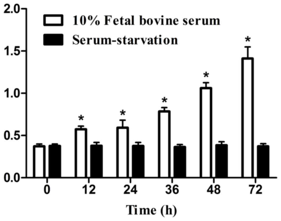
Growth inhibitory effects on cellular proliferation,
measured by MTT assay, are shown in Fig. 1 Compared with the 0 h control, no
significant differences were observed following serum-starvation
for 12, 24, 36, 48 and 72 h, cell proliferation levels (P>0.05;
Fig. 1). Conversely, cell
proliferation significantly increased compared with the 0 h control
in medium containing 10% FBS (P<0.05; Fig. 1).
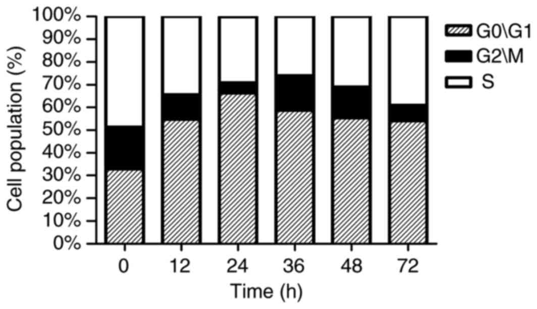
Serum-starvation induces cell cycle
arrest at G1 phase
A significant and time-dependent G1 phase
arrest was noted in serum-starved cells, compared with 0 h
(P<0.05; Table I and Fig. 2). A peak in this effect was reached
at 24 h of treatment, when the number of cells at G1
phase had significantly increased from 32.84% (untreated control)
to 66.40%.
 | Table I.Serum-starvation induces cell cycle
arrest at G1 phase. |
Table I.
Serum-starvation induces cell cycle
arrest at G1 phase.
|
| Time (h) |
|---|
|
|
|
|---|
| Cell population
(%) | 0 | 12 | 24 | 36 | 48 | 72 |
|---|
| G0/G1 | 32.84 | 54.82a | 66.40a | 58.79a | 55.49a | 54.10a |
| G2/M | 18.43 | 10.80 | 4.44 | 15.13 | 13.43 | 6.96 |
| S | 48.73 | 34.39 | 29.15 | 26.08 | 31.08 | 38.94 |
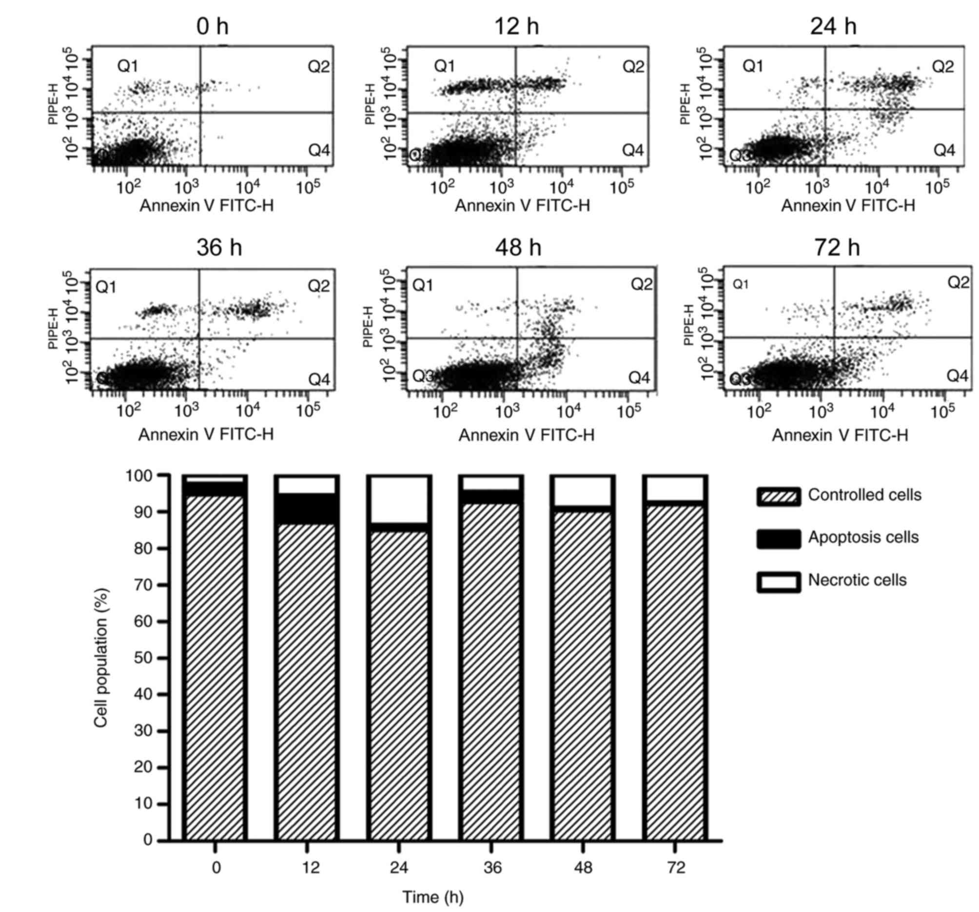
Effect of serum-starvation on cellular
apoptosis
Apoptotic cells were examined by counting the
percentage of early apoptotic cells
(Annexin-V+/PI−) and late apoptotic cells
(Annexin-V+/PI+). Under conditions of
serum-starvation, the percentages of both early and late apoptotic
cells were significantly elevated in a time-dependent fashion,
compared with 0 h (P<0.05; Table
II and Fig. 3). However,
apoptosis did not continue to increase with treatment time:
Following 24 h of treatment, the percentage of late apoptotic cells
(Q2) peaked at 8.97% and then declined. The early apoptosis (Q4)
rates continued to rise, dipped at 36 h, before reaching an apex at
48 h and then gradually declining.
| Table II.Effect of serum-starvation on cellular
apoptosis. |
Table II.
Effect of serum-starvation on cellular
apoptosis.
|
| Time (h) |
|---|
|
|
|
|---|
|
| 0 | 12 | 24 | 36 | 48 | 72 |
|---|
| Late-apoptotic
cells (Q2) | 0.87±0.34 |
4.33±1.96a |
8.97±2.72a |
4.67±1.45a |
2.50±0.45a |
2.77±0.34a |
| Early apoptotic
cells (Q4) | 0.23±0.09 |
2.63±1.25a |
5.57±1.61a |
1.00±0.42a |
7.00±0.99a |
3.90±1.27a |
Hoechst staining
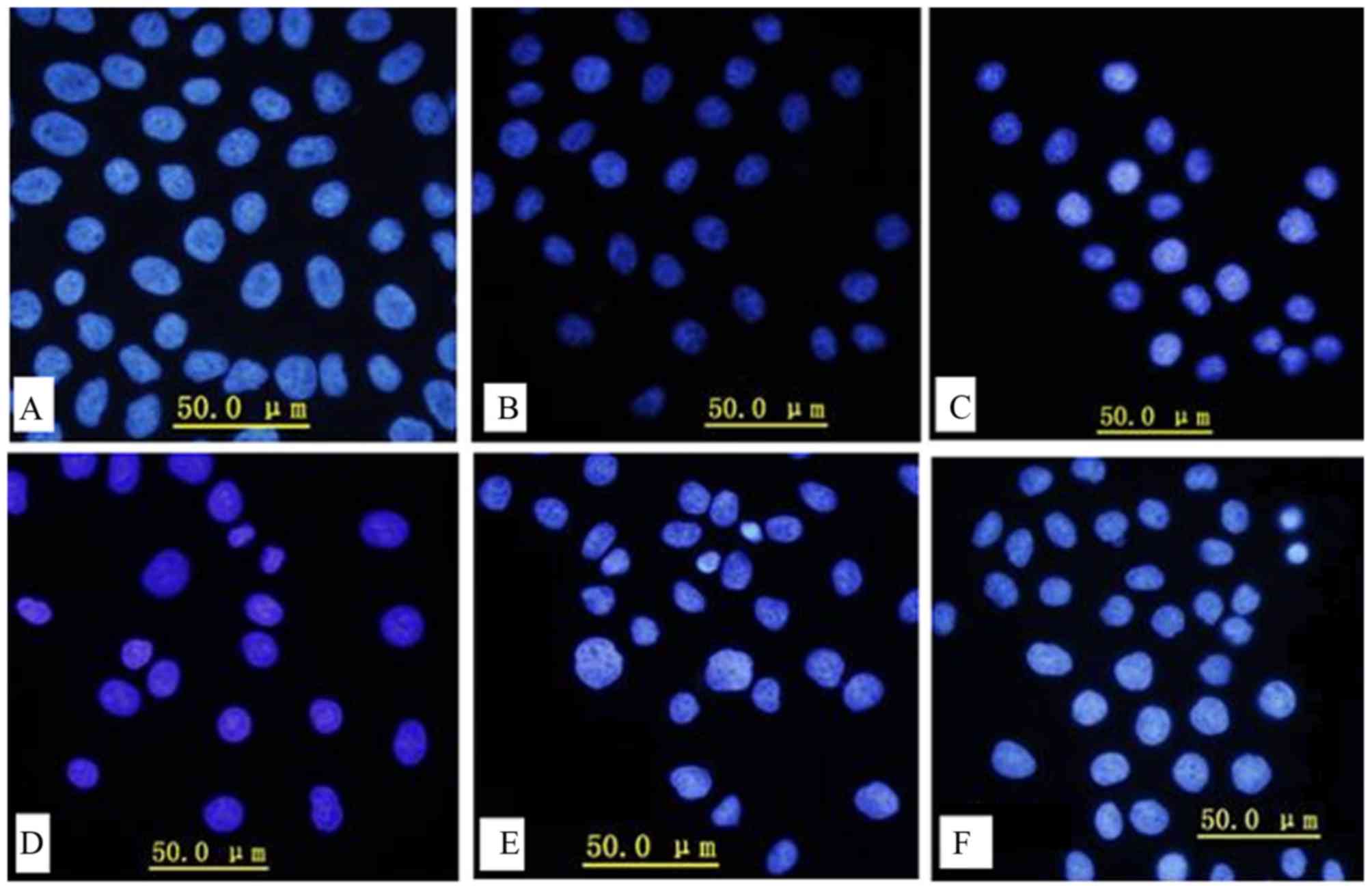
Apoptosis is one of the major pathways that leads to
cell death, therefore the effects of serum-starvation on apoptosis
were further examined using the Hoechst staining method. Manual
observation of apoptotic cells based on cytoplasmic condensation,
karyopyknosis and nuclear fragmentation revealed that the
percentage of apoptotic cells increased as the duration of serum
starvation increased (Fig. 4A-F).
Serum-starvation for 36 h resulted in karyopyknosis (Fig. 4D), and as serum-starvation time
increased further, classic characteristics of apoptosis were
observed, including chromatin condensation and nuclei
fragmentation. However, no nucleosomes were observed.
Effect of serum-starvation on cell
surface morphological characteristics and ultrastructure
SEM was then used to study the effect of
serum-starvation on cell surface morphological characteristics
(Fig. 5). Control cells exhibited
a uniform distribution of palpate and microvilli on their surfaces
(Fig. 5A and D), while after 24 h
of serum-starvation, the microvilli numbers were reduced and a
smoothening of the cell surface was observed (Fig. 5B and E). After 72 h of
serum-starvation, the microvilli were absent and the cell membrane
showed breakage (Fig. 5C and
F).
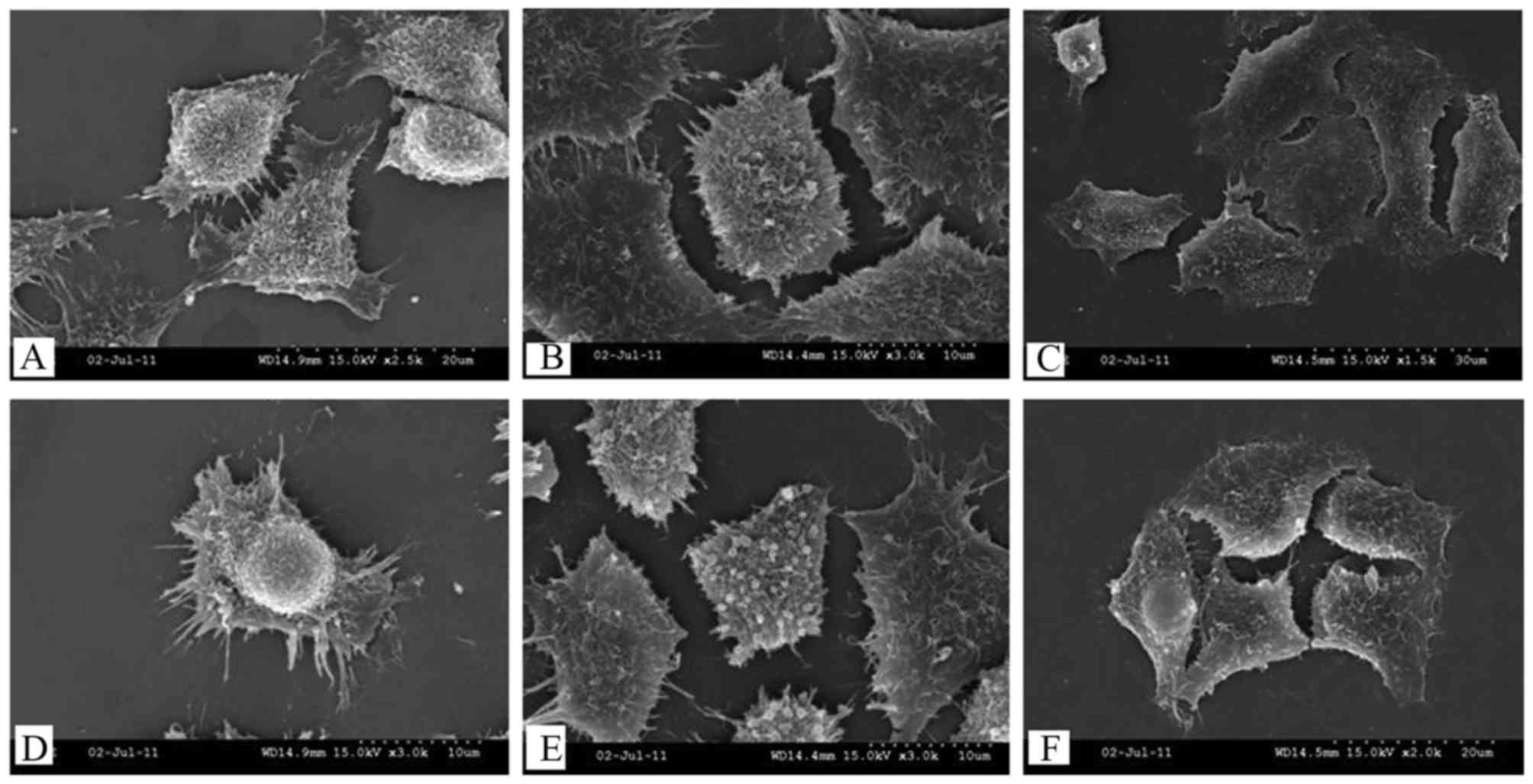 | Figure 5.Surface morphological characteristics
of cells following (A and D) 0, (B and E) 24 and (C and F) 72 h of
serum-starvation, detected by scanning electron microscopy. (A)
Synapse connections (magnification, ×2500) and (D) a uniform
distribution of microvilli on their surfaces (magnification,
×3,000) among control cells. After 24 h of serum-starvation, (B)
the microvilli numbers were reduced (magnification, ×3,000) and (E)
a smoothening of the cell surface was observed (magnification,
×3,000). After 72 h of serum-starvation, (C) the cell membrane
showed breakage (magnification, ×1,500) and (F) the microvilli were
absent (magnification, ×2,000). |
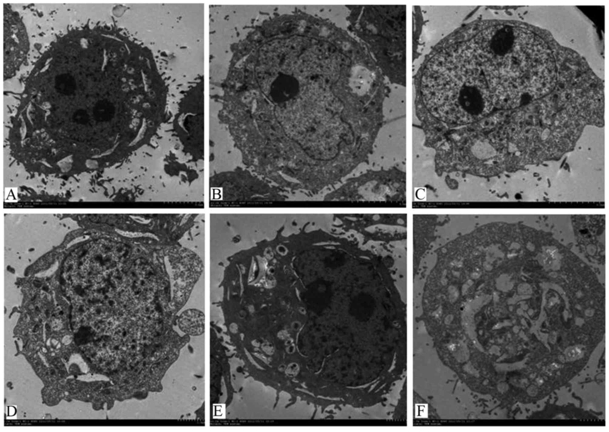
To study the effects of serum-starvation on cell
ultrastructures, TEM was then performed. Control cells appeared
round, had abundant organelles and normal double-membraned nuclei
(Fig. 6A). Following
serum-starvation, the nuclear membrane was domed outward with a
sharp angle, the chromatin within the nuclei was concentrated and
clustered on the inner border of cells and cell blebbing became
visible (Fig. 6B-F).
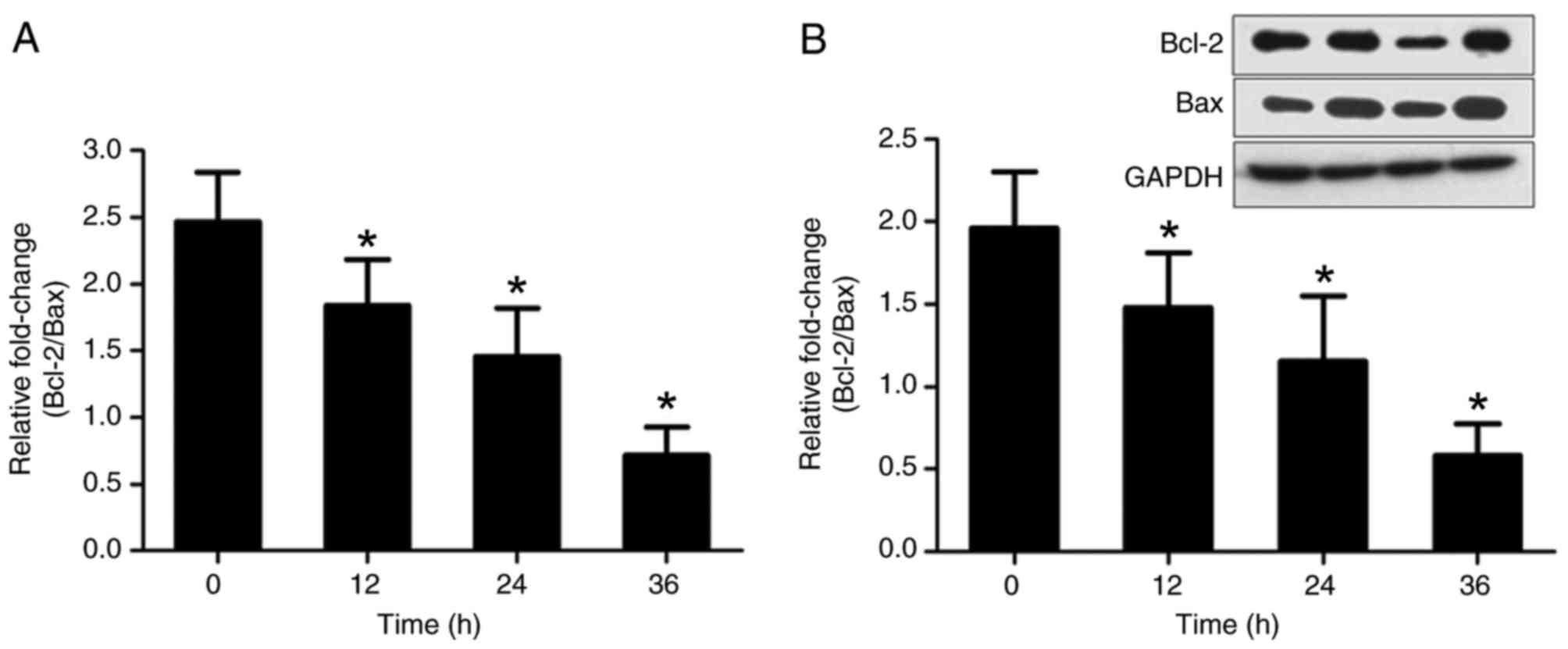
Effects of serum starvation on Bcl-2
and Bax expression
In order to investigate the effects of
serum-starvation on cellular apoptosis, Bcl-2 and Bax mRNA and
protein expression was examined. Owing to the opposite effects of
Bcl-2 and Bax on apoptosis, the ratio of Bcl-2/Bax was calculated.
Relative to the 0 h control group, the ratio of Bcl-2/Bax was
increasingly decreased after 12, 24 and 36 h serum-starvation
(P<0.05; Fig. 7). These results
suggest that serum-deprivation induces cellular apoptosis in a
time-dependent manner.
Discussion
Changes to the tongue coating may reflect the
condition of vitality and pathogen status in the body. Tongue
squamous epithelial cells are the main component of the tongue
coating, with proliferation, differentiation and apoptosis being
the root cause of the formation and maintenance of tongue coating
(21). Therefore, examination of
the mechanisms of the formation of tongue coating is meaningful at
the cellular level.
However, very little research pertaining to cell
function related to tongue coating formation has been examined,
mainly due to: i) The oral environment being a contaminated
environment, with a low cultivation success rate; ii) it is
generally more difficult to obtain normal human tongue back mucosal
epithelial materials because submucous tissue cannot be completely
cleaned, which causes the epithelial cells to develop synchronously
inside fibroblasts to different degrees (22); and iii) epithelial cells are
challenging to grow in general synthetic media because they have
extremely complex nutritional requirements which are not fully
achievable in a uniform culturing condition (23).
Current research has found that tongue carcinoma
squamous cells, which share a source in common with tongue
papillary squamous epithelial cells, cultured in vitro can
imitate the biological traits of the epithelial cell in the tongue
dorsum (24). Therefore, the
present study aimed to simulate tongue coating cell apoptosis by
culturing tongue carcinoma squamous cells in a serum-starvation
environment in vitro, to provide a theoretical basis for
clarification of the molecular mechanisms relating to the formation
of tongue coating.
There are an increasing number of reports detailing
the influence of serum-starvation on cells. An increase in cell
apoptosis has been observed in some cell types following serum
deprivation. For example, goat skin fibroblast apoptosis was
enhanced by 3- and 10-fold following 48 and 120 h serum starvation,
respectively (25). In the present
study, the cellular proliferative capability was estimated by the
MTT method. In medium containing 10% serum, tongue carcinoma
squamous cells continued to proliferate, but stopped proliferating
in serum-free medium.
Based on these findings, cell cycle and apoptosis
were examined by flow cytometry. As the duration of
serum-starvation increased, G0/G1 cell cycle
arrest was induced, with an apex reached at 24 h and then gradually
reduced, suggesting that there could be other factors at work.
Tongue squamous cancer cells showed both early and late apoptosis
following serum-starvation. Early apoptotic rates increased to a
peak at 24 h and then decreased, while late apoptotic rates
continued to rise, but with a slight decline at 36 h, and reached
an apex at 48 h, followed by a gradual decline. Examination of
cells under an inverted microscope showed cellular shrinkage,
rounding and a gradual loss of adherence, with the majority of
cells floating after 36 h of serum-starvation. This could be due to
the late apoptotic cell disruption between 24–36 h, which is
consistent with the G0 cell percentage gradually
declining after 24 h, with G0 arrest beginning to be
relieved.
Morphological observations are considered the golden
standard for identification of apoptotic cells, with indicators of
apoptotic cells including chromatin condensation, nuclear membrane
dissociation, chromatin dividing into blocks and the formation of
apoptotic bodies. In the present experiments, Hoechst fluorescence
microscope, TEM and SEM were applied to observe structural changes
in tongue carcinoma squamous cells between experimental and control
groups. These results showed that over time the percentage of
apoptotic cells increased significantly, with characteristic
changes noted. TEM observations demonstrated half-moon-shaped
vacuoles appearing within the cells following 48 and 72 h of
serum-starvation, which is due to the cells crimping when scraped
with a cell knife. The results of this study concur with previous
findings and visually confirmed that serum-starvation does have a
role in inducing tongue squamous cancer cell apoptosis in a
time-dependent manner.
In addition, the present study observed that the
ratio of Bcl-2/Bax was altered in favor of apoptosis in
serum-starved samples. This experimental result is consistent with
the result of Papa et al (26), who observed that a down-regulation
in the ratio of Bcl-2/Bax was associated with apoptosis resistance
as compared to normal keratinocytes. Therefore, we speculate that
the apoptosis-inducing effects might be due to a change in the
mitochondrial membrane potential through the inhibition of Bcl-2
expression and/or increased expression of Bax. These results,
combined with the results of MTT and flow cytometric analysis,
indicate that serum-starvation could reduce the Bcl-2/Bax ratio and
promote apoptosis, which is consistent with the noted inhibition of
cellular proliferation.
Taken together, tongue squamous cell carcinoma cells
in a serum-free medium may be used to simulate apoptosis related to
the formation of tongue coating, which can offer guidance for
future investigations about other factors.
Acknowledgements
This work was financially supported by National
Natural Science Foundation of China (81473458), Qing Lan Project
and a Project funded by the Priority Academic Program Development
of Jiangsu Higher Education Institutions (PAPD).
References
|
1
|
Jiang B, Liang X, Chen Y, Ma T, Liu L, Li
J, Jiang R, Chen T, Zhang X and Li S: Integrating next-generation
sequencing and traditional tongue diagnosis to determine tongue
coating microbiome. Sci Rep. 2:9362012. View Article : Google Scholar : PubMed/NCBI
|
|
2
|
Zhao Y, Gou XJ, Dai JY, Peng JH, Feng Q,
Sun SJ, Cao HJ, Zheng NN, Fang JW, Jiang J, et al: Differences in
metabolites of different tongue coatings in patients with chronic
hepatitis B. Evid Based Complement Alternat Med. 2013:2049082013.
View Article : Google Scholar : PubMed/NCBI
|
|
3
|
Singha KB, Konar S, Mondal MK and Das J:
Scanning electron microscopic study of the human fungiform
papillae. J Anat Soc India. 59:154–157. 2010. View Article : Google Scholar
|
|
4
|
Jackowiak H and Godynicki S: The scanning
electron microscopic study of lingual papillae in the silver fox
(Vulpes vulpes fulva, Desmarest, 1820). Ann Anat. 186:179–183.
2004. View Article : Google Scholar : PubMed/NCBI
|
|
5
|
Kawasaki K, Porntaveetus T, Oommen S,
Ghafoor S, Kawasaki M, Otsuka-Tanaka Y, Blackburn J, Kessler JA,
Sharpe PT and Ohazama A: Bmp signalling in filiform tongue papillae
development. Arch Oral Biol. 57:805–813. 2012. View Article : Google Scholar : PubMed/NCBI
|
|
6
|
Da Silva AE, Rados PV, Da Silva Lauxen I,
Gedoz L, Villarinho EA and Fontanella V: Nuclear changes in tongue
epithelial cells following panoramic radiography. Mutat Res.
632:121–125. 2007. View Article : Google Scholar : PubMed/NCBI
|
|
7
|
Gonçalves AS, Arantes DA, Bernardes VF,
Jaeger F, Silva JM, Silva TA, Aguiar MC and Batista AC:
Immunosuppressive mediators of oral squamous cell carcinoma in
tumour samples and saliva. Hum Immunol. 76:52–58. 2015. View Article : Google Scholar : PubMed/NCBI
|
|
8
|
Zhang A, Sun H, Wang P and Wang X:
Salivary proteomics in biomedical research. Clin Chim Acta.
415:261–265. 2013. View Article : Google Scholar : PubMed/NCBI
|
|
9
|
Sun ZM, Zhao J, Qian P, Wang YQ, Zhang WF,
Guo CR, Pang XY, Wang SC, Li FF and Li Q: Metabolic markers and
microecological characteristics of tongue coating in patients with
chronic gastritis. BMC Complement Altern Med. 13:2272013.
View Article : Google Scholar : PubMed/NCBI
|
|
10
|
Pérez-Garijo A and Steller H: Spreading
the word: Non-autonomous effects of apoptosis during development,
regeneration and disease. Development. 142:3253–3262. 2015.
View Article : Google Scholar : PubMed/NCBI
|
|
11
|
Martin LJ: Neuronal cell death in nervous
system development, disease, and injury (Review). Int J Mol Med.
7:455–478. 2001.PubMed/NCBI
|
|
12
|
Mosmann T: Rapid colorimetric assay for
cellular growth and survival: Application to proliferation and
cytotoxicity assays. J Immunol Methods. 65:55–63. 1983. View Article : Google Scholar : PubMed/NCBI
|
|
13
|
Ozeki N, Mogi M, Nakamura H and Togari A:
Differential expression of the Fas-Fas ligand system on
cytokine-induced apoptotic cell death in mouse osteoblastic cells.
Arch Oral Biol. 47:511–517. 2002. View Article : Google Scholar : PubMed/NCBI
|
|
14
|
Tano T, Okamoto M, Kan S, Nakashiro K,
Shimodaira S, Koido S, Homma S, Sato M, Fujita T, Kawakami Y and
Hamakawa H: Prognostic impact of expression of Bcl-2 and Bax genes
in circulating immune cells derived from patients with head and
neck carcinoma. Neoplasia. 15:305–314. 2013. View Article : Google Scholar : PubMed/NCBI
|
|
15
|
Jiang W, Wang Q, Chen S, Gao S, Song L,
Liu P and Huang W: Influenza A virus NS1 induces G0/G1 cell cycle
arrest by inhibiting the expression and activity of RhoA protein. J
Virol. 87:3039–3052. 2013. View Article : Google Scholar : PubMed/NCBI
|
|
16
|
Zhang W, Wang F, Xu P, Miao C, Zeng X, Cui
X, Lu C, Xie H, Yin H, Chen F, et al: Perfluorooctanoic acid
stimulates breast cancer cells invasion and up-regulates matrix
metalloproteinase-2/-9 expression mediated by activating NF-κB.
Toxicol Lett. 229:118–125. 2014. View Article : Google Scholar : PubMed/NCBI
|
|
17
|
Livak KJ and Schmittgen TD: Analysis of
relative gene expression data using real-time quantitative PCR and
the 2(-Delta Delta C(T)) method. Methods. 25:402–408. 2001.
View Article : Google Scholar : PubMed/NCBI
|
|
18
|
Cerella C, Sobolewski C, Chateauvieux S,
Henry E, Schnekenburger M, Ghelfi J, Dicato M and Diederich M:
COX-2 inhibitors block chemotherapeutic agent-induced apoptosis
prior to commitment in hematopoietic cancer cells. Biochem
Pharmacol. 82:1277–1290. 2011. View Article : Google Scholar : PubMed/NCBI
|
|
19
|
Brunk UT, Dalen H, Roberg K and Hellquist
HB: Photo-oxidative disruption of lysosomal membranes causes
apoptosis of cultured human fibroblasts. Free Radic Biol Med.
23:616–626. 1997. View Article : Google Scholar : PubMed/NCBI
|
|
20
|
Zhang H, An X, Zhou Y, Zhang B, Zhang S,
Li D, Chen Z, Li Y, Bai S, Lv J, et al: Effect of oxidative stress
induced by Brevibacterium sp. BS01 on a HAB causing
species-Alexandrium tamarense. PLoS One. 8:e630182013. View Article : Google Scholar : PubMed/NCBI
|
|
21
|
Izumi K, Nakajima T, Maeda T, Cheng J and
Saku T: Adenosquamous carcinoma of the tongue: Report of a case
with histochemical, immunohistochemical, and ultrastructural study
and review of the literature. Oral Surg Oral Med Oral Pathol Oral
Radiol Endod. 85:178–184. 1998. View Article : Google Scholar : PubMed/NCBI
|
|
22
|
Görögh T, Lippert BM, Gottschlich S, Folz
B and Werner JA: Establishment and characterization of 2 cell lines
of squamous epithelial carcinoma of the mouth floor and tongue.
Laryngorhinootologie. 74:684–690. 1995. View Article : Google Scholar : PubMed/NCBI
|
|
23
|
Chen AK, Reuveny S and Oh SK: Application
of human mesenchymal and pluripotent stem cell microcarrier
cultures in cellular therapy: Achievements and future direction.
Biotechnol Adv. 31:1032–1046. 2013. View Article : Google Scholar : PubMed/NCBI
|
|
24
|
Wilmarth PA, Riviere MA, Rustvold DL,
Lauten JD, Madden TE and David LL: Two-dimensional liquid
chromatography study of the human whole saliva proteome. J Proteome
Res. 3:1017–1023. 2004. View Article : Google Scholar : PubMed/NCBI
|
|
25
|
Yu YS, Sun XS, Jiang HN, Han Y, Zhao CB
and Tan JH: Studies of the cell cycle of in vitro cultured skin
fibroblasts in goats: Work in progress. Theriogenology.
59:1277–1289. 2003. View Article : Google Scholar : PubMed/NCBI
|
|
26
|
Papa F, Scacco S, Vergari R, De Benedittis
M, Petruzzi M, Lo Muzio L and Serpico R: Expression and subcellular
distribution of Bcl-2 and BAX proteins in serum-starved human
keratinocytes and mouth carcinoma epidermoid cultures. Life Sci.
73:2865–2872. 2003. View Article : Google Scholar : PubMed/NCBI
|