Introduction
Ischemic stroke (IS), one of the major causes of
mortality and disability, and comprises >80% of stroke cases,
compared with hemorrhagic stroke accounting for the remainder
(1). Transient or permanent
reduction in middle cerebral artery flow leads to IS, which is
primarily caused by an embolus or local thrombosis (2). As carotid atherosclerotic plaques
represent one of the sources of emboli, they are associated with
~15% of IS cases and future stroke development (3), in addition to predisposing patients
to fainting and syncope. High socio-economic costs, persistent
neurological impairment and physical disabilities are usually
inevitable when IS occurs in an individual due to the narrow
therapeutic time window (4).
Therefore, there is motivation to improve the prevention of IS
through interference with etiological factors, including carotid
atherosclerotic plaques, rather than treating IS passively.
Carotid atherosclerotic plaques are derived from
atherosclerosis, which is a complex process characterized by
inflammation of the vascular wall and accumulation of lipids
(5). From a pathophysiological
point of view, plaque composition alters dynamically during the
course of atherosclerosis. Therefore, there are a number of
differences between early and advanced plaques, including the
thickness of the fibrous cap, the size of the necrotic core and
intraplaque hemorrhage (6). The
advanced plaque is more likely to rupture, leading to IS (7); therefore, it is a reasonable approach
to prevent the progression of carotid atherosclerotic plaques.
Identifying the factors associated with progression can provide a
foundation for further manipulation.
Several studies have suggested factors involved in
plaque progression from different points of view and have led to
different decisions. For example, endothelial dysfunction due to
injury initiates atherosclerosis (8). High plasma concentrations of
low-density lipoprotein and homocysteine promote arterial
inflammation, leading to the aggregation of inflammatory cells
under the endothelium, including macrophages and lymphocytes
(9). Several cytokines,
particularly chemokines have been identified as causal factors in
plaque progression (10). However,
as carotid atherosclerotic plaque formation is a multifactorial
disease, a comprehensive and multi-dimensional perspective is
required. Therefore, it is more meaningful to analyze the
progression at a higher level, including the biological processes
and pathways involved, rather than determining the fold changes of
specific genes or behavioral changes of cells.
Advances in molecular genetics have enabled the
simultaneous screening of differentially expressed genes (DEGs)
between samples using microarrays. Therefore, information regarding
how to understand the complex nature of carotid atherosclerotic
plaques has been obtained, which is based on data on the temporal
and spatial variation of key genes, and the use of bioinformatics
methods to perform systematic analysis (11–15).
Regarding the available relevant datasets in the Gene Expression
Omnibus (GEO), the present study examined the gene expression
dataset, GSE28829, which includes gene expression profiles of early
and advanced carotid atherosclerotic plaques. Through in-depth
analysis of the dataset, the present study aimed to provide a
comprehensive perspective for understanding the molecular mechanism
underlying carotid atherosclerotic plaques, and for identifying
novel therapeutic targets for the prevention and treatment of
carotid atherosclerotic plaques.
Materials and methods
Microarray datasets
The GSE28829 dataset was downloaded from the GEO
(https://www.ncbi.nlm.nih.gov/geo/)
for analysis in the present study. The gene expression profiles of
16 advanced and 13 early carotid atherosclerotic plaques from
autopsy were determined using the Affymetrix Human Genome U133 Plus
2.0 Array (Affymetrix, Inc., Santa Clara, CA, USA). As it not
ethical or possible to obtain early carotid atherosclerotic plaques
in clinical practice, these specimens were collected during autopsy
and stored at the Maastricht Pathology Tissue Collection
(Maastricht, the Netherlands). An advanced carotid atherosclerotic
plaque was defined as a plaque with a thin or thick fibrous cap,
whereas an early carotid atherosclerotic plaque was identified as
intimal thickening and intimal xanthoma.
Data preprocessing
The gene expression profiles were assessed using
Expression Console 1.4.1.46 (Affymetrix, Inc.). Data preprocessing,
including background adjustment, normalization and log
transformation of the values, was performed using the robust
multiarray average method (12).
In doing so, the probe-level data were converted to gene expression
values. When several probes corresponded to one gene symbol, the
mean of the probe-level data was taken to represent the gene
expression value.
Differential expression analysis and
clustering
An unpaired t-test was used to identify the DEGs
between the two groups using R (version 3.2.3; Statistics
Department of the University of Auckland). To avoid an excess false
positive results due to multi-test problems, the raw P-values were
adjusted into the false discovery rate (FDR) using the Benjamin and
Hochberg method (16). An
FDR<0.05 and a |log2Fold Change (FC)|>0.58
(17) were used as the cut-off
criterion to identify the final DEGs. Cluster analysis was then
used to group the cases into clusters according to the DEGs using R
(version 3.2.3; Statistics Department of the University of
Auckland). The cluster analysis was able to group the cases based
on the similarity in their gene expression profiles. The cases
assigned to the same cluster were more closely associated with one
another, compared with cases assigned to separate clusters.
GO and pathway enrichment
analyses
GO (18), a tool
for the unification of biology is comprised of biological process,
molecular function and cellular component. The Reactome pathway
database is a curated, peer-reviewed pathway database of human
biological processes, which is used for the classification of
correlating gene sets into their respective pathways (19). To analyze the DEGs at the
functional level, GO and Reactome pathway enrichment analyses were
performed using the Database for Annotation, Visualization and
Integrated Discovery (DAVID; https://david.ncifcrf.gov) online tool to obtain the
enriched GO terms and pathways via a clustering algorithm (20). P<0.05 was set as the threshold
value.
PPI network construction
In the present study, the online tool of the Search
Tool for the Retrieval of Interacting Genes (STRING) database 10.0
was used to screen the PPIs of the DEGs, with a required confidence
(combined score) >0.4 (21). To
assist in visualizing the biological networks and integrating the
data generated by the STRING database, the PPI networks were
constructed and visualized using Cytoscape (version 3.3.0;
http://cytoscape.org/) (22). As the majority of the networks were
scale-free, the hub genes were then selected with a connectivity
degree >15 (23).
Results
Data preprocessing and identification
of DEGs
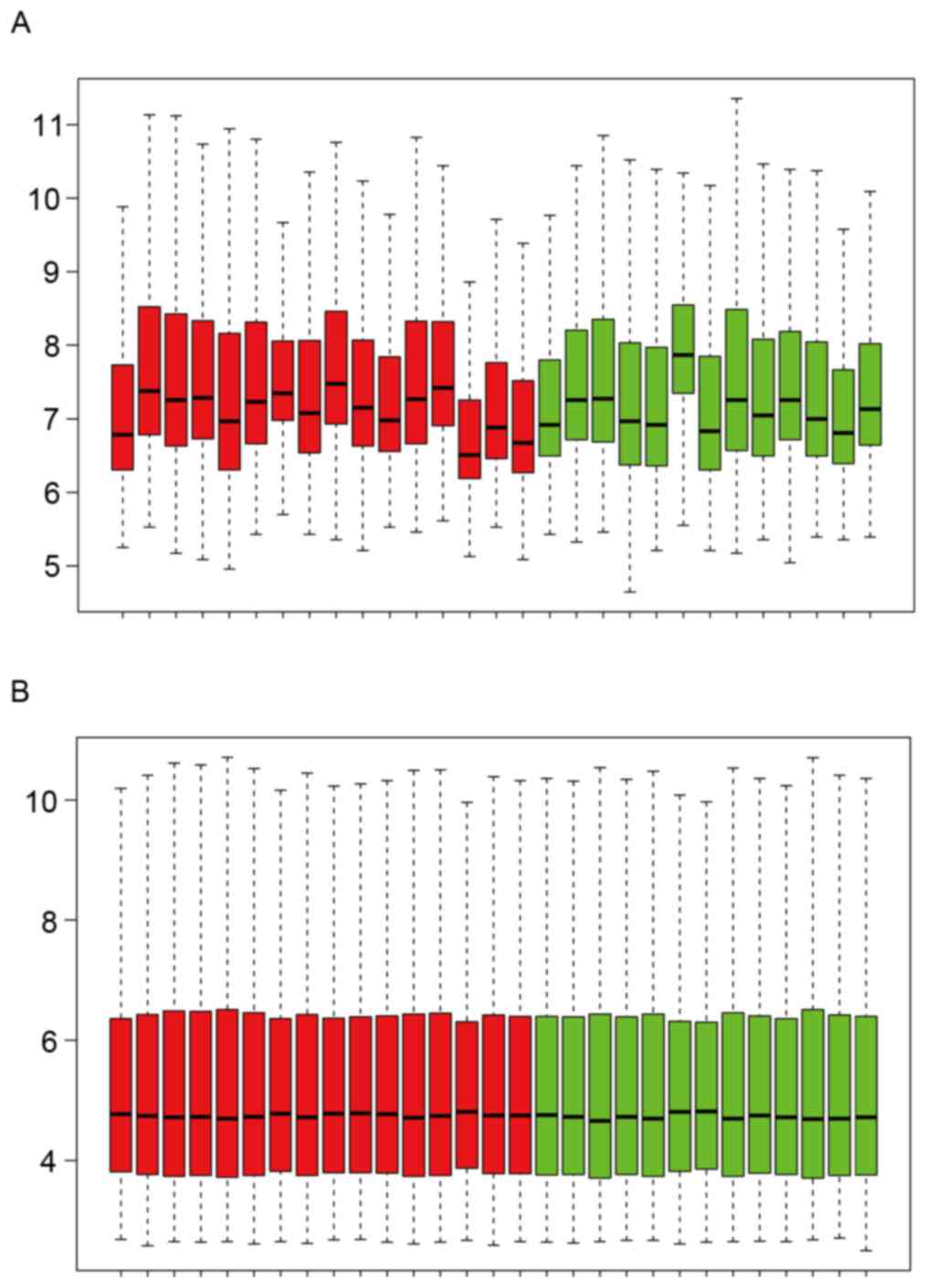
The expression distribution characteristics of the
dataset prior to and following data pretreatment are shown in
Fig. 1A and B. Their median values
were almost on a straight line, indicating that the raw data were
normalized successfully. The DEGs, including the upregulated and
downregulated genes, were identified from the two groups with an
FDR<0.05 and a |log2FC|>0.58. As a result, a total
of 758 DEGs were obtained following data processing. Among these,
there were 515 upregulated genes and 243 downregulated genes.
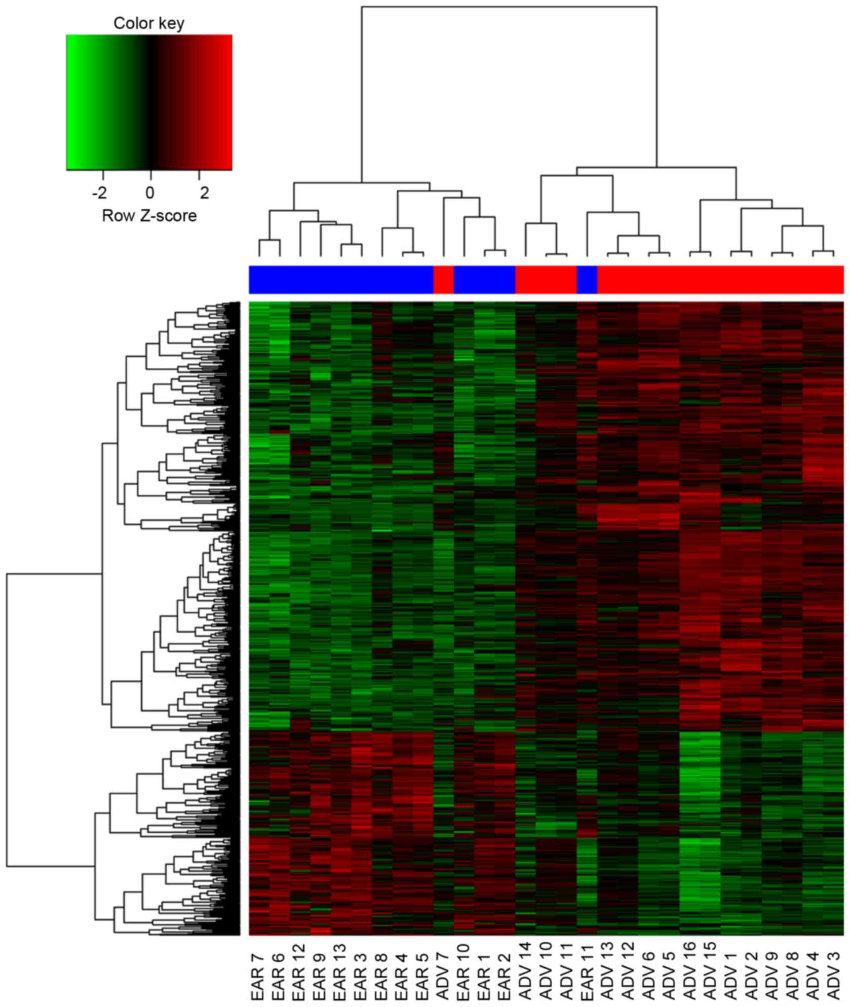
Cluster analysis
The clustering of the DEGs showed that the signature
of the majority of the advanced carotid atherosclerotic plaques
resembled one another, rather than one of the early plaques, and
vice versa (Fig. 2). There were
notable differences between the advanced and early carotid
atherosclerotic plaques according to their gene expression
profiles. However, there was one advanced carotid atherosclerotic
plaque clustered in the early group, and one early carotid
atherosclerotic plaque clustered in the advanced group, suggesting
that the gene expression profiles of those two patients were
different from their own group.
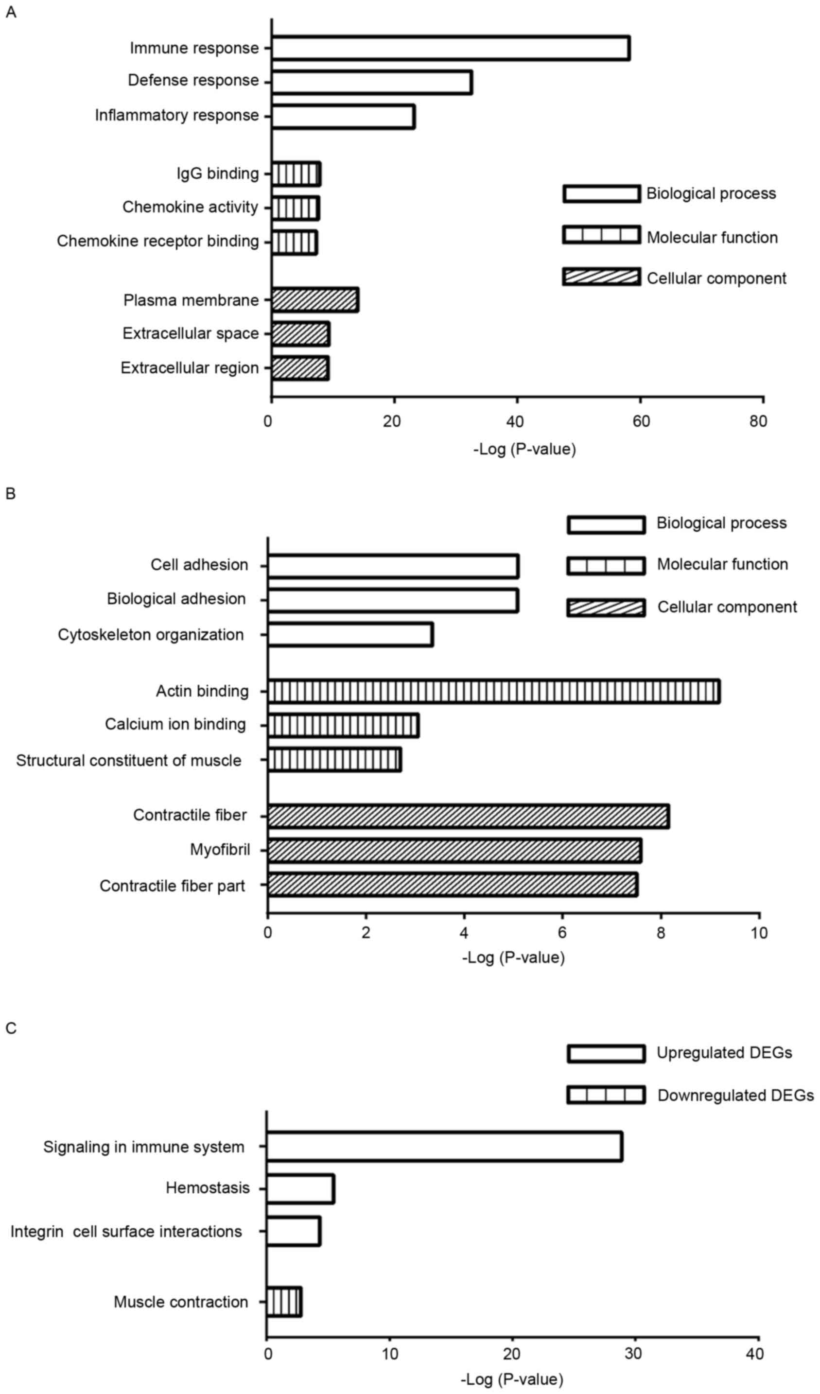
GO enrichment analyses
The DEGs were analyzed using the DAVID tool and
categorized into the three major terms in GO, the biological
process, molecular function and cellular component. The top three
GO terms of the three categories are shown in Fig. 3. The enriched terms of the
upregulated genes were significantly associated with immune
response, IgG binding and plasma membrane in the three categories,
respectively (Fig. 3A). By
contrast, the enriched terms identified among the downregulated
genes were associated with cell adhesion, actin binding and
contractile fiber in the three categories, respectively (Fig. 3B).
Pathway enrichment analysis
To examine the biological pathways associated with
the DEGs in the progression of carotid atherosclerotic plaques, the
DAVID tool was also used. The upregulated genes were intrinsically
linked with signaling in immune system, hemostasis and integrin
cell surface interactions (Fig.
3C). By contrast, the downregulated genes were significantly
associated with muscle contraction only (Fig. 3C).
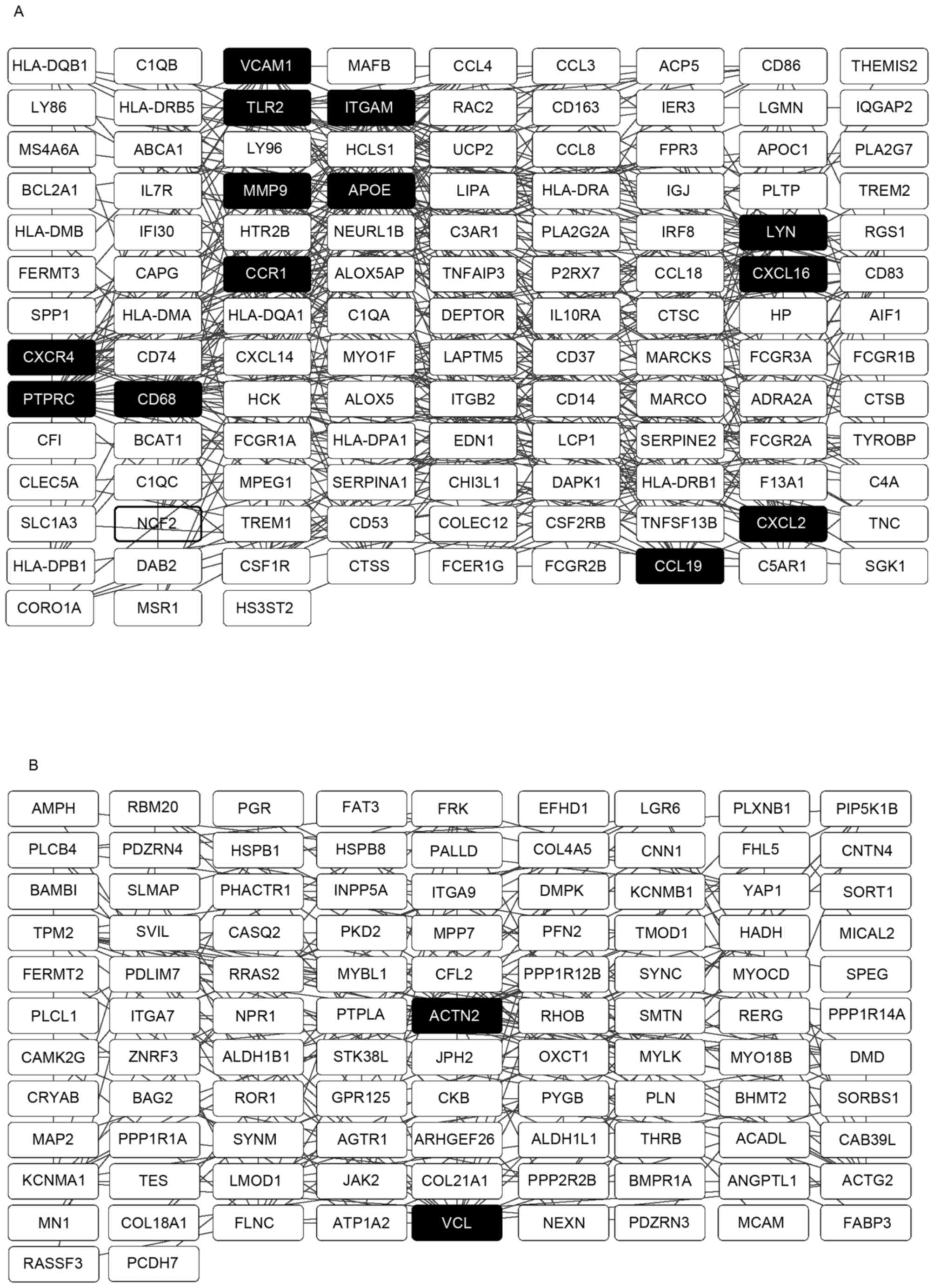
PPI network construction
The PPI networks with the hub genes possessing a
connectivity degree of >15 were constructed using Cytoscape
software, based on the results from the analysis using the STRING
database. Networks with 120 and 101 nodes were obtained using the
upregulated and downregulated genes, respectively (Fig. 4A and B). Within a PPI network, each
node indicates a gene, whereas the edges represent the interactions
between nodes. The degree represents the number of edges linked to
a specific node. Therefore, nodes with a high degree are defined as
hub genes, which assume important biological functions. A total of
13 hub genes were selected from the upregulated PPI network, which
included ITGAM, PTPRC and APOE, which had
connectivity degrees of 37, 27 and 26, respectively. From the
downregulated PPI network, two hub genes, ACTN2 and
VCL, with respective connectivity degrees of 19 and 17, were
identified.
Discussion
A number of previous studies have used the GSE28829
dataset to examine the DEGs involved in the progression of carotid
atherosclerotic plaques (12,14).
However, one of these two studies bridged the gap between the gene
expression of M1/M2 macrophage phenotypes and antioxidant genes in
atherosclerosis, which only included a subset of the dataset
(14). The other study adopted a
stricter standard, compared with that used in the present study, to
judge the DEGs, resulting in relatively fewer results returned from
the functional and pathway enrichment analysis, compared with the
present study (12). As carotid
atherosclerotic plaques formation is a multifactorial disease,
identifying additional potential biological processes and pathways
offers potential in palliation or curing of the disease. It can be
difficult to directly counteract the effects of each key gene;
therefore, it is necessary to abate the effects through the
biological processes and pathways enriched by these key genes.
Therefore, the results of the present study provide a more
comprehensive perspective towards understanding the molecular
mechanism and towards discovering novel therapeutic targets in this
field.
In the present study, 758 DEGs were obtained
following analysis of the microarray data deposited in the GSE28829
dataset. This gene expression signature indicated that the
expression of the selected genes was altered during the progression
of carotid atherosclerotic plaque formation. Although expression
analyses from the dataset cannot confirm a causal association
between altered gene expression and plaque state, there is merit in
performing an integrative systematic bioinformatics approach,
including functional and pathway analyses, and in the construction
of a PPI network based on DEGs. This may assist in understanding
the molecular mechanisms underlying carotid atherosclerotic plaques
at higher levels, providing an insight into the prevention and
treatment of carotid atherosclerotic plaques.
The results of the cluster analysis suggested marked
differences between the advanced and early carotid atherosclerotic
plaques, and also indicated that the selected genes were associated
with disease progression. Of note, the samples did not perfectly
group together in the two groups in the heatmap as expected. There
are several factors, which may have contributed to the presence of
one advanced plaque within the early plaque group and one early
plaque within the advanced plaque group. Experimental variance,
including RNA isolation, and biological variance, including
incorrect classification of samples during autopsy, may affect the
interpretation of datasets (24).
However, it is possible that the group inconsistency was due to
other genes not considered in the present study.
The results of the functional analysis from GO
enabled the translation of DEGs into the underlying biological
mechanism. In the present study, the enriched GO terms from the
upregulated DEGs were primarily associated with immune response and
IgG binding, which occurred in the plasma membrane. This suggested
the important effects of immune cells on the initiation and
progression of the atherosclerotic process (25). The enriched GO terms from the
downregulated DEGs were primarily linked to cell adhesion and actin
binding, which existed in the contractile fiber. This is concordant
with the suggestion that dysregulated cytoskeletal proteins can
abate their ability to promote endothelial repair through
alterations in cell shape and through regulation of cell migration
following wounding of the endothelium (26). Therefore, the upregulated and
downregulated DEGs acted in concert to accelerate the
atherosclerotic process.
Pathway analysis using the Reactome database
identifies differential expression patterns based on gene groups
rather than individual genes; in cases where an individual genes
shows subtle biological function or property changes, they are
omitted by the usual individual gene analysis (27). The results of the present study
showed that the enriched pathways from the upregulated genes were
predominantly associated with signaling in immune system, whereas
the downregulated genes were associated with muscle contraction.
These results overlapped with the GO enrichment analyses,
therefore, the data supported the findings of previous studies,
which reported immunity to be involved in the process of the
initiation and progression of carotid atherosclerotic plaques
intertwined with blood vessel dysfunction (28,29).
PPI networks are critical for understanding
underlying mechanisms as these vital activities are ascribed to
interactions between proteins to form a signal transduction network
system when confronted with external and internal environmental
stimuli. As several vital activities are associated with the
combination and dissociation of proteins, hub genes are important
in this process. The top hub genes identified in the present study
were ITGAM and ACTN2 from the upregulated and downregulated genes,
respectively. ITGAM encodes the integrin αM chain. An α
chain and a β chain combine to form a heterodimeric integral
membrane protein, integrin, which mediates the adhesion of cells to
extracellular matrix-associated proteins. The αM β2 integrin is
involved in the adherence of neutrophils and monocytes to
stimulated endothelium in cases of inflammation and thrombosis
(30), and also in CD40L-mediated
inflammation during atherogenesis as an alternative pathway
(31). ACTN2 is identical
to one of the key interaction nodes reported by Perisic et
al (13), which encodes a
cytoskeletal protein belonging to the spectrin gene superfamily.
This actin-binding protein has multiple roles in different cell
types. In smooth muscle, the protein assists in anchoring the
myofibrillar actin filaments to the Z-disc and analogous dense
bodies. Together with VCL, which also encodes a cytoskeletal
protein, this cytoskeletal deregulation supports the hypothesis
that medial vascular smooth muscle cells switch from a contractile
to a synthetic phenotype during the development of atherosclerosis
at the molecular level (32). This
is in accordance with the above-mentioned dysregulated muscle
contraction identified from the pathway analyses of the
downregulated DEGs.
There were a number of limitations in the present
study. The samples were collected from autopsy, affecting the
quality of isolated RNA and downstream analysis of the dataset. In
addition, the results from the array and bioinformatics analyses
require further biological experiments. Therefore, genetic and
experimental investigations with strict protocols are required
based on the results of the present study in the future.
In conclusion, based on comprehensive bioinformatics
analyses of the dataset, the data obtained in the present study
elucidated the DEGs, which are potentially associated with the
common molecular mechanism underlying the progression of carotid
atherosclerotic plaques. Furthermore, deregulation of the immune
system and smooth muscle cell cytoskeleton were associated with the
progression of carotid atherosclerotic plaques. These findings
provide novel insights into the biological properties of carotid
atherosclerotic plaques, and provide clues and direction for the
prevention and treatment of atherosclerosis in the carotid artery.
Further experiments are required in order to confirm these
results.
Acknowledgements
The authors would like to thank Dr. Marco Manca for
uploading data to the GEO database., as well as Professor Xueyuan
Liu and Dr. Yanyan Zhao (Department of Neurology, Shanghai Tenth
People's Hospital, School of Medicine, Tongji University, Shanghai,
China) for revising the article.
Funding
No funding was received.
Availability of data and materials
The analyzed data sets generated during the study
are available from the corresponding author on reasonable
request.
Authors' contributions
JW conceived the study. WL and YZ performed the
analysis and wrote the paper. All authors read and approved the
manuscript.
Ethics approval and consent to
participate
Not applicable.
Consent for publication
Not applicable.
Competing interests
All authors declared that they have no conflict of
interest with regard to this work.
References
|
1
|
Dichgans M: Genetics of ischaemic stroke.
Lancet Neurol. 6:149–161. 2007. View Article : Google Scholar : PubMed/NCBI
|
|
2
|
Dirnagl U, Iadecola C and Moskowitz MA:
Pathobiology of ischaemic stroke: An integrated view. Trends
Neurosci. 22:391–397. 1999. View Article : Google Scholar : PubMed/NCBI
|
|
3
|
Palm F, Dos Santos M, Urbanek C, Greulich
M, Zimmer K, Safer A, Grau AJ and Becher H: Stroke seasonality
associations with subtype, etiology and laboratory results in the
Ludwigshafen Stroke Study (LuSSt). Eur J Epidemiol. 28:373–381.
2013. View Article : Google Scholar : PubMed/NCBI
|
|
4
|
Schellinger PD, Kaste M and Hacke W: An
update on thrombolytic therapy for acute stroke. Curr Opin Neurol.
17:69–77. 2004. View Article : Google Scholar : PubMed/NCBI
|
|
5
|
Ross R: Atherosclerosis-an inflammatory
disease. N Engl J Med. 340:115–126. 1999. View Article : Google Scholar : PubMed/NCBI
|
|
6
|
Teng Z, Sadat U, Brown AJ and Gillard JH:
Plaque hemorrhage in carotid artery disease: Pathogenesis, clinical
and biomechanical considerations. J Biomech. 47:847–858. 2014.
View Article : Google Scholar : PubMed/NCBI
|
|
7
|
McNally JS, McLaughlin MS, Hinckley PJ,
Treiman SM, Stoddard GJ, Parker DL and Treiman GS: Intraluminal
thrombus, intraplaque hemorrhage, plaque thickness, and current
smoking optimally predict carotid stroke. Stroke. 46:84–90. 2015.
View Article : Google Scholar : PubMed/NCBI
|
|
8
|
Gimbrone MA Jr and García-Cardeña G:
Endothelial cell dysfunction and the pathobiology of
atherosclerosis. Circ Res. 118:620–636. 2016. View Article : Google Scholar : PubMed/NCBI
|
|
9
|
Luque MC, Gutierrez PS, Debbas V, Kalil J
and Stolf BS: CD100 and plexins B2 and B1 mediate
monocyte-endothelial cell adhesion and might take part in
atherogenesis. Mol Immunol. 67:559–567. 2015. View Article : Google Scholar : PubMed/NCBI
|
|
10
|
Wong HS, Jaumouille V, Freeman SA,
Doodnauth SA, Schlam D, Canton J, Mukovozov IM, Saric A, Grinstein
S and Robinson LA: Chemokine signaling enhances CD36 responsiveness
toward oxidized low-density lipoproteins and accelerates foam cell
formation. Cell Rep. 14:2859–2871. 2016. View Article : Google Scholar : PubMed/NCBI
|
|
11
|
Wang Z, Guo D, Yang B, Wang J, Wang R,
Wang X and Zhang Q: Integrated analysis of microarray data of
atherosclerotic plaques: Modulation of the ubiquitin-proteasome
system. PLoS One. 9:e1102882014. View Article : Google Scholar : PubMed/NCBI
|
|
12
|
Wang J, Wei B, Cao S, Xu F, Chen W, Lin H,
Du C and Sun Z: Identification by microarray technology of key
genes involved in the progression of carotid atherosclerotic
plaque. Genes Genet Syst. 89:253–258. 2014. View Article : Google Scholar : PubMed/NCBI
|
|
13
|
Perisic L, Aldi S, Sun Y, Folkersen L,
Razuvaev A, Roy J, Lengquist M, Åkesson S, Wheelock CE,
Maegdefessel L, et al: Gene expression signatures, pathways and
networks in carotid atherosclerosis. J Intern Med. 279:293–308.
2016. View Article : Google Scholar : PubMed/NCBI
|
|
14
|
da Rocha RF, De Bastiani MA and Klamt F:
Bioinformatics approach to evaluate differential gene expression of
M1/M2 macrophage phenotypes and antioxidant genes in
atherosclerosis. Cell Biochem Biophys. 70:831–839. 2014. View Article : Google Scholar : PubMed/NCBI
|
|
15
|
Cipollone F, Felicioni L, Sarzani R,
Ucchino S, Spigonardo F, Mandolini C, Malatesta S, Bucci M,
Mammarella C, Santovito D, et al: A unique microRNA signature
associated with plaque instability in humans. Stroke. 42:2556–2563.
2011. View Article : Google Scholar : PubMed/NCBI
|
|
16
|
Reiner A, Yekutieli D and Benjamini Y:
Identifying differentially expressed genes using false discovery
rate controlling procedures. Bioinformatics. 19:368–375. 2003.
View Article : Google Scholar : PubMed/NCBI
|
|
17
|
Sun J, Wen X, Jin F, Li Y, Hu J and Sun Y:
Bioinformatics analyses of differentially expressed genes
associated with bisphosphonate-related osteonecrosis of the jaw in
patients with multiple myeloma. Onco Targets Ther. 8:2681–2688.
2015. View Article : Google Scholar : PubMed/NCBI
|
|
18
|
Ashburner M, Ball CA, Blake JA, Botstein
D, Butler H, Cherry JM, Davis AP, Dolinski K, Dwight SS, Eppig JT,
et al: Gene ontology: Tool for the unification of biology. The Gene
Ontology Consortium. Nat Genet. 25:25–29. 2000. View Article : Google Scholar : PubMed/NCBI
|
|
19
|
Croft D, O'Kelly G, Wu G, Haw R, Gillespie
M, Matthews L, Caudy M, Garapati P, Gopinath G, Jassal B, et al:
Reactome: A database of reactions, pathways and biological
processes. Nucleic Acids Res. 39(Database Issue): D691–D697. 2011.
View Article : Google Scholar : PubMed/NCBI
|
|
20
|
Huang da W, Sherman BT and Lempicki RA:
Systematic and integrative analysis of large gene lists using DAVID
bioinformatics resources. Nat Protoc. 4:44–57. 2009. View Article : Google Scholar : PubMed/NCBI
|
|
21
|
von Mering C, Huynen M, Jaeggi D, Schmidt
S, Bork P and Snel B: STRING: A database of predicted functional
associations between proteins. Nucleic Acids Res. 31:258–261. 2003.
View Article : Google Scholar : PubMed/NCBI
|
|
22
|
Smoot ME, Ono K, Ruscheinski J, Wang PL
and Ideker T: Cytoscape 2.8: New features for data integration and
network visualization. Bioinformatics. 27:431–432. 2011. View Article : Google Scholar : PubMed/NCBI
|
|
23
|
Kou Y, Zhang S, Chen X and Hu S: Gene
expression profile analysis of colorectal cancer to investigate
potential mechanisms using bioinformatics. Onco Targets Ther.
8:745–752. 2015.PubMed/NCBI
|
|
24
|
Hatfield GW, Hung SP and Baldi P:
Differential analysis of DNA microarray gene expression data. Mol
Microbiol. 47:871–877. 2003. View Article : Google Scholar : PubMed/NCBI
|
|
25
|
Pende A, Artom N, Bertolotto M, Montecucco
F and Dallegri F: Role of neutrophils in atherogenesis: An update.
Eur J Clin Invest. 46:252–263. 2016. View Article : Google Scholar : PubMed/NCBI
|
|
26
|
Lee JS and Gotlieb AI: Understanding the
role of the cytoskeleton in the complex regulation of the
endothelial repair. Histol Histopathol. 18:879–887. 2003.PubMed/NCBI
|
|
27
|
Nam D and Kim SY: Gene-set approach for
expression pattern analysis. Brief Bioinform. 9:189–197. 2008.
View Article : Google Scholar : PubMed/NCBI
|
|
28
|
Ammirati E, Moroni F, Magnoni M and Camici
PG: The role of T and B cells in human atherosclerosis and
atherothrombosis. Clin Exp Immunol. 179:173–187. 2015. View Article : Google Scholar : PubMed/NCBI
|
|
29
|
Zhou Q and Liao JK: Rho kinase: An
important mediator of atherosclerosis and vascular disease. Curr
Pharm Des. 15:3108–3115. 2009. View Article : Google Scholar : PubMed/NCBI
|
|
30
|
Mazzone A, Mazzucchelli I, Fossati G,
Gritti D, Girola S, Canale C, Cusa C and Ricevuti G: Iloprost
effects on phagocytes in patients suffering from ischaemic
diseases: In vivo evidence for down-regulation of alpha M beta 2
integrin. Eur J Clin Invest. 26:860–866. 1996. View Article : Google Scholar : PubMed/NCBI
|
|
31
|
Zirlik A, Maier C, Gerdes N, MacFarlane L,
Soosairajah J, Bavendiek U, Ahrens I, Ernst S, Bassler N, Missiou
A, et al: CD40 ligand mediates inflammation independently of CD40
by interaction with Mac-1. Circulation. 115:1571–1580. 2007.
View Article : Google Scholar : PubMed/NCBI
|
|
32
|
de la Cuesta F, Zubiri I, Maroto AS,
Posada M, Padial LR, Vivanco F, Alvarez-Llamas G and Barderas MG:
Deregulation of smooth muscle cell cytoskeleton within the human
atherosclerotic coronary media layer. J Proteomics. 82:155–165.
2013. View Article : Google Scholar : PubMed/NCBI
|