Introduction
Transient ischemic attack may result in damage to
local brain tissues and decreased brain function (1). The sequelae of cerebral ischemia may
lead to serious physical disabilities in patients, which may exert
a great influence on patient quality of life and bring heavy
economic burden on society and family (2). Historically, treatment of ischemic
brain injury was focused on reducing neuronal death; however, an
increasing number of studies have suggested that angiogenesis
following cerebral ischemia may serve an important role in
neurological function reconstruction and the repair process
(3,4).
Dexmedetomidine (DEX) is a specific and short-acting
agonist of the α2 adrenergic receptor (5); it is a potent drug that may produce
severe physiological changes. A previous study confirmed that
intrathecal administration of DEX prolonged motor and sensory
block, enhanced hemodynamic stability and reduced the requirement
for rescue analgesics in 24 h (6).
Brain-derived neurotrophic factor (BDNF) is the most
abundant neurotrophic factor in brain tissue and serves a role in
inhibiting apoptosis, promoting neuronal survival, activating
neural stem cells (NSCs) following ischemic brain injury, and
regulating neural plasticity. BDNF belongs to a class of proteins
that are synthesized by brain tissues and are widely expressed in
the central nervous system (CNS); it serves a significant role in
CNS development and neuronal growth and differentiation (7). In addition, BDNF has been reported to
prevent neuronal death, improve pathology and facilitate the
regeneration and differentiation of damaged neurons (8). Previous studies have demonstrated
that the administration of BDNF following brain artery occlusion
improved the survival rate of neurons and decreased the volume of
cerebral infarction, and it was revealed that the expression level
of BDNF was positively correlated with the ability of local neurons
to tolerate ischemic injury (9,10).
However, further studies are required to investigate whether DEX
post-treatment promoted the survival and repair of the neurons by
increasing BDNF expression levels.
Vascular endothelial growth factor (VEGF) belongs to
a class of heparin-binding proteins that have a highly conserved
structure and they function to promote angiogenesis, maintain
vascular permeability and endothelial proliferation (11). Previous studies have reported that
VEGF was widely expressed in the brain tissues of adult rats, and
this expression was increased following brain ischemia (12,13).
It may be possible that VEGF expression promotes the maturation of
newborn neurons and the generation of the striatum nerve in adult
rats by inhibiting potassium channel activity, which may contribute
to neural protection.
A number of studies have confirmed that BDNF
expression promoted the survival of neurons and upregulated the
release of neurotransmitters by combining with tropomyosin-related
kinase B (TrkB) and subsequently activating the
phosphatidylinositol 3-kinase (PI3K)/Akt (also known as protein
kinase B) signaling pathway, which served a significant role in
neural protection (14–16). In addition, VEGF was revealed to
regulate the PI3K/Akt signaling to inhibit the activity of
Caspase-3 and to regulate voltage-gated potassium channels,
thereby, reducing ischemia-induced neuronal death. DEX
post-treatment was reported to be beneficial to neuroprotection by
activating PI3K/Akt signaling pathway (17). The present study aimed to further
explore whether the post-treatment with DEX increased the
expression levels of BDNF and VEGF following ischemia/reperfusion
(I/R) injury. The I/R injury and the morbidity rate, was also
described and investigated the association between BDNF and VEGF.
These results may provide the scientific basis for elucidating the
mechanism of post-treatment with DEX on I/R injury.
Materials and methods
Experimental design
A total of 30 healthy, clean male Wistar rats (6
weeks old, 160–180 g) were randomly divided into three experimental
groups (10 rats/group) under the circumstance of SPF level as
follows: i) The Control group, which received surgical procedure to
expose the external carotid artery, without ischemia induction; ii)
the I/R group, which received an intraperitoneal injection of
sterile saline (1 ml) in reperfusion 60 min following brain
ischemia surgery (18); iii) the
DEX group, which received an intraperitoneal injection of DEX (50
µg/kg; Jinzhou Jiutai Pharmaceutical Co., Ltd, Liaoning, China) in
reperfusion 60 min following brain ischemia surgery.
Breeding environment was as follows: 12-h light and
dark cycle, temperature in 18–26°C, relative humidity in 40–70%,
and ventilation for 8–12 times/h. The water was supplied by a water
bottle and changed for three times a week. Sufficient food was
supplied. The study protocol was approved by The Institutional
Review Board of the First Affiliated Hospital of Zhengzhou
University (Henan, China). All animal handling procedures were
carried out in accordance with the protocols of the animal care
guidelines of the Institutional Animal Care Committee.
Rats in each group were anesthetized with 10%
chloral hydrate at 6 h, 1 day, 3 days and 7 days following I/R
surgery, and immediately decapitated 2, 2, 3 and 3 rats at the
respective time points. Whole brain tissue samples were extracted
and placed on ice-cold glass. The ischemic penumbra in the parietal
cortex was quickly separated and placed into vials; an example of
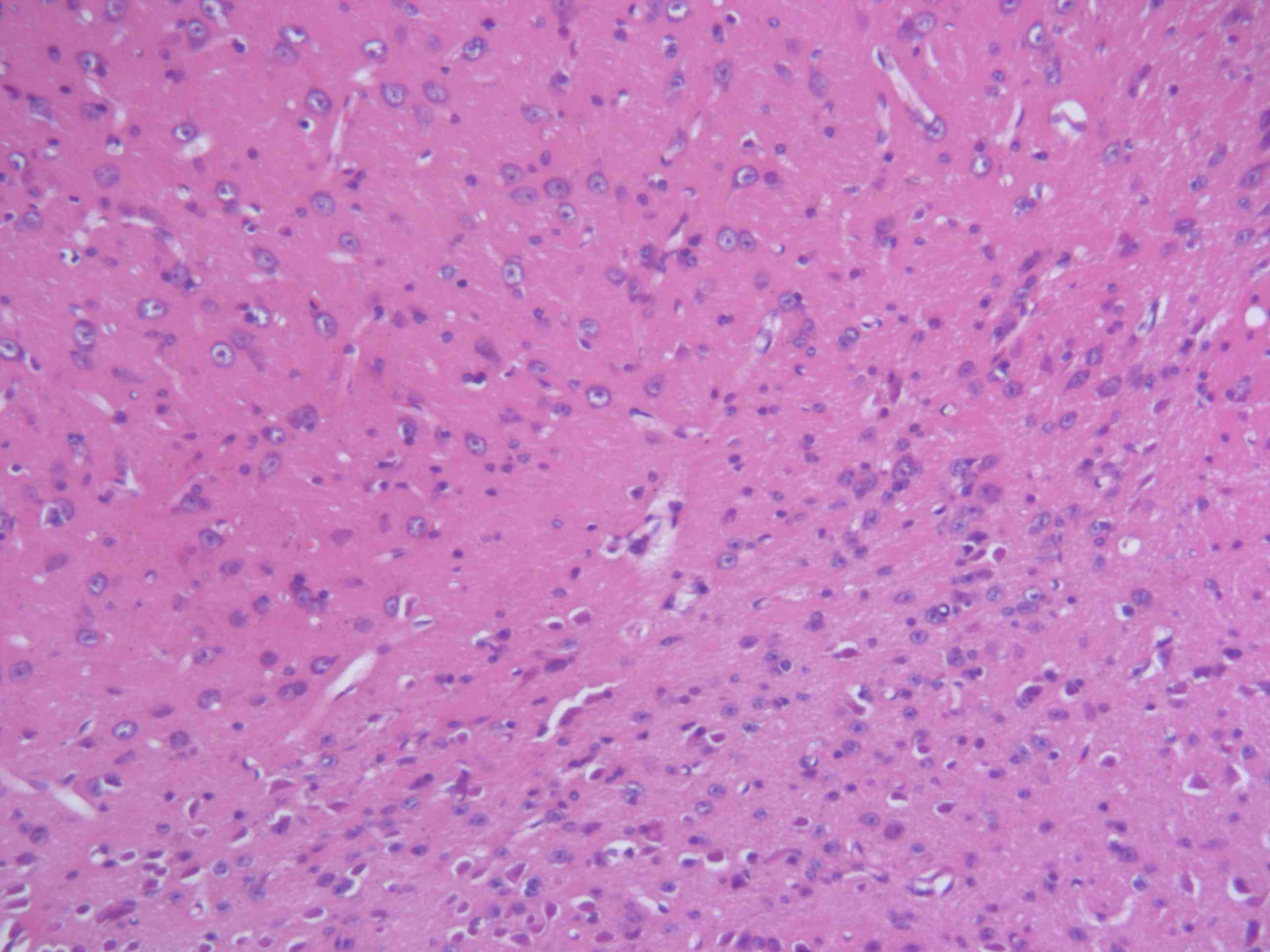
the typical histology of cerebral tissues is provided in Fig. 1. Brain tissues were frozen in
liquid nitrogen and stored at −70°C until use.
BDNF and VEGF mRNA expression
analysis
Total RNA was extracted from the 200 mg brain
samples. The RNA concentration and purity were detected using a
Nanodrop 2000 (Thermo Fisher Scientific, Inc., Wilmington, DE,
USA). Total RNA was reverse transcribed into cDNA using the M-MLV
Reverse Transcription kit (Kangweishiji, Beijing, China; cat no.
20219) at 37°C for 10 min according to the manufacturer's protocol.
Primers were designed to detect BDNF and VEGF mRNA by polymerase
chain reaction (PCR); a list of primer sequences is provided in
Table I. The Cq value for each
gene was measured, and the ΔΔCq values were determined from the
average of three parallel experiments. The values were calculated
as follows: ΔCq=Cq(Target Gene)-Cq(Reference
gene); ΔΔCq=ΔCq(sample)-ΔCq(control).
The relative expression level of target genes was calculated using
the 2−ΔΔCq method (19). The relative expression level of the
control (β-actin) was 20=1. The qPCR reactions
contained: 10 µl 2X SYBR Fast qPCR mix; 0.8 µl PCR forward/reverse
Primers (10 mM); 0.4 µl 50X ROX reference dye II; 2 µl cDNA
template. The reaction parameters were as follows: Initial
denaturation at 95°C for 30 sec, followed by 40 cycles of 95°C for
3 sec and 58°C for 30 sec. PCR products were detected by 1.5%
agarose gel electrophoresis stained by ethidium bromide. β-actin
was used as the internal reference and for densitometric analysis
by Quantity One 4.62 (Bio-Rad Laboratories, Inc., Hercules, CA,
USA). Every experiment repeated three times.
 | Table I.List of primer sequences used for
reverse transcription-polymerase chain reaction. |
Table I.
List of primer sequences used for
reverse transcription-polymerase chain reaction.
| Gene | Sequence (5′-3′) | Expected size
(bp) |
|---|
| BDNF | F:
CGTGGGGAGCTGAGCGTGTG | 192 |
|
| R:
GCCCCTGCAGCCTTCCTTC |
|
| VEGF | F:
GCCGCAGGAGGCAAACCGAT | 242 |
|
| R:
TGGCGGGCTCCTCTCCCTT |
|
| β-actin | F:
CTCCATCCTGGCCTCGCTG | 250 |
|
| R:
GCTGTCACCTTCACCGTTCC |
|
BDNF and VEGF protein expression
analysis
Lysis buffer (C1053+; Beijing Pulilai Gene
Technology, Co., Ltd., Beijing, China) were added to 200 µg brain
samples. Following 30 min of lysis, the cells were centrifuged at
11,180 × g for 20 min at 4°C. The supernatant was collected and the
total protein was extracted. Total protein concentration was
quantified by the bicinchoninic acid method. Proteins were
separated by SDS-PAGE and transferred to a polyvinylidene
difluoride membrane (Pierce; Thermo Fisher Scientific, Inc.). A
total of ~20 µl protein was loaded per lane. Following blocking for
2 h at 37°C, the primary (Abcam, Cambridge, UK; cat. no. ab108319
for BDNF; cat. no. ab32152) and incubated at room temperature for 2
h. The membrane was rinsed three times (5–10 min each) with TBST
and incubated with horseradish peroxidase-labeled secondary
antibody (Wuhan BOSHIDE Biological engineering Co. Ltd., Wuhan,
China; cat. no. SV0002), which was diluted (1:10,000) with TBST
containing 0.05% of skim milk and incubated at room temperature for
2 h. The membrane was rinsed three times with TBST (5–10 min) and
the experimental results were preserved by exposure and
photographic processing. The optical density of the immunoblotting
zone was measured by laser light density scanner. Quantity One
software version 4.62 (Bio-Rad Laboratories, Inc.) was used for
densitometric analysis, and the semi-quantitative values of target
protein/reference protein were used for statistical analysis
(20). Every experiment was
repeated three times.
Statistical analysis
Statistical analysis was performed with SPSS 13.0
statistical software (SPSS, Inc., Chicago, IL, USA). All
experimental results are presented as the mean ± standard
deviation. Multiple groups were compared by analysis of variance
followed by Student-Newman-Keuls the post hoc test. P<0.05 was
considered to indicate a statistically significant difference.
Results
BDNF and VEGF mRNA expression
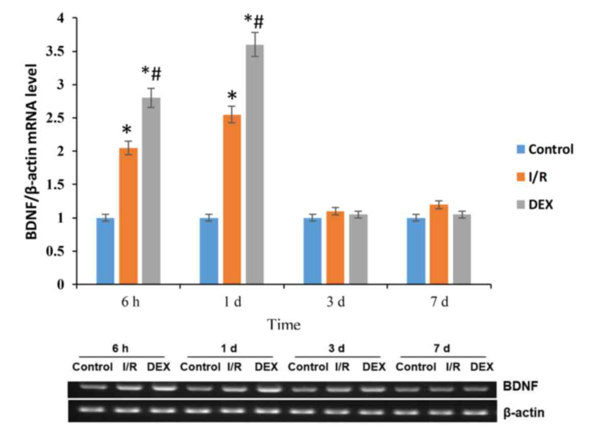
The mRNA expression levels of BDNF in cortical
ischemic penumbra of rats in each group were examined (Fig. 2). The results indicated that BDNF
mRNA expression levels were higher in both the I/R and DEX groups
at 6 h and 1 day post-surgery compared with the expression level in
the Control group (P<0.05). The levels of BNDF mRNA expression
were higher in the DEX-treated group at 6 h and 1 day post-surgery
compared with the levels of expression in the I/R group
(P<0.05). Compared with the Control group, BDNF mRNA expression
in the I/R and DEX groups increased at 6 h post-surgery, reached
the peak on day 1, decreased Day 3 and reached Control group levels
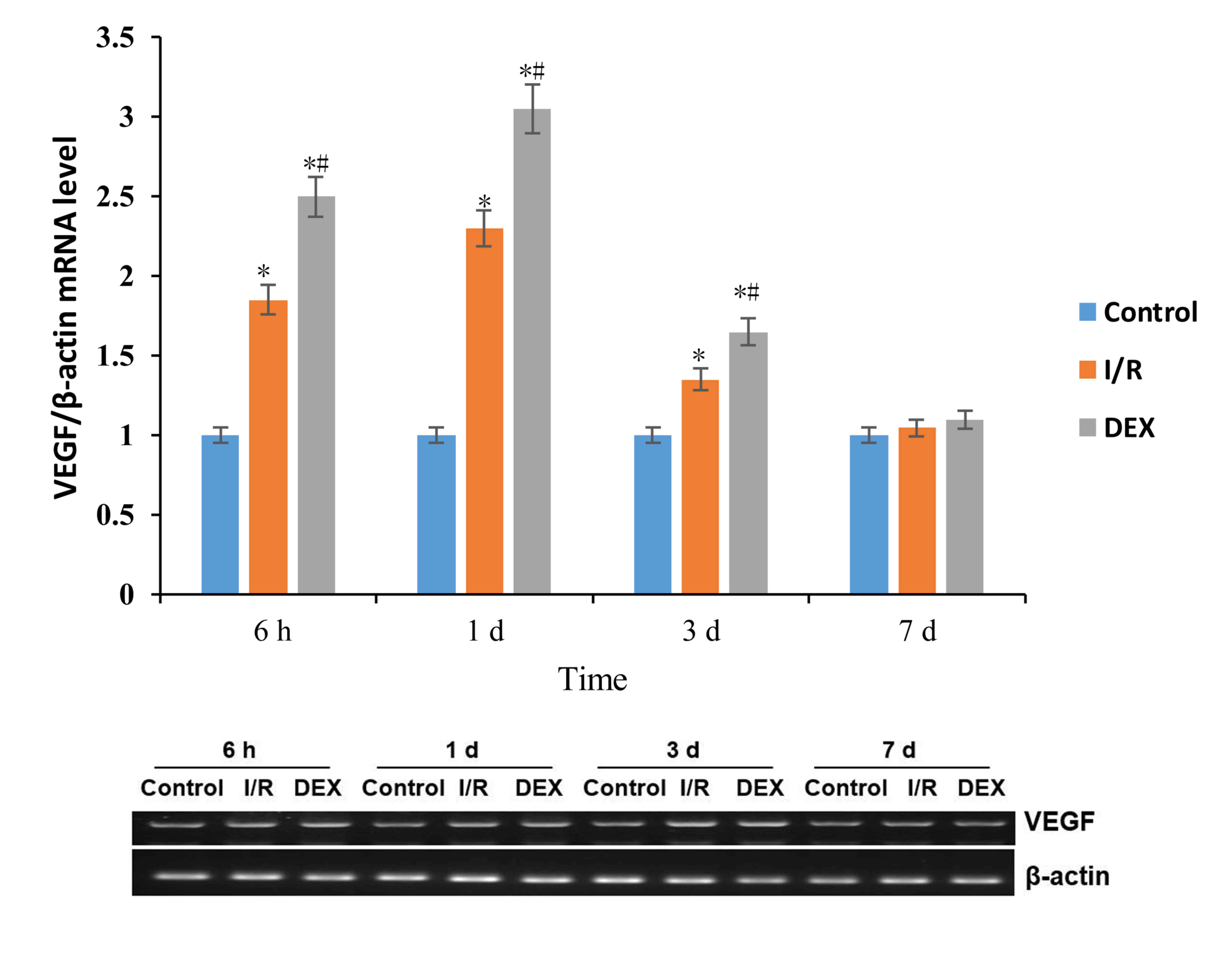
at day 7. Similar expression levels and a similar trend over time
were observed for VEGF mRNA in the three groups (Fig. 3). However, the significant
differences among the three groups were observed at Day 3 for
VEGF.
BDNF and VEGF protein expression
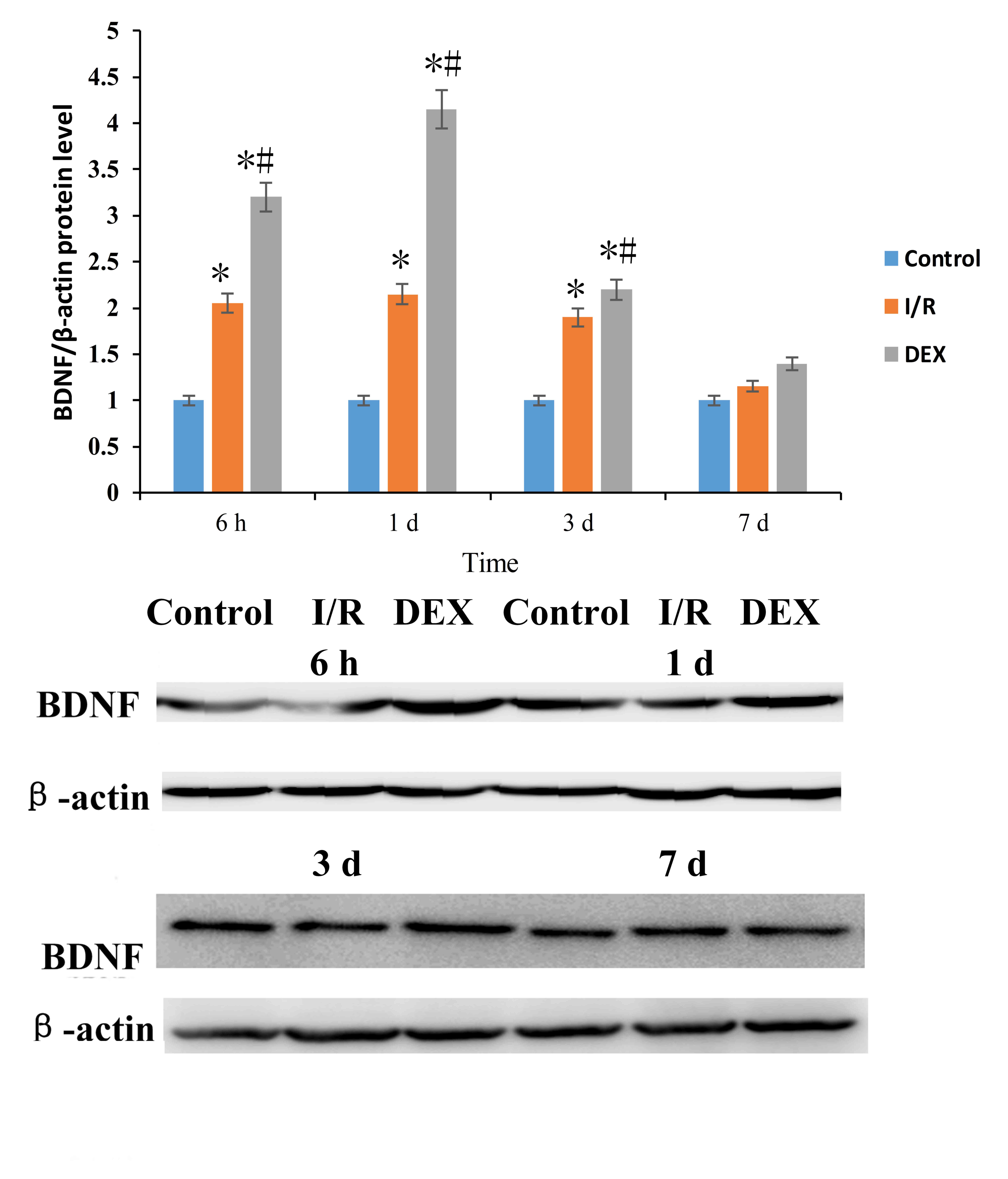
The protein expression levels of BDNF were detected
in cortical ischemic penumbra tissues of rats in each group
(Fig. 4). The result demonstrated
that the expression levels of BDNF proteins were significantly
higher in the I/R and DEX groups compared with protein expression
levels in the Control group at 6 h, 1 day and 3 days post-surgery
(P<0.05). Similar to the miRNA expression levels, the protein
expression levels of BDNF in the I/R and DEX groups increased at 6
h, reached the peak in day 1, and declined at day 3, and reached a
similar level of expression as the Control group at day 7. Compared
with the I/R group, rats in the DEX group exhibited a significantly
higher expression level of BDNF protein at 6 h, 1 day and 3 days
following I/R surgery (P<0.05). However, there was no
significant difference between two groups at day 7 (P>0.05).
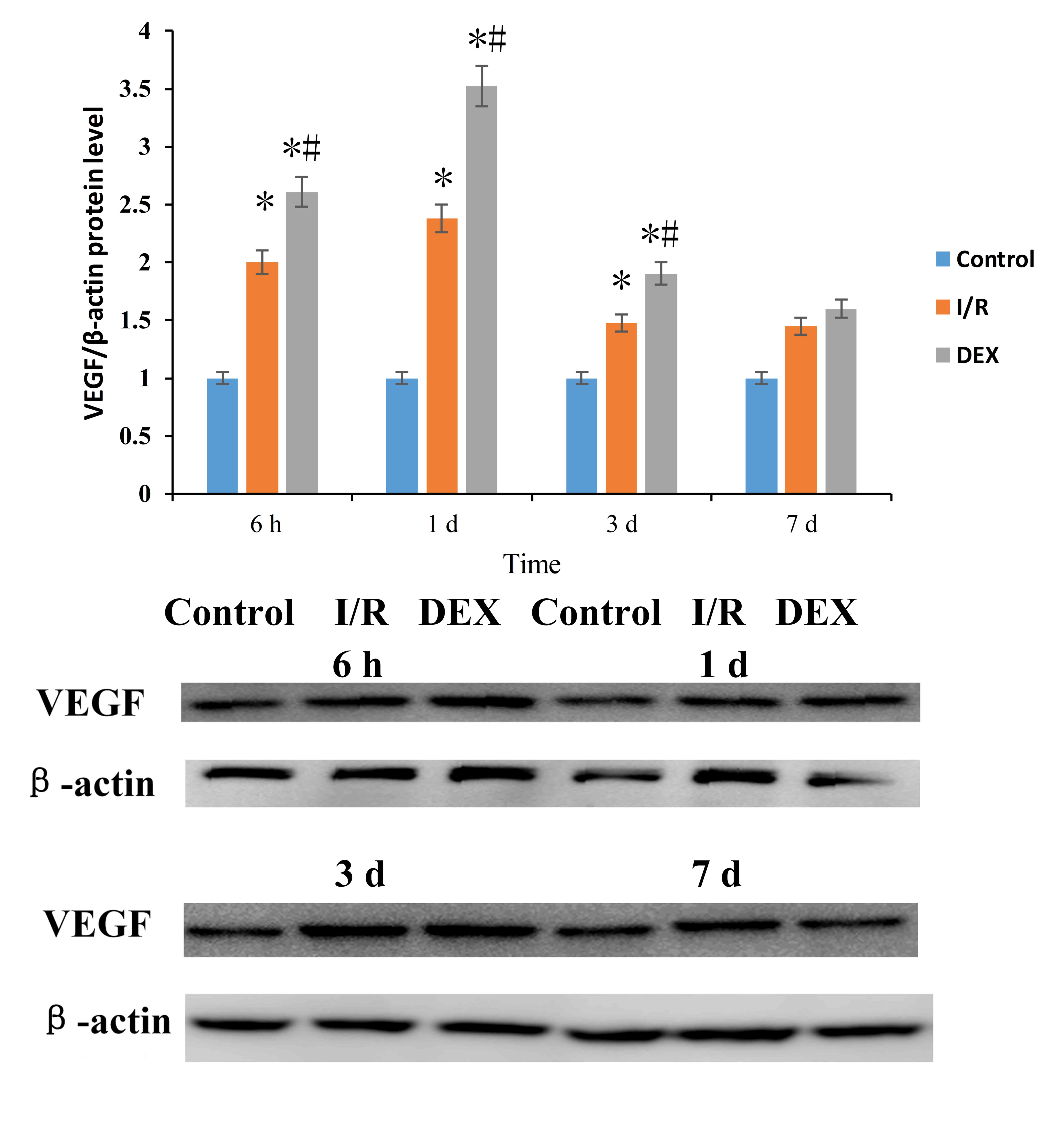
The expression levels of VEGF proteins in cortical
ischemic penumbra of rats in the three groups were also examined
(Fig. 5). The expression levels of
VEGF protein in the I/R and DEX groups increased at 6 h
post-surgery, reached a peak at day 1, declined at Day 3 reaching
similar levels as the Control group at day 7. The expression levels
of VEGF protein in the I/R and DEX groups were significantly higher
compared with the Control group at 6 h, 1 day and 3 days
(P<0.05). Compared with the I/R group, DEX-treated rats
exhibited a significantly higher expression level of BDNF protein
at 6 h, 1 day and 3 days post-I/R surgery (P<0.05).
Discussion
BDNF is an important member of the family of
neurotrophic factors and is widely expressed in the brain cells of
adult mammals. BDNF combines with TrkB and activates intracellular
signaling pathways, such as mitogen activated protein kinase
(MAPK)/extracellular signal-regulated kinase, phospholipase Cγ
(PLCγ) and PI3K (21,22). BDNF serves an important role in
promoting the survival and functional repair of neuronal cells by
regulating these signaling pathways. In addition, BDNF is essential
in neural transmission and in activity-dependent plasticity
(23). Long-term potentiation is
the major form of synaptic plasticity, which is induced and
maintained by BDNF (24). BDNF was
previously reported to upregulate the depolarization of isolated
cortical and hippocampal neurons, which induced the release of
glutamic acid (25–27). Therefore, BDNF may have a vital
effect on the survival and functional recovery of neurons in the
CNS. In the present study, cerebral ischemia led to the increased
expression of BDNF mRNA and protein, which is an endogenous
protective mechanism of the body. Compared to the I/R group, DEX
treatment post-I/R surgery upregulated the expression levels of
BDNF, which suggested that post-treatment with DEX may have a
cerebral protective effect by increasing the expression of BDNF,
thus promoting the survival of neurons and the activation of NSC.
However, these results also suggested that the DEX-induced increase
in BDNF expression may only be maintained for three days
post-reperfusion. This may be due to the early expression of the
protective protein BDNF. Therefore, the effects of single DEX
post-treatment may by limited. Future studies should consider
conducting multiple DEX treatments to maintain these effects.
VEGF is a highly conserved heparin-binding protein.
In ischemic brain tissues, the expression of endogenous VEGF was
upregulated, which was reported to have a protective effect on the
brain tissue during the pathological and physiological processes
induced by ischemia (28).
Previous in vitro and in vivo studies have
demonstrated that the administration of VEGF antisense
oligonucleotides increased the sensitivity to ischemic and anoxic
injuries in brain tissue or neurons, which led to an increase of
the number of apoptotic cells and cerebral infarction volume, and
subsequently reduced the survival rate of nerve cells, aggravating
the nerve injury caused by ischemia (29–31).
Results from the present study demonstrated that DEX-treatment
post-I/R surgery significantly increased the expression levels of
VEGF mRNA and protein compared with rats in the untreated I/R
group. These data suggested that post-treatment with DEX may
promote angiogenesis and maintain vascular permeability by
increasing the expression levels of VEGF, thereby promoting the
maturation of new neurons and the generation of striatal
neurons.
Cerebral ischemia may lead to the activation of
static NSC in the injured brain tissue, which may be beneficial to
repairing the structure and function of damaged brain tissue.
Proliferation of neural cells requires angiogenesis to provide
nutrition, and the formation of new blood vessels requires the
proliferation of endothelial cells (32). BDNF and VEGF serve essential roles
in the activation of NSC, the reduction of apoptosis and the
promotion of angiogenesis. The present study demonstrated that DEX
post-treatment significantly increased BDNF and VEGF expression
levels in rats following I/R injury in the cortical ischemic
penumbra. However, the mechanism of neuronal proliferation and
angiogenesis require further investigation. The results indicated
that post-treatment with DEX increased the expression levels of
BDNF and VEGF, which may serve a role in nerve protection.
Acknowledgements
The present study was supported by The Fundamental
and Advanced Research from Science and Technology Agency of Henan
Province (grant no. 122300410407).
References
|
1
|
Gaur V and Kumar A: Effect of nonselective
and selective COX-2 inhibitors on memory dysfunction, glutathione
system, and tumor necrosis factor alpha level against cerebral
ischemia reperfusion injury. Drug Chem Toxicol. 35:218–224. 2012.
View Article : Google Scholar : PubMed/NCBI
|
|
2
|
Fróes KS, Valdés MT, Lopes Dde P and Silva
CE: Factors associated with health-related quality of life for
adults with stroke sequelae. Arq Neuropsiquiatr. 69:371–376. 2011.
View Article : Google Scholar : PubMed/NCBI
|
|
3
|
Wang J, Wei D and Xie Y: Effects of
electroacupuncture on angiogenesis after ischemia and reperfusion.
Chin J Phys Med Rehabilit. 37:503–507. 2015.
|
|
4
|
Vegetarianism, . Addition by subtraction.
An increasing number of studies are finding health benefits from a
low- or no-meat diet. Harv Health Lett. 29:62004.
|
|
5
|
Khan ZP, Ferguson CN and Jones RM: Alpha-2
and imidazoline receptor agonists. Their pharmacology and
therapeutic role. Anaesthesia. 54:146–165. 1999. View Article : Google Scholar : PubMed/NCBI
|
|
6
|
Mahendru V, Tewari A, Katyal S, Grewal A,
Singh MR and Katyal R: A comparison of intrathecal dexmedetomidine,
clonidine, and fentanyl as adjuvants to hyperbaric bupivacaine for
lower limb surgery: A double blind controlled study. J Anaesthesiol
Clin Pharmacol. 29:496–502. 2013. View Article : Google Scholar : PubMed/NCBI
|
|
7
|
Kowiański P, Lietzau G, Czuba E, Waśkow M,
Steliga A and Moryś J: BDNF: A key factor with multipotent impact
on brain signaling and synaptic plasticity. Cell Mol Neurobiol. Jun
16–2017.(Epub ahead of print).
|
|
8
|
Miao JT, Li ZY, Lei GS, et al: Expression
of NGF and BDNF after amyloid-β protein injected into meynert
nucleus in rat. Chin J Neuroimmunol Neurol. 2:67–70. 2003.
|
|
9
|
Cascante A, Klum S, Biswas M,
Antolin-Fontes B, Barnabé-Heider F and Hermanson O: Gene-specific
methylation control of H3K9 and H3K36 on neurotrophic BDNF versus
astroglial GFAP genes by KDM4A/C regulates neural stem cell
differentiation. J Mol Biol. 426:3467–3477. 2014. View Article : Google Scholar : PubMed/NCBI
|
|
10
|
Xu H and Heilshorn SC: Microfluidic
investigation of BDNF-enhanced neural stem cell chemotaxis in
CXCL12 gradients. Small. 9:585–595. 2013. View Article : Google Scholar : PubMed/NCBI
|
|
11
|
Brown LF, Detmar M, Claffey K, Nagy JA,
Feng D, Dvorak AM and Dvorak HF: Vascular permeability
factor/vascular endothelial growth factor: A multifunctional
angiogenic cytokine. EXS. 79:233–269. 1997.PubMed/NCBI
|
|
12
|
Jin K, Mao XO, Batteur SP, McEachron E,
Leahy A and Greenberg DA: Caspase-3 and the regulation of hypoxic
neuronal death by vascular endothelial growth factor. Neuroscience.
108:351–358. 2001. View Article : Google Scholar : PubMed/NCBI
|
|
13
|
Neufeld G, Cohen T, Gengrinovitch S and
Poltorak Z: Vascular endothelial growth factor (VEGF) and its
receptors. FASEB J. 13:9–22. 1999. View Article : Google Scholar : PubMed/NCBI
|
|
14
|
Chang J, Yao X, Zou H, Wang L, Lu Y, Zhang
Q and Zhao H: BDNF/PI3K/Akt and Nogo-A/RhoA/ROCK signaling pathways
contribute to neurorestorative effect of Houshiheisan against
cerebral ischemia injury in rats. J Ethnopharmacol. 194:1032–1042.
2016. View Article : Google Scholar : PubMed/NCBI
|
|
15
|
Luo L, Liu XL, Li J, Mu RH, Liu Q, Yi LT
and Geng D: Macranthol promotes hippocampal neuronal proliferation
in mice via BDNF-TrkB-PI3K/Akt signaling pathway. Eur J Pharmacol.
762:357–363. 2015. View Article : Google Scholar : PubMed/NCBI
|
|
16
|
Wang W, Lu Y, Xue Z, Li C, Wang C, Zhao X,
Zhang J, Wei X, Chen X, Cui W, et al: Rapid-acting
antidepressant-like effects of acetyl-l-carnitine mediated by
PI3K/AKT/BDNF/VGF signaling pathway in mice. Neuroscience.
285:281–291. 2015. View Article : Google Scholar : PubMed/NCBI
|
|
17
|
Cheng XY, Gu XY, Gao Q, Zong QF, Li XH and
Zhang Y: Effects of dexmedetomidine postconditioning on myocardial
ischemia and the role of the PI3K/Akt-dependent signaling pathway
in reperfusion injury. Mol Med Rep. 14:797–803. 2016. View Article : Google Scholar : PubMed/NCBI
|
|
18
|
Yu PL, Sui LM, Han S, Zhao HY, Jiang J and
Li JF: Evaluation of middle cerebral artery occlusion induced focal
cerebral ischemia model in mice. J Cap Med Univ. 31:586–590.
2010.
|
|
19
|
Livak KJ and Schmittgen TD: Analysis of
relative gene expression data using real-time quantitative PCR and
the 2(-Delta Delta C(T)) method. Methods. 25:402–408. 2001.
View Article : Google Scholar : PubMed/NCBI
|
|
20
|
Siegel EM, Riggs BM, Delmas AL, Koch A,
Hakam A and Brown KD: Quantitative DNA methylation analysis of
candidate genes in cervical cancer. PLoS One. 10:e01224952015.
View Article : Google Scholar : PubMed/NCBI
|
|
21
|
Numakawa T, Richards M, Adachi N, Kishi S,
Kunugi H and Hashido K: MicroRNA function and neurotrophin BDNF.
Neurochem Int. 59:551–558. 2011. View Article : Google Scholar : PubMed/NCBI
|
|
22
|
Soumen R, Johnston AH, Moin ST, Dudas J,
Newman TA, Hausott B, Schrott-Fischer A and Glueckert R: Activation
of TrkB receptors by NGFβ mimetic peptide conjugated polymersome
nanoparticles. Nanomedicine. 8:271–274. 2012. View Article : Google Scholar : PubMed/NCBI
|
|
23
|
Lepack AE, Fuchikami M, Dwyer JM, Banasr M
and Duman RS: BDNF release is required for the behavioral actions
of ketamine. Int J Neuropsychopharmacol. 18(pii):
pyu0332014.PubMed/NCBI
|
|
24
|
Patterson SL, Abel T, Deuel TA, Martin KC,
Rose JC and Kandel ER: Recombinant BDNF rescues deficits in basal
synaptic transmission and hippocampal LTP in BDNF knockout mice.
Neuron. 16:1137–1145. 1996. View Article : Google Scholar : PubMed/NCBI
|
|
25
|
Li J, Che YQ, Kang ZW and Lin Q: The
dynamic expression of Nogo-A mRNA of cerebellar cortex after the
infarction of rats middle cerebral artery supply area. Chin J
Hemorheol. 20:246–2249. 2010.
|
|
26
|
Li YP, Guo RF, Li YC and Li HY: The
expression and its significance of brain-derived neurotrophic
factor in different hippocampal subfields after focal cerebral
ischemia in rats. Chin J Gerontol. 24:1180–1182. 2004.
|
|
27
|
Wang ZJ, Lu BX and Ji Z: Effect on
Fluoxetine pretreatment hippocampus BDNF and the BCL-2 expression
in ischemia-reperfusion rats. Chin J Minima Invas Neruosurg.
10:414–416. 2005.
|
|
28
|
Zhang HL and Li SY: Progress on the Hif-1,
vascular endothelial growth facor and cerebral ischemia tolerance.
J Clin Neurol. 28:69–70. 2015.
|
|
29
|
Foxton RH, Finkelstein A, Vijay S,
Dahlmann-Noor A, Khaw PT, Morgan JE, Shima DT and Ng YS: VEGF-A is
necessary and sufficient for retinal neuroprotection in models of
experimental glaucoma. Am J Pathol. 182:1379–1390. 2013. View Article : Google Scholar : PubMed/NCBI
|
|
30
|
Winther H, Ahmed A and Dantzer V:
Immunohistochemical localization of vascular endothelial growth
factor (VEGF) and its two specific receptors, Flt-1 and KDR, in the
porcine placenta and non-pregnant uterus. Placenta. 20:35–43. 1999.
View Article : Google Scholar : PubMed/NCBI
|
|
31
|
Xia DY, Li W, Qian HR, Yao S, Liu JG and
Qi XK: Ischemia preconditioning is neuroprotective in a rat
cerebral ischemic injury model through autophagy activation and
apoptosis inhibition. Braz J Med Biol Res. 46:580–588. 2013.
View Article : Google Scholar : PubMed/NCBI
|
|
32
|
Liu YP, Lang BT, Baskaya MK, Dempsey RJ
and Vemuganti R: The potential of neural stem cells to repair
stroke-induced brain damage. Acta Neuropathol. 117:469–480. 2009.
View Article : Google Scholar : PubMed/NCBI
|


