Introduction
Capsaicin, which is a principal component of red
peppers and hot chili peppers, has traditionally been used to treat
a variety of neuropathic pain conditions, including rheumatoid
arthritis, diabetic neuropathy, cluster headaches and herpes zoster
(1–3). The anticancer activity of capsaicin
has also been identified in various types of tumor. Capsaicin has
been demonstrated to exhibit inhibitory activity against tumor
growth in human leukemia cells (4), lung cancer (5), colon cancer (6), gastric cancer (7), prostate cancer (8) and hepatocellular carcinoma cells
(9). Investigation into the
underlying mechanisms has demonstrated that treatment with
capsaicin induced tumor cells to undergo cell cycle arrest and
apoptosis (10). Previous studies
have reported that capsaicin inhibited the translocation of nuclear
factor-κB and activator protein-1 (11), and the signal transducer and
activator of transcription-3 signaling pathway (12), which were required for cancer
development. However, the effect of capsaicin on tumor glycolysis
remains unclear.
In mammalian tissues, as a source of cellular energy
and precursor carbon source for biosynthesis, glucose is an
indispensable metabolite. The majority of tissues metabolize
glucose to pyruvate and, in the presence of oxygen, harness the
energy within this molecule in the form of ATP via oxidative
phosphorylation, in which pyruvate is converted into
CO2. By contrast, tumor tissues exhibit an increase in
the less-efficient process of anoxic regeneration of
NAD+, in which pyruvate is converted into lactate even
in oxygen-rich conditions; this is termed aerobic glycolysis or the
Warburg effect, in order to separate it from the normal glycolysis.
Tumor glycolysis supplies energy for tumor rapid growth, and the
secretion of lactate provides an appropriate microenvironment for
tumor cells to evade apoptosis and metastasize (13). The conversion of glucose to
glucose-6-phosphate, an essential and irreversible step in tumor
glycolysis, is catalyzed by hexokinases (HK). A total of four
different HK isoforms, termed HK-1-4, have been identified
(14). HK-1 is ubiquitously
expressed, whereas HK-2 is expressed in limited types of tissues.
In malignant tumors, particularly in tumors with a highly
glycolytic phenotype, HK-2 is overexpressed, whereas HK-1 is
expressed to a lesser extent, suggesting a predominant role of HK-2
in the regulation of tumor glycolysis (15). 2-Deoxy-D-glucose, an analogue of
glucose, is able to be phosphorylated by HK-2 and not metabolized
further; it may be labeled with the positron emitter
18F-fluorodeoxyglucose (18F-FDG) and used for
positron emission tomography (PET) scanning to detect cancers in a
non-invasive way (16). Numerous
clinical studies have demonstrated that the overexpression of HK-2
was associated with poor prognosis in patients with various types
of cancer, including pancreatic cancer (17), ovarian cancer (18), hepatocellular carcinoma (19) and esophageal adenocarcinoma
(20).
In the present study, the effect of capsaicin on
tumor growth and glycolysis in esophageal squamous cell carcinoma
(ESCC) was investigated. The potential mechanism by which capsaicin
may inhibit glycolysis was investigated. The results of the present
study demonstrated that capsaicin-meditated glycolysis inhibition
in ESCC cells was associated with its effect on phosphatase and
tensin homolog (PTEN) and subsequent PTEN-mediated HK-2
inhibition.
Materials and methods
Cell line and reagents
Het-1A cell was purchased from American Type Culture
Collection (ATCC), 293T, KYSE150, KYSE410 and KYSE510 cells were
obtained from the Cell Bank of the Chinese Academy of Sciences
(Shanghai, China). Het-1A and HEK 293T cells were cultured with
DMEM (Gibco; Thermo Fisher Scientific, Inc., Waltham, MA, USA) and
KYSE150, KYSE410 and KYSE510 cells were cultured with RPMI 1640
medium (Gibco; Thermo Fisher Scientific, Inc.) supplemented with
10% fetal bovine serum (Gibco; Thermo Fisher Scientific, Inc.), 100
U/ml penicillin and 100 mg/ml streptomycin in a 37°C incubator with
5% CO2. Capsaicin (cat. no. 03813) and anti-β-actin
(cat. no. A5316) antibody were obtained from Sigma-Aldrich (Merck
KGaA, Darmstadt, Germany). Anti-rabbit immunoglobulin
(Ig)G-horseradish peroxidase (HRP) (cat. no. sc-2004) and
anti-mouse IgG-HRP (cat. no. sc-2005) were purchased from Santa
Cruz Biotechnology, Inc. (Dallas, TX, USA). Anti-HK-2 (cat. no.
2867), anti-PTEN (cat. no. 9188), anti-phosphorylated (p)-RAC-α
serine threonine-protein kinase (Akt) (Ser473; cat. no. 4060) and
anti-p-Akt (Thr308; cat. no. 13038) antibodies were obtained from
Cell Signaling Technology, Inc. (Danvers, MA, USA). Lentiviral
plasmid pLKO.1-short hairpin green fluorescent protein (shGFP)
(cat. no. 30323) was obtained from Addgene, Inc. (Cambridge, MA,
USA); pLKO.1-shPTEN#1, (cat. no. TRCN0000028991) and
pLKO.1-shPTEN#2 (cat. no. TRCN0000028989) were obtained from Thermo
Fisher Scientific, Inc.
Cell proliferation assay
Cells were seeded (2,000 cells/well) in 96-well
plates and cultured for 24 h. Following treatment with different
concentrations of capsaicin (30, 60 and 120 µM), the plates were
cultured in a 5% CO2 incubator at 37°C. At various time
points (0, 24, 48 or 72 h), 20 µl/well CellTiter96 Aqueous One
Solution (Promega Corporation, Madison, WI, USA) was added and
incubated at 37°C for 1 h, and the absorbance was measured at 490
nm by SpectraMax microplate reader (Molecular Devices, LLC,
Sunnyvale, CA, USA).
Western blotting
Cells were harvested by trypsinization and pelleted
by centrifugation at 300 × g for 5 min at room temperature. The
pellets were lysed in NP40 lysis buffer [50 mmol/l Tris-HCl (pH
8.0); 150 mmol/l NaCl; 0.5% NP40] supplemented with protease
cocktail (Roche Diagnostics GmbH, Mannheim, Germany). Protein
concentrations were determined using the Bradford assay (Bio-Rad
Laboratories, Inc., Hercules, CA, USA). Protein samples (10
µg/lane) were subjected to 10% SDS-PAGE and subsequently
electrically transferred to a polyvinylidene fluoride membrane (EMD
Millipore, Billerica, MA, USA). Following blocking in 5% non-fat
dry milk in TBS at room temperature for 1 h., the membranes were
probed with specific primary antibodies (1:100 dilution) overnight
at 4°C, washed three times with TBS-Tween 20, and incubated with
HRP-conjugated secondary antibodies (1:2,000 dilution) at room
temperature for 1 h. The membranes were washed with TBS-Tween-20
and the protein bands were visualized using enhanced
chemiluminescence reagents (Pierce; Thermo Fisher Scientific,
Inc.), according to the manufacturer's protocol.
Measurement of glucose uptake and
lactate production
Tumor cells were exposed to varying concentrations
of capsaicin for 24 h, and subsequently trypsinized and seeded in
6-well plates (5×10⁵ cells/well). Following incubation
for 4 h at 37°C, media were discarded and cells were incubated in
fresh culture medium for a further 8 h at 37°C. Glucose and lactate
levels were measured using the Automatic Biochemical Analyzer
(AU680, Beckman Coulter International, Brea, CA, USA). The relative
glucose consumption rate and lactate production rate were
normalized by the protein concentration of the samples.
Lentiviral infection
KYSE150 cells (2×10⁶) were seeded in
10-cm dishes and pLKO.1-shp53 was co-transfected into 293T
(5×10⁶) cells together with PSPAX2 and PMD2-G at 37°C. A
total of 48 h subsequent to transfection, viral supernatant
fractions were collected and infected into KYSE150 cells with 10
µg/ml polybrene. 24 h subsequent to infection, the medium was
replaced with fresh medium containing 0.5 µg/ml puromycin (cat. no.
S7417; Selleck Chemicals, Shanghai, China). Further experiments
were performed with these cells until the control cells (without
infection) completely died (2–3 days) in the puromycin medium.
Statistical analysis
All statistical analysis was performed using SPSS
software (version 13.0; SPSS, Inc., Chicago, IL, USA). The
experiments were performed in triplicate. All the quantitative data
are expressed as the mean ± standard deviation. The significant
differences between two groups were assessed using a two-tailed
Student's t-test. P<0.05 was considered to indicate a
statistically significant difference.
Results
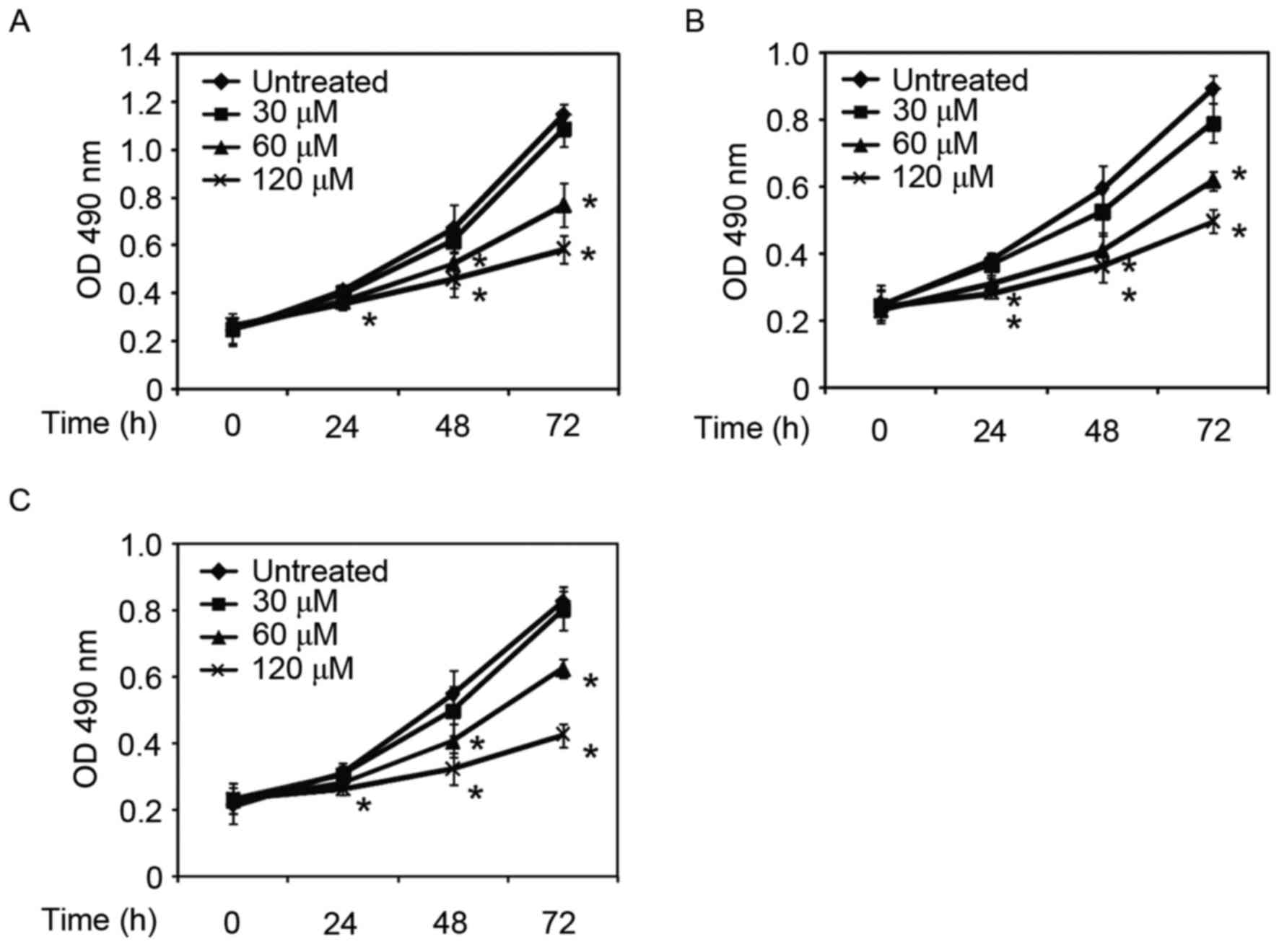
Capsaicin inhibits ESCC cell
proliferation in vitro
The efficacy of capsaicin against ESCC cell
proliferation was investigated in vitro. In three ESCC cell
lines, KYSE150, KYSE510 and KYSE410, capsaicin demonstrated an
inhibitory effect on cell growth. At a low concentration (30 µM),
exposure to capsaicin led to little growth inhibition in these
three cell lines. With an increase of capsaicin concentration and
the duration of treatment with capsaicin, cell growth was markedly
repressed. As presented in Fig. 1,
in all tested cell lines, at a high concentration (120 µM) for 72
h, cell proliferation was inhibited by >50% compared with the
control group. The results of the present study demonstrated that
capsaicin exhibited antitumor activity in ESCC cells in
vitro.
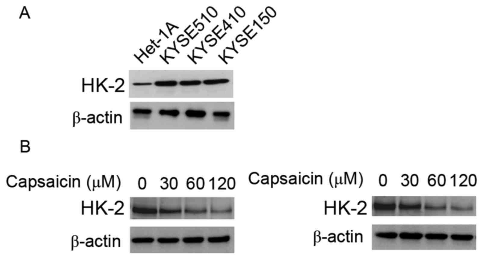
HK-2 decreases following treatment
with capsaicin in ESCC cells
Aerobic glycolysis is one of the metabolic
characteristics of tumor cells, and is important for the survival
and growth of cancer cells. In tumor cells, particularly those with
a highly glycolytic phenotype, HK-2 has been reported to be
overexpressed; however, its expression in ESCC cells was unknown.
Therefore, the expression of HK-2 was investigated in three ESCC
cell lines. As presented in Fig.
2A, compared with the normal esophageal epithelial cell line
Het-1A, the expression level of HK-2 in the three ESCC cells was
increased, suggesting that tumor glycolysis in these ESCC cells is
highly active. The effect of capsaicin on HK2 expression was
subsequently investigated. As presented in Fig. 2B, in KYSE150 and KYSE150 cells, the
expression of HK-2 was suppressed by capsaicin in a concentration
dependent manner. At a concentration of 120 µM, the expression of
HK-2 was markedly decreased compared with the control group.
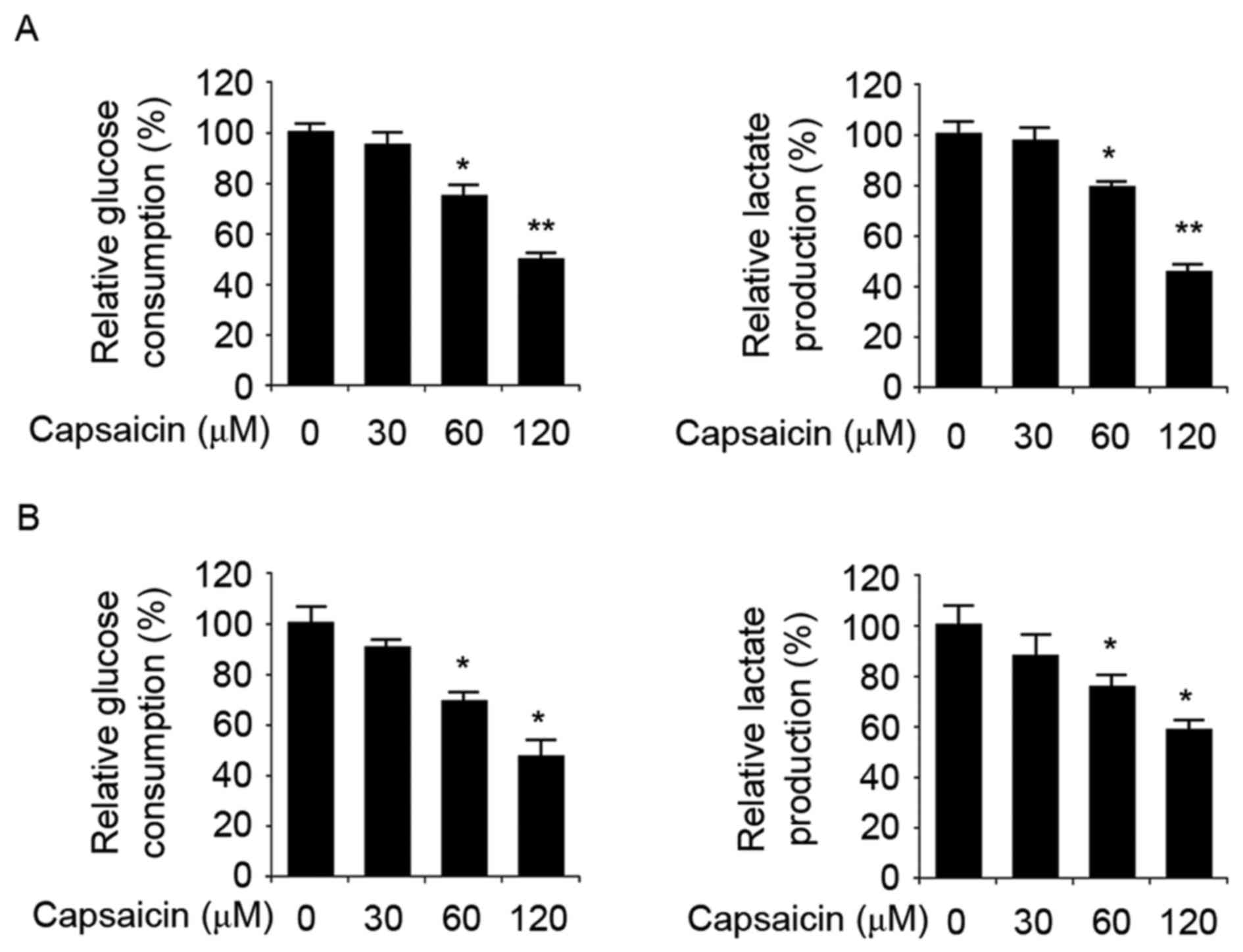
Capsaicin suppresses glucose uptake
and lactate secretion in ESCC cells
It is known that HK-2 serves a role in the process
of tumor glycolysis. Due to the effect of capsaicin on HK-2
expression, it was hypothesized that capsaicin may exhibit an
inhibitory effect on tumor glycolysis. Glucose uptake and lactate
secretion are indicators of tumor glycolysis; therefore, the effect
of capsaicin on glucose uptake and lactate secretion was
investigated. As presented in Fig.
3A, KYSE150 cells treated with capsaicin (60 µM) demonstrated
decreased glucose uptake compared with control cells. In KYSE510
cells, capsaicin repressed glucose consumption in a dose-dependent
manner. In addition to the suppression of glucose consumption, the
secretion of lactate, which is the product of tumor glycolysis, was
decreased. In KYSE150 and KYSE510 cells, treatment with 60 µM
capsaicin resulted in a notable reduction of lactate production in
the supernatant compared with the control group.
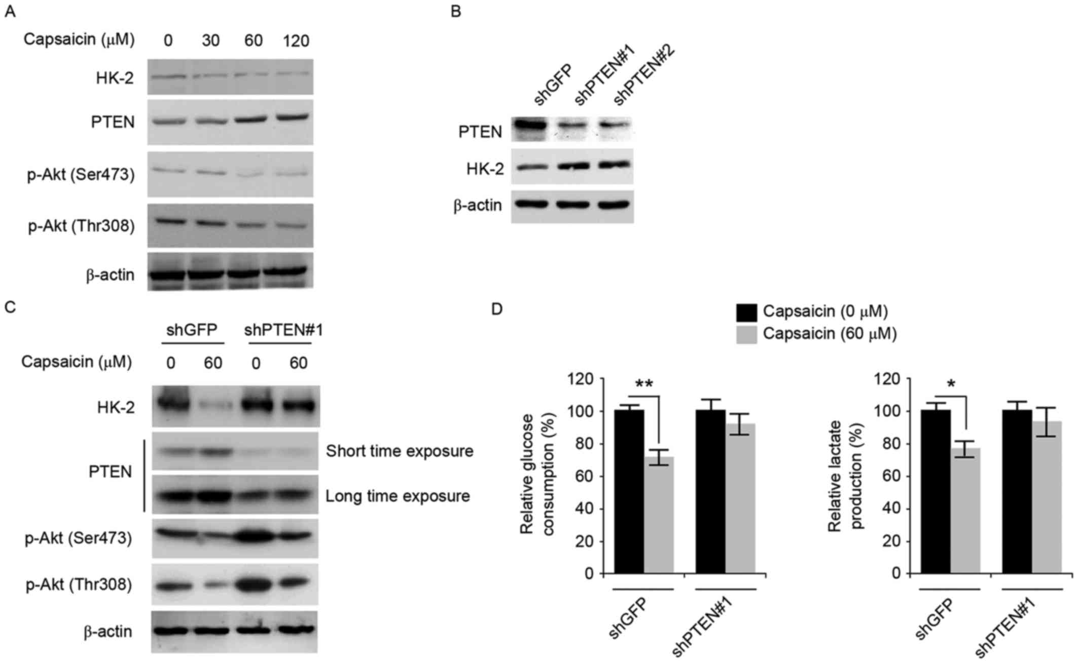
Capsaicin mediates glycolysis
inhibition in a PTEN dependent manner
As presented in Fig.
4A, in KYSE150 cells, the expression of PTEN, which negatively
regulates the phosphatidylinositol 3-kinase/Akt signaling pathway
in tumor cells, was increased in a dose-dependent manner following
treatment with capsaicin. In addition to an increase in PTEN
expression, phosphorylation of Akt at Ser473 and Thr308 was
suppressed. In order to investigate the role of PTEN in
facilitating capsaicin-mediated inhibition of glycolysis, PTEN
shRNA was developed to knock down the expression of PTEN in KYSE150
cells. Following transfection of KYSE-150 cells with PTEN shRNA,
the expression of PTEN was decreased compared with the control
group (shGFP group), which validated the efficiency of the shRNA
used (Fig. 4B). In addition to the
knockdown of PTEN, the expression of HK-2 was increased. The
efficacy of capsaicin was assessed in PTEN-knockdown KYSE-150
cells, as presented in Fig. 4C; in
PTEN-knockdown cells, the expression of HK-2 was unaltered
following treatment with capsaicin. In addition, the
phosphorylation of AKT at Ser473 and Thr308 was increased compared
with the control group. The effect of PTEN knockdown on glucose
uptake and lactate secretion was additionally investigated. As
presented in Fig. 4D, in
PTEN-knockdown KYSE150 cells, glycolysis inhibition caused by 60 µM
capsaicin was attenuated. Glucose uptake and lactate production in
PTEN shRNA cells was markedly recovered compared with the shGFP
group, which suggested an important role for PTEN in
capsaicin-mediated glycolysis inhibition.
Discussion
Esophageal cancer is the eighth most common cancer
and the sixth leading cause of mortality from cancer worldwide. In
Asia, particularly in China, ESCC is the predominant type. Despite
advances in cancer surgery and chemotherapy, the five-year survival
rate of patients with late-stage esophagus carcinoma is relatively
low (21). Therefore, there is a
requirement to discover and develop chemical entities with novel
antitumor mechanisms against esophagus carcinoma. Capsaicin, a
component of red peppers which is widely consumed, has been
reported to exert chemopreventive and chemotherapeutic activities
in various types of cancer; however, its effect on tumor glycolysis
remains unknown. The results of the present study demonstrated that
capsaicin exhibited an inhibitory effect on tumor glycolysis in
ESCC by downregulating HK-2 expression. Further investigations
revealed that PTEN was involved in the capsaicin-mediated
inhibition of tumor glycolysis in ESCC cells.
Clinical studies have demonstrated that the level of
glycolysis in tumor tissue, which can be detected with
18F-FDG-PET/computerized tomography technology, is a
useful prognostic factor for patients with ESCC (22,23).
The conversion of glucose to glucose-6-phosphate, an irreversible
step in glycolysis, is mediated by HK-2. HK-2 localizes to the
outer mitochondrial membrane protein voltage-dependent anion
channel, where it gains preferential access to the ATP generated by
the mitochondria and protection from inhibition by
glucose-6-phosphate (24).
Therefore, HK-2 is reported to be overexpressed in various types of
cancer, particularly those with a highly glycolytic phenotype. In
the present study, the overexpression of HK-2 was identified in
ESCC cells. Compared with normal esophageal epithelial cells, HK-2
expression in ESCC cells was increased, suggesting that the
glycolysis level in the ESCC cells was increased. In
capsaicin-treated ESCC cells, the expression of HK-2 was decreased.
In addition, the uptake of glucose and the secretion of lactate by
ESCC cells was reduced. The results of the present study
demonstrated that capsaicin effectively inhibited tumor glycolysis.
A previous study indicated that HK-2 was necessary for the
tumorigenicity of non-small cell lung cancer and breast cancer in
humans, whereas HK2 deletion resulted in rapid suppression of tumor
growth (25). The proliferation of
ESCC cells was suppressed by capsaicin in vitro in the
present study. Certain studies have demonstrated that the activity
of HK-2 was associated with chemoresistance (26). The sensitivity of tumor cells to
chemotherapy has been demonstrated to be enhanced through the
inhibition of glycolysis by targeting HK-2 (27,28).
Due to the effect of capsaicin on HK-2 and tumor glycolysis,
capsaicin may be able to increase the efficacy of other
chemotherapies.
Following treatment with capsaicin, the expression
of PTEN, which has been identified to be a tumor suppressor, was
increased in a dose-dependent manner. In addition, the
phosphorylation of Akt at Ser473 and Thr308 was decreased. As
reported by Wang et al (29), HK-2 was selectively upregulated by
the combined loss of PTEN and p53 in prostate cancer cells; the
PTEN deletion increased HK-2 mRNA translation through the
activation of the Akt-methylated target of rapamycin complex
1–4-Erb-binding protein 1 axis. In order to investigate the role of
PTEN in capsaicin-mediated glycolysis inhibition, PTEN expression
in KYSE150 cells was knocked down using shRNA in the present study.
In PTEN-knockdown cells, glycolysis inhibition mediated by
capsaicin was attenuated, suggesting that PTEN facilitates the
effect of capsaicin on tumor metabolism. As a tumor suppresser,
loss or mutations of PTEN have been identified in various types of
cancer. Clinical evidence has demonstrated that PTEN expression in
ESCC was an important prognostic indicator; the 5-year survival
rate in patients with PTEN-positive expression was 82%, compared
with 39% in patients with PTEN-negative expression (30). Consistent with previous reports, in
the present study, the expression of HK-2 was increased in
PTEN-knockdown cells compared with control cells. Therefore, in
patients with ESCC, aberrant tumor glycolysis may be a reason for
the poor prognosis associated with PTEN loss.
In conclusion, the present study demonstrated that
capsaicin exhibited an inhibitory effect on tumor glycolysis by
decreasing HK-2 expression in ESCC cells. Further investigation
demonstrated that the inhibition of glycolysis mediated by
capsaicin was associated with an increase of PTEN expression
following treatment with capsaicin. The present study provides a
novel mechanism to elucidate the antitumor activity of
capsaicin.
Acknowledgements
Not applicable.
Funding
The present study was funded by the Health Bureau of
Zhejiang Province (grant no. 2015KYB433).
Availability of data and materials
The analyzed data sets generated during the study
are available from the corresponding author on reasonable
request.
Authors' contributions
XLM, HYZ, DHL and YZ designed the experiments. XLM
and HYZ carried out the majority of the experimental work. LPY and
HFY performed the glucose and lactate assays. JSZ and YZ were
responsible for shRNA construction. XLM, HYZ, DHL and YZ analyzed
the data. XLM, HYZ and YZ wrote the manuscript.
Ethics approval and consent to
participate
Not applicable.
Consent for publication
Not applicable.
Competing interests
The authors declare that they have no competing
interests.
References
|
1
|
Richards BL, Whittle SL and Buchbinder R:
Neuromodulators for pain management in rheumatoid arthritis.
Cochrane Database Syst Rev. 1:CD0089212012.PubMed/NCBI
|
|
2
|
Burness CB and McCormack PL: Capsaicin 8%
Patch: A review in peripheral neuropathic pain. Drugs. 76:123–134.
2016. View Article : Google Scholar : PubMed/NCBI
|
|
3
|
Matharu M: Cluster headache. BMJ Clin
Evid. 2010:12122010.PubMed/NCBI
|
|
4
|
Ito K, Nakazato T, Yamato K, Miyakawa Y,
Yamada T, Hozumi N, Segawa K, Ikeda Y and Kizaki M: Induction of
apoptosis in leukemic cells by homovanillic acid derivative,
capsaicin, through oxidative stress: Implication of phosphorylation
of p53 at Ser-15 residue by reactive oxygen species. Cancer Res.
64:1071–1078. 2004. View Article : Google Scholar : PubMed/NCBI
|
|
5
|
Lau JK, Brown KC, Dom AM, Witte TR,
Thornhill BA, Crabtree CM, Perry HE, Brown JM, Ball JG, Creel RG,
et al: Capsaicin induces apoptosis in human small cell lung cancer
via the TRPV6 receptor and the calpain pathway. Apoptosis.
19:1190–1201. 2014. View Article : Google Scholar : PubMed/NCBI
|
|
6
|
Jin J, Lin G, Huang H, Xu D, Yu H, Ma X,
Zhu L, Ma D and Jiang H: Capsaicin mediates cell cycle arrest and
apoptosis in human colon cancer cells via stabilizing and
activating p53. Int J Biol Sci. 10:285–295. 2014. View Article : Google Scholar : PubMed/NCBI
|
|
7
|
Sarkar A, Bhattacharjee S and Mandal DP:
Induction of apoptosis by eugenol and capsaicin in human gastric
cancer AGS cells-elucidating the role of p53. Asian Pac J Cancer
Prev. 16:6753–6759. 2015. View Article : Google Scholar : PubMed/NCBI
|
|
8
|
Díaz-Laviada I: Effect of capsaicin on
prostate cancer cells. Future Oncol. 6:1545–1550. 2010. View Article : Google Scholar : PubMed/NCBI
|
|
9
|
Huang SP, Chen JC, Wu CC, Chen CT, Tang
NY, Ho YT, Lo C, Lin JP, Chung JG and Lin JG: Capsaicin-induced
apoptosis in human hepatoma HepG2 cells. Anticancer Res.
29:165–174. 2009.PubMed/NCBI
|
|
10
|
Clark R and Lee SH: Anticancer properties
of capsaicin against human cancer. Anticancer Res. 36:837–843.
2016.PubMed/NCBI
|
|
11
|
Hu F, Yang S, Zhao D, Zhu S, Wang Y and Li
J: Moderate extracellular acidification inhibits capsaicin-induced
cell death through regulating calcium mobilization, NF-kappaB
translocation and ROS production in synoviocytes. Biochem Biophys
Res Commun. 424:196–200. 2012. View Article : Google Scholar : PubMed/NCBI
|
|
12
|
Lee HK, Seo IA, Shin YK, Park JW, Suh DJ
and Park HT: Capsaicin inhibits the IL-6/STAT3 pathway by depleting
intracellular gp130 pools through endoplasmic reticulum stress.
Biochem Biophys Res Commun. 382:445–450. 2009. View Article : Google Scholar : PubMed/NCBI
|
|
13
|
Bhattacharya B, Mohd Omar MF and Soong R:
The Warburg effect and drug resistance. Br J Pharmacol.
173:970–979. 2016. View Article : Google Scholar : PubMed/NCBI
|
|
14
|
Wilson JE: Isozymes of mammalian
hexokinase: Structure, subcellular localization and metabolic
function. J Exp Biol. 206:2049–2057. 2003. View Article : Google Scholar : PubMed/NCBI
|
|
15
|
Mathupala SP, Ko YH and Pedersen PL:
Hexokinase II: Cancer's double-edged sword acting as both
facilitator and gatekeeper of malignancy when bound to
mitochondria. Oncogene. 25:4777–4786. 2006. View Article : Google Scholar : PubMed/NCBI
|
|
16
|
DeLaPaz RL, Patronas NJ, Brooks RA, Smith
BH, Kornblith PL, Milam H and Di Chiro G: Positron emission
tomographic study of suppression of gray-matter glucose utilization
by brain tumors. AJNR Am J Neuroradiol. 4:826–829. 1983.PubMed/NCBI
|
|
17
|
Ogawa H, Nagano H, Konno M, Eguchi H,
Koseki J, Kawamoto K, Nishida N, Colvin H, Tomokuni A, Tomimaru Y,
et al: The combination of the expression of hexokinase 2 and
pyruvate kinase M2 is a prognostic marker in patients with
pancreatic cancer. Mol Clin Oncol. 3:563–571. 2015. View Article : Google Scholar : PubMed/NCBI
|
|
18
|
Jin Z, Gu J, Xin X, Li Y and Wang H:
Expression of hexokinase 2 in epithelial ovarian tumors and its
clinical significance in serous ovarian cancer. Eur J Gynaecol
Oncol. 35:519–524. 2014.PubMed/NCBI
|
|
19
|
Guzman G, Chennuri R, Chan A, Rea B,
Quintana A, Patel R, Xu PZ, Xie H and Hay N: Evidence for
heightened hexokinase II immunoexpression in hepatocyte dysplasia
and hepatocellular carcinoma. Dig Dis Sci. 60:420–426. 2015.
View Article : Google Scholar : PubMed/NCBI
|
|
20
|
Schreurs LM, Smit JK, Pavlov K, Pultrum
BB, Pruim J, Groen H, Hollema H and Plukker JT: Prognostic impact
of clinicopathological features and expression of biomarkers
related to (18)F-FDG uptake in esophageal cancer. Ann Surg Oncol.
21:3751–3757. 2014. View Article : Google Scholar : PubMed/NCBI
|
|
21
|
Enzinger PC and Mayer RJ: Esophageal
cancer. N Engl J Med. 349:2241–2252. 2003. View Article : Google Scholar : PubMed/NCBI
|
|
22
|
Li YM, Lin Q, Zhao L, Wang LC, Sun L, Dai
MM, Luo ZM, Zheng H and Wu H: Pre-treatment metabolic tumor volume
and total lesion glycolysis are useful prognostic factors for
esophageal squamous cell cancer patients. Asian Pac J Cancer Prev.
15:1369–1373. 2014. View Article : Google Scholar : PubMed/NCBI
|
|
23
|
Hong JH, Kim HH, Han EJ, Byun JH, Jang HS,
Choi EK, Kang JH and Yoo Ie R: Total lesion glycolysis using
(18)F-FDG PET/CT as a prognostic factor for locally advanced
esophageal cancer. J Korean Med Sci. 31:39–46. 2016. View Article : Google Scholar : PubMed/NCBI
|
|
24
|
Mathupala SP, Ko YH and Pedersen PL:
Hexokinase-2 bound to mitochondria: Cancer's stygian link to the
‘Warburg Effect’ and a pivotal target for effective therapy. Semin
Cancer Biol. 19:17–24. 2009. View Article : Google Scholar : PubMed/NCBI
|
|
25
|
Patra KC, Wang Q, Bhaskar PT, Miller L,
Wang Z, Wheaton W, Chandel N, Laakso M, Muller WJ, Allen EL, et al:
Hexokinase 2 is required for tumor initiation and maintenance and
its systemic deletion is therapeutic in mouse models of cancer.
Cancer Cell. 24:213–228. 2013. View Article : Google Scholar : PubMed/NCBI
|
|
26
|
Suh DH, Kim MA, Kim H, Kim MK, Kim HS,
Chung HH, Kim YB and Song YS: Association of overexpression of
hexokinase II with chemoresistance in epithelial ovarian cancer.
Clin Exp Med. 14:345–353. 2014. View Article : Google Scholar : PubMed/NCBI
|
|
27
|
Peng Q, Zhou J, Zhou Q, Pan F, Zhong D and
Liang H: Silencing hexokinase II gene sensitizes human colon cancer
cells to 5-fluorouracil. Hepato-gastroenterology. 56:355–360.
2009.PubMed/NCBI
|
|
28
|
Jiang JX, Gao S, Pan YZ, Yu C and Sun CY:
Overexpression of microRNA-125b sensitizes human hepatocellular
carcinoma cells to 5-fluorouracil through inhibition of glycolysis
by targeting hexokinase II. Mol Med Rep. 10:995–1002. 2014.
View Article : Google Scholar : PubMed/NCBI
|
|
29
|
Wang L, Xiong H, Wu F, Zhang Y, Wang J,
Zhao L, Guo X, Chang LJ, You MJ, Koochekpour S, et al: Hexokinase
2-mediated Warburg effect is required for PTEN- and
p53-deficiency-driven prostate cancer growth. Cell Reports.
8:1461–1474. 2014. View Article : Google Scholar : PubMed/NCBI
|
|
30
|
Chang D, Wang TY, Li HC, Wei JC and Song
JX: Prognostic significance of PTEN expression in esophageal
squamous cell carcinoma from Linzhou City, a high incidence area of
northern China. Dis Esophagus. 20:491–496. 2007. View Article : Google Scholar : PubMed/NCBI
|