Introduction
Chronic kidney disease (CKD) has been recognized as
a major public health problem that is associated with substantial
morbidity, mortality and financial cost to the healthcare system
(1,2). Diabetes is one of the most important
causes of CKD and diabetic kidney disease (DKD) is one of the most
important complications of diabetes (3,4). DKD
prevention is crucial for patients with diabetes.
Although hyperglycemia is an essential requirement
for DKD, the pathogenic pathway initiated and maintained in the
kidney by elevated glucose levels can be enhanced by a number of
different factors. These factors include excess fatty acids,
oxidative stress and hemodynamic factors. These factors do not
themselves contribute to DKD, but in the presence of diabetes they
feed back and reinforce common pathogenic mechanisms, including
increased levels of inflammatory cytokines and fibrosis-related
growth factors in the kidney (5).
In addition, the peroxisome proliferator-activated receptors
(PPARs; α, β/δ, γ) are a family of ligand-activated transcription
factors of the nuclear receptor superfamily that regulates cellular
metabolic homeostasis. Mutations in the PPARγ gene have been
demonstrated to be associated with dysfunctional lipid and glucose
homeostasis (6). In preclinical
and/or clinical studies, activation of PPARγ has been shown to have
a positive effect on insulin resistance, diabetic nephrotic
syndrome, and renal fibrosis (7).
Therefore, PPARγ may serve an important role in DKD.
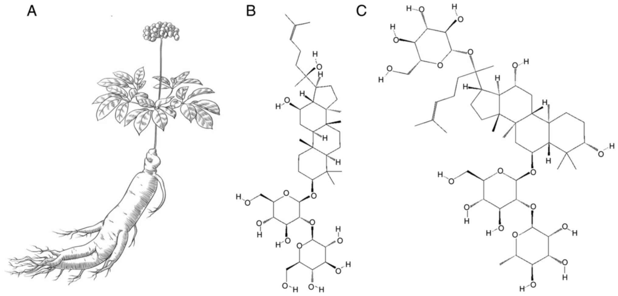
Ginsenosides are major active ingredients extracted
from the roots, stems, leaves or fruits of Panax ginseng
C.A. Meyer (Ginseng; Fig. 1A),
which is widely cultivated in Korea and Northeast China.
Ginsenosides are widely used to develop drugs for adjuvant therapy
for cancer and chronic cardiovascular and metabolic diseases,
especially the complications of these disorders (8–12).
These natural products do not have sufficiently strong biological
activities to become first-line drugs for those diseases.
Ginsenoside Rg3 (Rg3; chemical structure shown in
Fig. 1B) is one of the major
active ingredients extracted from Korean red ginseng. It can also
be chemically or biologically converted from other ginsenosides
(13–15). Rg3 is well known for its antitumor
effects (16–20) and has been developed into an
adjuvant antitumor drug called Shenyi capsule (SYC) in China
(11,21–23).
Ginsenoside Re (Re; chemical structure shown in
Fig. 1C) is the essential
component of total saponins from ginseng fruits and was developed
into a drug called Zhenyuan capsule (ZYC) in China (10). According to the quality control
standard documents for ZYC, the content of Re is the only technical
standard in its quality control. Re alleviates the complications of
cardiovascular and metabolic diseases (24–26)
and ZYC is widely used for adjunctive treatment of type II diabetes
(T2DM) and coronary heart disease in the clinic in China (10,27,28).
Our group is currently undertaking a long-term study
assessing the effects of various ginsenosides (including Rg3 and
Re) in various animal models of chronic diseases, such as
hypertension, hyperlipidemia and T2DM (21–24,29,30).
Notably, our previous studies demonstrated that Rg3, the essential
component of SYC, has various beneficial effects on the target
organs of those chronic diseases (21–23,29),
although SYC is used for adjunctive cancer treatment in the clinic
(11,18). In particular, Rg3 attenuated
atherosclerosis in hyperlipidemic mice (29) and nonalcoholic steatohepatitis
(NASH) in T2DM mice (22), both of
which are chronic metabolic diseases. In these two studies, the
mechanisms of the protective effects included regulation of
inflammation, fibrosis and PPARγ.
In another study, the authors demonstrated that Re
attenuated NASH in T2DM mice, also via the regulation of
inflammation, fibrosis and PPARγ (24). In contrast to Rg3/SYC, Re/ZYC has
already been widely used for T2DM treatment in the clinic. The
present study observed the renoprotective effects of Rg3 on db/db
mice (BKS-Leprem2Cd479/Gpt), and Re was used as the
control. The leptin receptors are defective in this type of mice,
leading to obesity and disorders of glucose-lipids metabolism.
Hyperleptinemia, hyperglycemia and hyperlipidemia also lead to DKD
and non-alcoholic fatty liver disease (NAFLD) in db/db mice >20
weeks old, with abnormal serum renal and liver function indicators.
The symptoms of T2DM in db/db mice are similar to those in humans,
and it is one of the best-known gene-deficient animal models of
T2DM with DKD (31,32). The main features of DKD in humans
are renal hypertrophy, enlarged glomeruli, albuminuria and dilated
thylakoid stroma and the db/db mice simulates these pathological
features successfully.
Materials and methods
Reagents
Rg3 (95% purity) was obtained from Dr Li Fu at the
Institute of Dalian Fusheng Natural Medicine, Dalian Fusheng
Pharmaceutical Co., Ltd. (Dalian, China). Re (95% purity) was
obtained from Dr Yanping Chen at the Department of Natural
Medicinal Chemistry, School of Chemistry, Jilin University
(Changchun, China). Rg3 and Re were dissolved in 0.5% sodium
carboxymethyl cellulose solution (0.5% CMC-Na) for use. All other
chemicals were analytical reagents.
Animals
A total of 24 db/db mice (25–27 g) and 8 wild-type
(WT) mice (45–47 g) were purchased from GemPharmatech Co., Ltd. All
the mice were male and 12–13 weeks old. The experimental animal
house was of specific pathogen-free (SPF) grade, and the mice were
kept in individually ventilated cages (four mice per cage). The
house was maintained at a constant temperature of 22–24°C relative
humidity of 45–55%. All mice had free access to water and food and
were maintained in a 12-h light/dark cycle. All feeding conditions
were the same as described in previous studies (22,24).
The mouse chow diet (cat. no. SZS9126-1010082) was purchased from
Jiangsu Xietong Pharmaceutical Bioengineering Co., Ltd.
Experimental protocols
A total of eight WT mice were designated as Group
WT. The 24 db/db mice were randomly divided into three groups with
eight mice in each: Group db/db, 8 db/db mice; Group Rg3, 8 db/db
mice; and Group Re, eight db/db mice. Mice in Rg3 and Re were
orally administered Rg3 and Re, respectively, at a dose of 30
mg·kg−1day−1, the same dose used in previous
studies (22,24). Mice in Group WT and Group db/db
were orally administered 0.5% CMC-Na as a placebo in the same
volume given to those in Group Rg3 and Group Re. The administration
of Rg3, Re and placebo was carried out for eight weeks. During
these eight weeks, the blood glucose and body weight of the mice
were measured weekly while general health and behavior states of
mice were monitored daily during oral administration.
After the eight week treatment, the mice in the four
groups were sacrificed (carbon dioxide euthanasia, the carbon
dioxide concentration in the container rose from 0.03–99% in 2 min)
for renal tissue sample collection immediately after collecting
blood samples from the ophthalmic venous. All the samples were
processed as described in previous studies (22,24).
Serum was isolated from blood samples for biochemical assays. Renal
tissue samples were fixed in 4% formaldehyde (20°C overnight) for
histopathological and immunohistochemistry (IHC) assessment,
snap-frozen and kept at −80°C for biochemical assays and reverse
transcription-quantitative (RT-q) PCR.
Fasting blood glucose measurement
Throughout the eight weeks of treatment, blood from
the lateral tail vein of the WT and db/db mice was collected, and
the fasting blood glucose level was monitored weekly using a blood
glucose test meter and strips (GlucoLab, Infopia Co., Ltd.)
according to the manufacturer's protocol as previously described
(22,24).
Serum biochemical assays
Biochemical assay kits were purchased from Nanjing
Jiancheng Bioengineering Institute: Creatinine (CRE; cat. no.
C011-2-1), blood urea nitrogen (BUN; cat. no. C013-2-1),
triglyceride (TG; cat. no. A110-1-1), total cholesterol (TC; cat.
no. A111-1-1), high-density lipoprotein cholesterol (HDL; cat. no.
A112-1-1), and low-density lipoprotein cholesterol (LDL; cat. no.
A113-1-1). Levels of CRE, BUN, TG, TC, HDL and LDL in serum were
assayed in accordance with the manufacturer's protocols as
previously described (22–24).
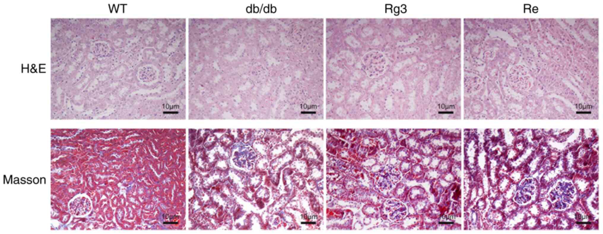
Histopathological assessment
Renal tissue specimens fixed in 4% formalin were
embedded in paraffin, cut into 4-µm-thick sections and then stained
with hematoxylin and eosin (H&E) at 5 and 1 min respectively at
room temperature or Masson's trichrome stain (Masson) as previously
described (22–24). A Masson kit (cat. no. BSBA-4079B)
was purchased from OriGene Technologies, Inc. Sections stained with
H&E and Masson were examined, and images were captured using a
Nikon E100 light microscope (Nikon Corporation).
IHC
Primary antibodies against PPARγ (cat. no.
bs-4590R), tumor necrosis factor-α (TNF-α; cat. no. bs-10802R),
transforming growth factor β1 (TGF-β1; cat. no. bs-0103R) and
connective tissue growth factor (CTGF; cat. no. bs-0743R) were
purchased from BIOSS. A DAB kit (cat. no. ZLI-9017) and a two-step
rabbit IHC kit (cat. no. PV-9001) were purchased from OriGene
Technologies, Inc. IHC was performed in accordance with the
manufacturer's protocols for the IHC kit and DAB kit as previously
described (22–24). Photomicrograph images were then
captured with a light microscope as aforementioned and were further
analyzed using Image-Pro Plus 6.0 (Media Cybernetics, Inc.).
RNA purification and RT-qPCR
Isolation of total RNA was carried out using
TRIzol® reagent (Thermo Fisher Scientific, Inc.)
according to the manufacturer's protocol as previously described
(22–24). RT and qPCR were performed were
performed according to the manufacturer's protocol with TransScript
Green TwoStep RT-qPCR SuperMix (Beijing Transgen Biotech Co., Ltd.)
on a Stratagene Mx3000P Real-Time PCR System (Agilent Technologies,
Inc.). The thermocycling conditions were denaturation 94°C for 5
sec, annealing 60°C for 15 sec and extension 72°C for 10 sec, 40
cycles. The 2−ΔΔCq method (33) was employed for analysis of the
expression of genes of interest, and β-actin was used as a
housekeeping gene. All primers are listed in Table I.
 | Table I.Primer sequence for reverse
transcription-quantitative PCR. |
Table I.
Primer sequence for reverse
transcription-quantitative PCR.
| Gene | Sequence
(5′-3′) |
|---|
| β-actin |
|
|
Forward |
GGCTGTATTCCCCTCCATCG |
|
Reverse |
CCAGTTGGTAACAATGCCATGT |
| CTGF |
|
|
Forward |
GTAACCGGGGAGGGAAATTA |
|
Reverse |
GCTTTATCACCTGCACAGCA |
| IL-6 |
|
|
Forward |
GTCCTTCAGAGAGATACAGAAACT |
|
Reverse |
AGCTTATCTGTTAGGAGAGCATTG |
| Col-I |
|
|
Forward |
CTTCACCTACAGCACCCTTGTG |
|
Reverse |
TGACTGTCTTGCCCCAAGTTC |
| Col-III |
|
|
Forward |
TGTCCTTTGCGATGACATAATCTG |
|
Reverse |
AATGGGATCTCTGGGTTGGG |
Statistical analysis
SPSS 16.0 (SPSS, Inc.) was employed for all
statistical analyses. Data are presented the mean ± standard
deviation (SD). One-way analysis of variance (ANOVA) with Tukey's
post-hoc test was employed for group comparisons. P<0.05 was
considered to indicate a statistically significant difference.
Results
Rg3 and Re had no significant effects
on body weight, blood glucose or lipids
Prior to and following eight weeks of treatment, the
mice in the db/db group had significantly higher body weight
(Fig. 2A) and blood glucose levels
(Fig. 2B) compared with the mice
in the WT group, which is the basic feature of db/db mice (31,32).
The body weights of the mice in the WT and db/db groups after eight
weeks of treatment were significantly elevated compared to those
before eight weeks of treatment, while their blood glucose levels
were slightly elevated but not significantly different. The body
weight and blood glucose levels in Groups Rg3 and Re were similar
to those in Group db/db, and there was no significant difference
between them either before or after the eight week treatment.
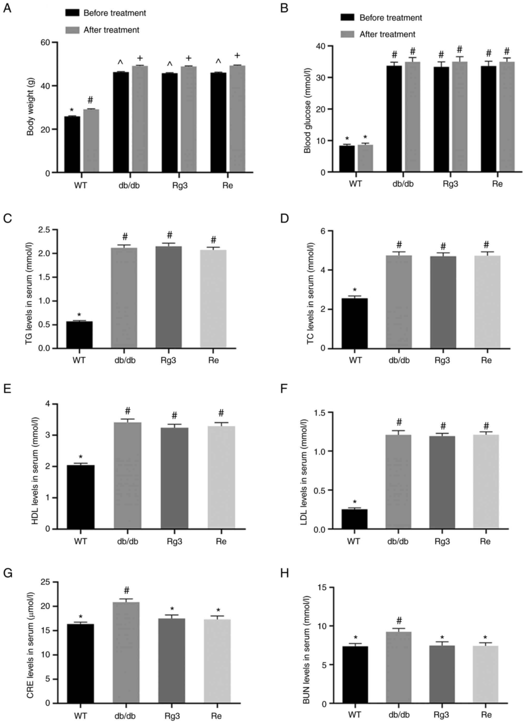 | Figure 2.Body weight, blood glucose, lipids
and renal function index in serum. (A) Body weight and (B) blood
glucose of mice prior to and following 8 weeks of treatment; serum
(C) TG, (D) TC, (E) HDL, (F) LDL, (G) CRE and (H) BUN levels in
mice after treatment. Data are presented as the mean ± standard
deviation, n=8. The same superscript symbols indicate no
significant difference between groups (P>0.05); a significant
difference existed between groups that do not have the same
superscript symbol (P<0.05). TG, triglyceride; TC, total
cholesterol; HDL, high-density lipoprotein cholesterol; LDL,
low-density lipoprotein cholesterol; CRE, creatinine; BUN, blood
urea nitrogen; WT, wild type; Rg3, ginsenoside Rg3; Re, ginsenoside
Re. |
The levels of blood lipids (TG, TC, HDL and LDL) in
the mice in Group db/db were all significantly higher than those in
Group WT after eight weeks of treatment (Fig. 2C-F). Similar to body weight and
blood glucose, lipid levels in Groups Rg3 and Re were similar to
those in Group db/db.
In summary, all the db/db mice presented significant
abnormalities and glucose and lipid metabolism disorders, which are
all typical symptoms of T2DM. Treatment with Rg3 and Re were not
able to significantly attenuate these symptoms, which is similar to
our previous studies (22–24).
Rg3 and Re improved early renal injury
in T2DM
According to the histology images of kidney
H&E-stained sections from the mice in Group WT, the outer
cortical glomerulus was of normal size and configuration (Fig. 3). The capillary tuft was fully
expanded with patent capillary loops and the glomerular basement
membrane appeared thin without matrix expansion, inflammation, or
sclerosis. The histology images from mice in Group db/db showed
striking differences in some glomeruli. The visceral epithelial
cells were swollen and appeared prominent. The glomerular capillary
basement membranes appeared thickened, and the peripheral capillary
loop appeared to be collapsed; 30–40% of glomeruli had a similar
appearance and the remaining had a lesser degree of mesangial
matrix expansion. The histology images from mice in Groups Rg3 and
Re showed that treatments with these two ginsenosides significantly
attenuated the pathological changes; only 10–20% of glomeruli had
an appearance similar to that aforementioned and most of the
glomeruli had a lesser degree of mesangial matrix expansion.
According to the histology images of Masson-stained kidney
sections, mice from Group db/db presented slight tubulointerstitial
fibrosis, absent in the mice from the other three groups.
The levels of CRE and BUN supported the results of
the pathological examination (Fig. 2G
and H). The two were significantly higher in the mice from
Group db/db compared with the mice from Group WT. The levels of the
other three groups did not present significant differences.
These results showed that mice from Group db/db were
in an early stage of DKD. There were slight pathological changes
according to the pathological examination and slight renal function
decompensation according to the blood biochemical examination.
Although Rg3 and Re had no significant effects on body weight,
blood glucose or lipids, their treatment prevented early-stage DKD
in db/db mice from Groups Rg3 and Re. Their protective effects were
comparable, and both were able to reduce the levels of CRE and BUN
to those in WT mice. These findings were also consistent with our
previous studies (22–24), in which ginsenoside treatment
improved renal or hepatic early-stage injury independent of
reducing the levels of blood pressure or blood glucose.
Rg3 and Re upregulated PPARγ
expression and inhibited inflammation and fibrosis in renal
tissue
In our two previous studies, Rg3 (22) and Re (24) were demonstrated to ameliorate
hepatic injury in db/db mice by upregulating PPARγ expression and
inhibiting inflammation and fibrosis in hepatic tissue. The present
study also performed IHC and RT-qPCR to assess the levels of PPARγ
expression and the inhibition of inflammation and fibrosis in renal
tissue.
According to the integrated optical density (IOD)
analysis of the IHC images, PPARγ expression in renal tissue from
mice in Group db/db was significantly higher than that in Group WT
(Fig. 4A and B). This increase was
a compensatory upregulation related to the high blood glucose
levels in db/db mice (34). The
PPARγ expression in the renal tissue was further upregulated by Rg3
and Re treatment, which was similar to the findings in hepatic
tissue in the previous aforementioned studies (22,24).
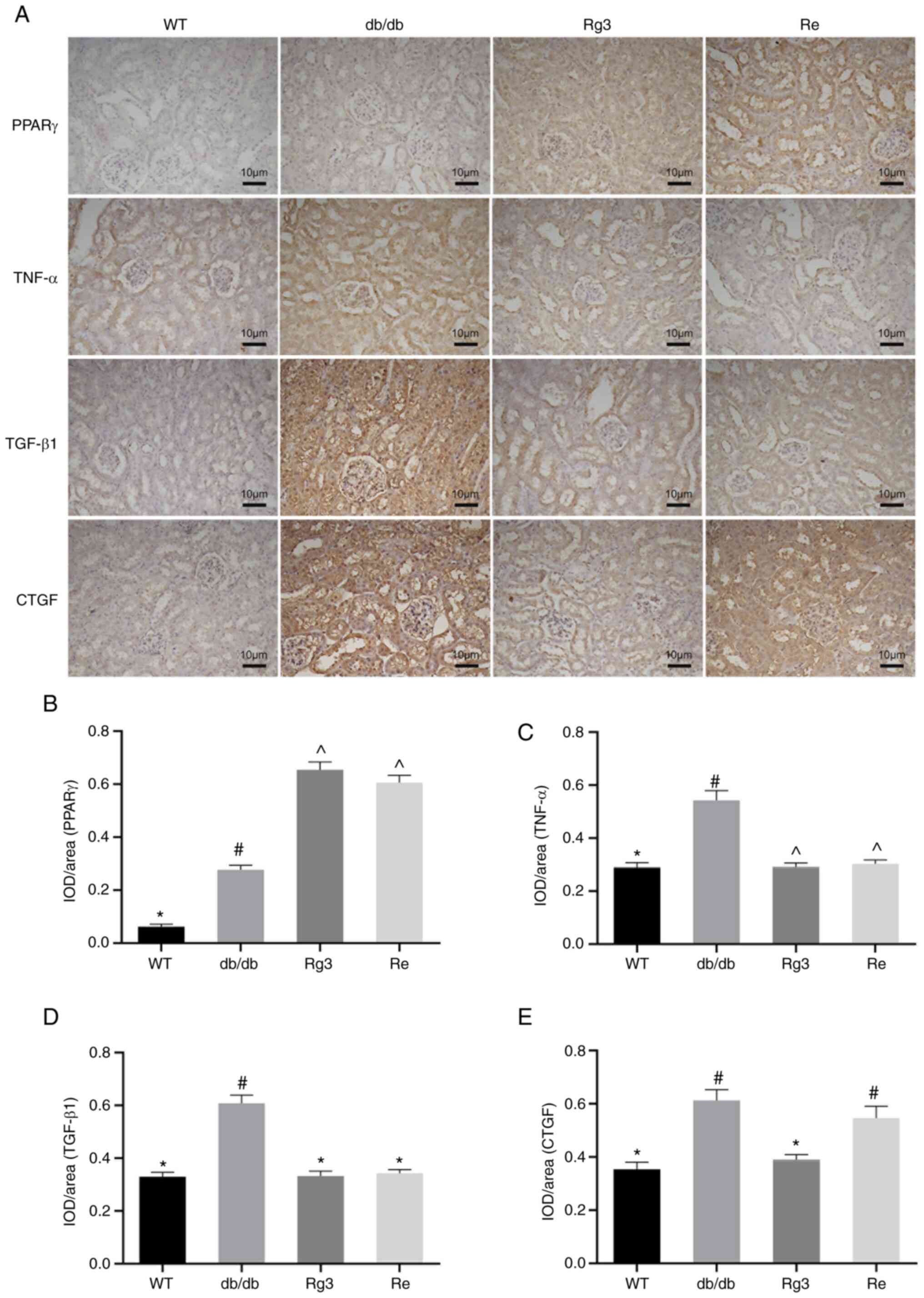 | Figure 4.IHC staining images of the renal
tissue in mice and analyses. (A) Representative IHC staining images
of the renal tissue in mice and quantitative results of IHC
staining, which are presented as IOD/area and are proportional to
the levels of (B) PPARγ, (C) TNF-α, (D) TGF-β1 and (E) CTGF. Scale
bars, 10 µm. Data are presented as the mean ± standard deviation,
n=4. The same superscript symbols indicate no significant
difference between groups (P>0.05); a significant difference
existed between groups that do not have the same superscript symbol
(P<0.05). IHC, immunohistochemistry; PPARγ, peroxisome
proliferator-activated receptor γ; CTGF, connective tissue growth
factor; WT, wild type; Rg3, ginsenoside Rg3; Re, ginsenoside
Re. |
TNF-α is one of the most important markers of
inflammation. The IHC results of TNF-α showed significantly higher
levels of inflammation in renal tissue from mice in Group db/db
compared with those in Group WT (Fig.
4A and C). The levels of TNF-α were significantly decreased in
Groups Rg3 and Re and were not significantly different from those
in Group WT.
TGF-β1 and CTGF are both important profibrotic
factors and markers of fibrosis. According to the IHC results, the
levels of TGF-β1 and CTGF in renal tissue were significantly higher
in the db/db group compared with the WT group. The levels of both
TGF-β1 and CTGF in Group Rg3 were significantly reduced and were
similar to those in Group WT (Fig. 4A,
D and E). Notably, only TGF-β1 levels were significantly
decreased in Group Re compared to those in Group db/db, while CTGF
levels were not significantly different from those in Group
db/db.
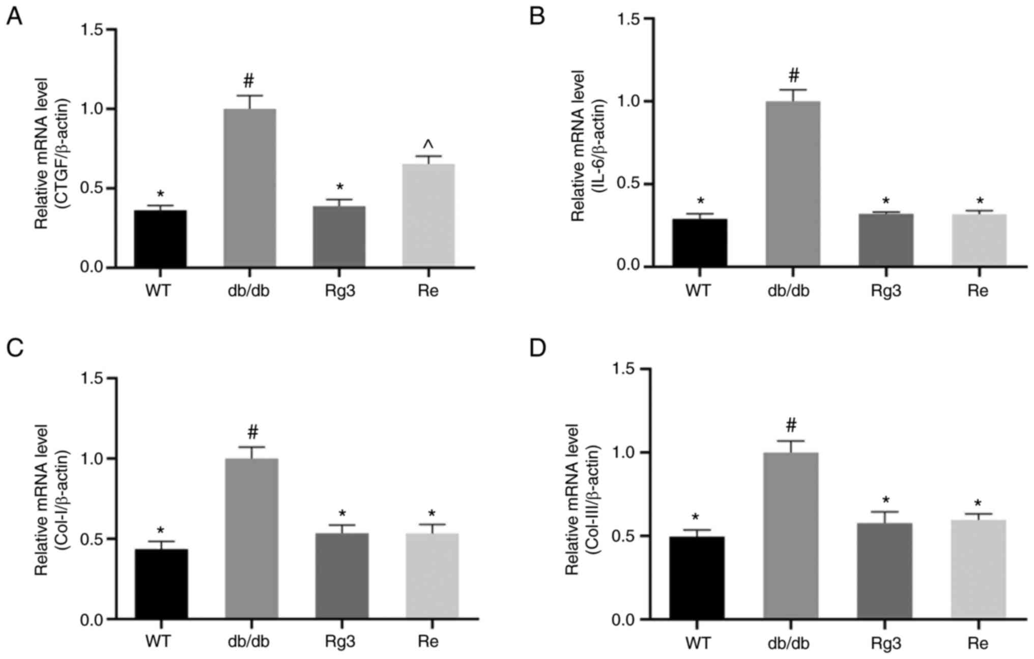
Thus, the present study measured the levels of CTGF
repeatedly using RT-qPCR. The relative mRNA levels of CTGF
supported the IHC results (Fig.
5A). The levels of CTGF in Group Re were significantly lower
than those in Group db/db but significantly higher than those in
Group WT. The levels of CTGF in Group Rg3 were not significantly
different from those in Group WT.
Levels of collagen type (Col)-I, Col-III, markers of
collagen deposition, and IL-6, another marker of inflammation, were
also measured using RT-qPCR (Fig.
5B-D). The relative mRNA levels of all three markers were
higher in the renal tissue of mice from the db/db group than in
that of mice from the WT group. Rg3 and Re treatment were both able
to significantly downregulate these mRNA levels in db/db mice.
Moreover, the degree of downregulation was similar.
In summary, Rg3 and Re both upregulated the
expression of PPARγ in the renal tissue of db/db mice and
downregulated inflammatory and fibrotic factors, such as TNF-α,
TGF-β1 and CTGF. These regulatory effects all contributed to the
renoprotective effects of Rg3 and Re. Notably, Rg3 had a stronger
effect of downregulating CTGF than Re. However, the overall effects
of Rg3 on inhibiting inflammation and fibrosis in renal tissue were
comparable to those of Re in this research.
Discussion
DKD has become the leading cause of end-stage renal
disease worldwide, which increases the risk of premature death and
represents a serious financial burden. Although DKD is controllable
with different drugs, such as angiotensin-converting enzyme
inhibitors (ACEIs), angiotensin receptor blockers (ARBs) and
hypoglycemic agents, such as thiazolidinediones (TZDs), the
preventive and therapeutic merits of natural medicines, such as
ginsenosides, have been widely recognized (4).
ACEIs and ARBs, blockers of the
renin-angiotensin-aldosterone system, are widely used to control
the progression of DKD, especially in patients with both diabetes
and hypertension. However, as ACEIs and ARBs also have a strong
biological ability to reduce blood pressure, they are not
recommended for diabetic patients without hypertension or any DKD
symptoms (35,36).
TZDs are agonists of the PPARγ pathway and have
anti-inflammatory and antifibrotic biological activities, increase
insulin sensitivity and reduce blood glucose; they are also used to
control DKD. However, TZDs have recently fallen into disuse for
glycemic control due to concerns over side effects and adverse
events. These drugs are recommended only for diabetic patients with
complications such as DKD and NASH to control their progression
(37,38).
In traditional Chinese medicine (TCM) theory, the
main causes of diabetic kidney disease are congenital deficiencies,
dietary disorders and emotional disorders. Insufficient innate
endowment (a type of birth defect) and weakness of the internal
organs by birth make these individuals more susceptible to DKD. The
best way to manage the complications of diabetes is ‘Zhi Wei Bing’
(39,40). Zhi Wei Bing means taking
appropriate measures to prevent the development of the disease. The
main idea in TCM theory is to prevent disease before it occurs and
to prevent the development of disease after it has occurred.
Correctly anticipating the development of the disease can stop its
exacerbation or transformation in time, which is why TCM, such as
Re/ZYC, is widely used in China for patients with T2DM. According
to our previous studies, ginsenosides such as Rg3 and Re exerted
anti-inflammatory and anti-fibrotic effects on vital organs such as
the heart, liver and kidneys, although they do not significantly
lower blood pressure, blood glucose and lipids in animals (21–24).
As aforementioned, natural products such as Rg3 and Re do not have
strong enough biological activity to be first-line antihypertensive
or hypoglycemic agents. However, it is also the mild biological
activity of Rg3 and Re that makes it possible for them to be
developed as safe drugs with few side effects for use in
early-stage diabetic patients without hypertension or any symptoms
of DKD.
As aforementioned, natural products such as Rg3 and
Re do not have sufficiently strong biological activities to become
first-line hypotensive or hypoglycemic agents. On the other hand,
Rg3 and Re have the potential to be used for early-stage diabetes
patients without hypertension or any DKD symptoms because of their
mild biological activities. Ginseng has been widely consumed as a
food/diet supplement from natural sources, and the safety of using
ginseng and ginsenosides has been widely recognized (4,41,42).
The situation of Rg3/SYC is different. Rg3 was
widely used for cancer patients in China at first (11,18).
Indeed, 10 years ago most studies focused on the role of Rg3 in
inducing apoptosis of tumor cells and inhibiting their growth
(43–45). More recently, an increasing number
of antitumor effects of Rg3 have been discovered (18–20,46–48)
and include inhibition of drug resistance, angiogenesis and
epithelial-mesenchymal transformation. The mechanisms include
regulation of the Wnt/β-catenin pathway, Hippo pathway, VEGF
pathway and TGF-β signaling pathway (18–20,46–48),
all of which have a close connection to inflammation, fibrosis and
the PPARγ pathway (49–51).
Although the use of TZDs in T2DM has been
controversial in recent years (37,38),
increasing evidence shows that activation of the PPARγ pathway is
beneficial to tumor treatments in most fields (52). Activation of the PPARγ pathway has
a positive correlation with the Hippo pathway (51) and a negative correlation with the
Wnt/β-catenin pathway (50). These
interactions occur not only in diabetes but also in tumors. The
Hippo pathway and Wnt/β-catenin pathway are also popular tumor
therapeutic targets. Moreover, activation of PPARγ usually has a
negative correlation with insulin resistance, inflammation and
fibrosis (37,38,50–52),
which are all important for the treatment of diabetes and its
complications.
Although Rg3 and Re belong to the ginsenosides
family, their aglycone structures are different, with Rg3 being a
protopanaxadiol saponin and Re being a protopanaxatriol saponin.
Nevertheless, both Rg3 and Re are protective against DKD or other
complications of T2DM, as well as several kinds of other
protopanaxadiol or protopanaxatriol saponins, all of which have
anti-inflammatory and anti-fibrosis effects. In addition, comparing
the effects of Rg3 and Re in the present study, the major
difference was that Rg3 had a stronger effect of regulating CTGF in
renal tissue. These results were consistent with our previous
studies of Rg3 and Re in db/db mice with NASH (22,24).
As CTGF has a close connection to the Hippo pathway (49), these results were also consistent
with the report of Rg3 activating the Hippo pathway in antitumor
research (48). These studies
might explain why Rg3 has more antitumor effects than Re while the
renoprotective effects are comparable. More experiments are needed
to confirm this hypothesis in the future.
Although neither has significant effects on body
weight, blood glucose and lipids, Rg3 and Re both have effects in
the prevention of DKD in db/db mice. The mechanisms of their
renoprotective effects include alleviation of inflammation and
fibrosis and upregulation of PPARγ expression in renal tissue. The
effects of Rg3 and Re were comparable, as they were both able to
reduce the CRE and BUN levels of db/db mice to levels similar to
those of WT mice, as well as the inhibition of pathological changes
in the renal tissue of those mice. The only difference was that Rg3
had a stronger effect of regulating CTGF in renal tissue. The
present study showed that Rg3, an adjuvant antitumor drug, has a
potential similar to that of Re, which has already been widely used
in the clinic for the preventive treatment of T2DM complications,
such as DKD. The timing of using Rg3 or Re could be much earlier
than that of using chemical drugs, such as ACEIs, ARBs or TZDs.
Acknowledgements
The authors would like to thank Dr Li Fu (Institute
of Dalian Fusheng Natural Medicine, Dalian Fusheng Pharmaceutical
Co., Ltd.) and Dr Yanping Chen (Department of Natural Medicinal
Chemistry, School of Chemistry, Jilin University), for providing
Rg3 and Re, respectively.
Funding
The present study received financial support from the National
Natural Science Foundation of China (grant no. 81473378) and
Science and Technology Development Projects of Jilin Province
(grant no. 20170101002JC).
Availability of data and materials
All data generated or analyzed during this study are
included in this published article.
Authors' contributions
ZS completed the biochemistry and molecular biology
experiments. DS, the recipient of the funded projects, lead the
design of the experiment. ML completed the animal experiments. QY
processed the experimental data. HL and YJ designed the experiment
and wrote the manuscript. ZS and ML confirm the authenticity of all
the raw data. All authors read and approved the final
manuscript.
Ethics approval and consent to
participate
All the experimental procedures involving animals
were conducted in accordance with the Institutional Animal Care
Guidelines of Jilin University and approved by the Laboratory
Animal Ethics Committee of School of Pharmaceutical Sciences, Jilin
University (approval no. 20210010).
Patient consent for publication
Not applicable.
Competing interests
The authors declare that they have no competing
interests.
References
|
1
|
Murphy D, McCulloch CE, Lin F, Banerjee T,
Bragg-Gresham JL, Eberhardt MS, Morgenstern H, Pavkov ME, Saran R,
Powe NR, et al: Trends in prevalence of chronic kidney disease in
the United States. Ann Intern Med. 165:473–481. 2016. View Article : Google Scholar : PubMed/NCBI
|
|
2
|
Coresh J, Selvin E, Stevens LA, Manzi J,
Kusek JW, Eggers P, Van Lente F and Levey AS: Prevalence of chronic
kidney disease in the United States. Jama. 298:2038–2047. 2007.
View Article : Google Scholar : PubMed/NCBI
|
|
3
|
Barrera-Chimal J and Jaisser F:
Pathophysiologic mechanisms in diabetic kidney disease: A focus on
current and future therapeutic targets. Diabetes Obes Metab. 22
(Suppl 1):S16–S31. 2020. View Article : Google Scholar : PubMed/NCBI
|
|
4
|
Jin D, Zhang Y, Zhang Y, Duan L, Zhou R,
Duan Y, Sun Y, Lian F and Tong X: Panax Ginseng C.A.Mey. as
Medicine: The potential use of panax ginseng C.A.Mey. as a remedy
for kidney protection from a pharmacological perspective. Front
Pharmacol. 12:7341512021. View Article : Google Scholar : PubMed/NCBI
|
|
5
|
Thomas MC, Brownlee M, Susztak K, Sharma
K, Jandeleit-Dahm KAM, Zoungas S, Rossing P, Groop PH and Cooper
ME: Diabetic kidney disease. Nat Rev Dis Primers. 1:150182015.
View Article : Google Scholar : PubMed/NCBI
|
|
6
|
Barroso I, Gurnell M, Crowley VE, Agostini
M, Schwabe JW, Soos MA, Maslen GL, Williams TD, Lewis H, Schafer
AJ, et al: Dominant negative mutations in human PPARgamma
associated with severe insulin resistance, diabetes mellitus and
hypertension. Nature. 402:880–883. 1999. View Article : Google Scholar : PubMed/NCBI
|
|
7
|
Kökény G, Calvier L and Hansmann G: PPARγ
and TGFβ-major regulators of metabolism, inflammation, and fibrosis
in the lungs and kidneys. Int J Mol Sci. 22:104312021. View Article : Google Scholar : PubMed/NCBI
|
|
8
|
Li YQ, Jin M and Qiu SL: Effect of Chinese
herbal medicine for benefiting qi and nourishing yin to promote
blood circulation on ventricular wall motion of AMI patients after
revascularization. Zhongguo Zhong Xi Yi Jie He Za Zhi. 29:300–304.
2009.(In Chinese). PubMed/NCBI
|
|
9
|
Du JP, Wang CL, Wang PL, Wang SL, Gao ZY,
Zhang DW, Xu H and Shi DZ: Efficacy of Chinese herbs for
supplementing qi and activating blood circulation on patients with
acute coronary syndrome and type 2 diabetes mellitus after
percutaneous coronary intervention: A clinical observation.
Zhongguo Zhong Xi Yi Jie He Za Zhi. 35:563–567. 2015.(In Chinese).
PubMed/NCBI
|
|
10
|
Qiao Y, Zhang J, Liu Y, Liang Z, Wang Y,
Zheng W and Shi D: Efficacy and safety of zhenyuan capsule for
coronary heart disease with abnormal glucose and lipid metabolism:
Study protocol for a randomized, double-blind, parallel-controlled,
multicenter clinical trial. Evid Based Complement Alternative Med.
2018:17164302018. View Article : Google Scholar : PubMed/NCBI
|
|
11
|
Huang JY, Sun Y, Fan QX and Zhang YQ:
Efficacy of Shenyi Capsule combined with gemcitabine plus cisplatin
in treatment of advanced esophageal cancer: A randomized controlled
trial. Zhong Xi Yi Jie He Xue Bao. 7:1047–1051. 2009.(In Chinese).
View Article : Google Scholar : PubMed/NCBI
|
|
12
|
Zhang RR, Shao MY, Fu Y, Zhao RX, Wang JW,
Li M, Zhao YX and Shao FL: Network Meta-analysis of oral Chinese
patent medicine for adjuvant treatment of primary liver cancer.
Zhongguo Zhong Yao Za Zhi. 46:2333–2343. 2021.PubMed/NCBI
|
|
13
|
Vo HT, Cho JY, Choi YE, Choi YS and Jeong
YH: Kinetic study for the optimization of ginsenoside Rg3
production by heat treatment of ginsenoside Rb1. J Ginseng Res.
39:304–313. 2015. View Article : Google Scholar : PubMed/NCBI
|
|
14
|
Cheng LQ, Na JR, Bang MH, Kim MK and Yang
DC: Conversion of major ginsenoside Rb1 to 20 (S)-ginsenoside Rg3
by Microbacterium sp. GS514. Phytochemistry. 69:218–224. 2008.
View Article : Google Scholar : PubMed/NCBI
|
|
15
|
Quan LH, Min JW, Yang DU, Kim YJ and Yang
DC: Enzymatic biotransformation of ginsenoside Rb1 to 20 (S)-Rg3 by
recombinant β-glucosidase from Microbacterium esteraromaticum. Appl
Microbiol Biotechnol. 94:377–384. 2012. View Article : Google Scholar : PubMed/NCBI
|
|
16
|
Mohanan P, Subramaniyam S, Mathiyalagan R
and Yang DC: Molecular signaling of ginsenosides Rb1, Rg1, and Rg3
and their mode of actions. J Ginseng Res. 42:123–132. 2018.
View Article : Google Scholar : PubMed/NCBI
|
|
17
|
Jeong D, Irfan M, Kim SD, Kim S, Oh JH,
Park CK, Kim HK and Rhee MH: Ginsenoside Rg3-enriched red ginseng
extract inhibits platelet activation and in vivo thrombus
formation. J Ginseng Res. 41:548–555. 2017. View Article : Google Scholar : PubMed/NCBI
|
|
18
|
Sun M, Ye Y, Xiao L, Duan X, Zhang Y and
Zhang H: Anticancer effects of ginsenoside Rg3 (Review). Int J Mol
Med. 39:507–518. 2017. View Article : Google Scholar : PubMed/NCBI
|
|
19
|
Nakhjavani M, Smith E, Townsend AR, Price
TJ and Hardingham JE: Anti-angiogenic properties of ginsenoside
Rg3. Molecules. 25:49052020. View Article : Google Scholar : PubMed/NCBI
|
|
20
|
Liu Z, Liu T, Li W, Li J, Wang C and Zhang
K: Insights into the antitumor mechanism of ginsenosides Rg3. Mol
Biol Rep. 48:2639–2652. 2021. View Article : Google Scholar : PubMed/NCBI
|
|
21
|
Jiang Y, Li M, Lu Z, Wang Y, Yu X, Sui D
and Fu L: Ginsenoside Rg3 induces ginsenoside Rb1-comparable
cardioprotective effects independent of reducing blood pressure in
spontaneously hypertensive rats. Exp Ther Med. 14:4977–4985.
2017.PubMed/NCBI
|
|
22
|
Jiang Y, Sui D, Yu X, Wang Y, Xu H and Fu
L: Ginsenoside Rg3 attenuates early hepatic injury via inhibiting
PPARγ- and Ang II-related inflammation and fibrosis in type II
diabetic mice. Natural Product Commun. 16:1934578X2110096912021.
View Article : Google Scholar
|
|
23
|
Liu H, Jiang Y, Li M, Yu X, Sui D and Fu
L: Ginsenoside Rg3 attenuates angiotensin II-mediated renal injury
in rats and mice by upregulating angiotensin-converting enzyme 2 in
the renal tissue. Evid Based Complement Alternat Med.
2019:67410572019. View Article : Google Scholar : PubMed/NCBI
|
|
24
|
Jiang Y, Sui D, Li M, Xu H, Yu X, Liu J
and Yu Q: Ginsenoside re improves inflammation and fibrosis in
hepatic tissue by upregulating PPARγ expression and inhibiting
oxidative stress in db/db mice. Evid Based Complement Alternat Med.
2021:90036032021. View Article : Google Scholar : PubMed/NCBI
|
|
25
|
Xie W, Zhou P, Qu M, Dai Z, Zhang X, Zhang
C, Dong X, Sun G and Sun X: Ginsenoside re attenuates high
glucose-induced RF/6A injury via regulating PI3K/AKT inhibited
HIF-1α/VEGF signaling pathway. Front Pharmacol. 11:6952020.
View Article : Google Scholar : PubMed/NCBI
|
|
26
|
Liu YW, Zhu X, Li W, Lu Q, Wang JY, Wei YQ
and Yin XX: Ginsenoside Re attenuates diabetes-associated cognitive
deficits in rats. Pharmacol Biochem Behav. 101:93–98. 2012.
View Article : Google Scholar : PubMed/NCBI
|
|
27
|
Lin SS, Liu CX, Zhang JH, Wang XL and Mao
JY: Efficacy and safety of oral chinese patent medicine combined
with conventional therapy for heart failure: An overview of
systematic reviews. Evid Based Complement Alternat Med.
2020:86201862020. View Article : Google Scholar : PubMed/NCBI
|
|
28
|
Yan D, Xu XR, Qian YL, Peng HY, Qian H,
Yue BW, Zhao LL, Zhang ZH and Fang ZY: Chinese Patent medicine to
treat a 32-year-old man with sinus bradycardia and cardiac sinus
arrests: A case report. Medicine (Baltimore). 98:e155362019.
View Article : Google Scholar : PubMed/NCBI
|
|
29
|
Geng J, Fu W, Yu X, Lu Z, Liu Y, Sun M, Yu
P, Li X, Fu L, Xu H and Sui D: Ginsenoside Rg3 alleviates ox-LDL
induced endothelial dysfunction and prevents atherosclerosis in
ApoE (−/-) mice by regulating PPARγ/FAK signaling pathway. Front
Pharmacol. 11:5002020. View Article : Google Scholar : PubMed/NCBI
|
|
30
|
Wang Y, Fu W, Xue Y, Lu Z, Li Y, Yu P, Yu
X, Xu H and Sui D: Ginsenoside rc ameliorates endothelial insulin
resistance via upregulation of angiotensin-converting enzyme 2.
Front Pharmacol. 12:6205242021. View Article : Google Scholar : PubMed/NCBI
|
|
31
|
Sharma K, McCue P and Dunn SR: Diabetic
kidney disease in the db/db mouse. Am J Physiol Renal Physiol.
284:F1138–F1144. 2003. View Article : Google Scholar : PubMed/NCBI
|
|
32
|
Tesch GH and Lim AK: Recent insights into
diabetic renal injury from the db/db mouse model of type 2 diabetic
nephropathy. Am J Physiol Renal Physiol. 300:F301–F310. 2011.
View Article : Google Scholar : PubMed/NCBI
|
|
33
|
Livak KJ and Schmittgen TD: Analysis of
relative gene expression data using real-time quantitative PCR and
the 2 (−Delta Delta C (T)) method. Methods. 25:402–408. 2001.
View Article : Google Scholar : PubMed/NCBI
|
|
34
|
Sun Y, Jia Z, Liu G, Zhou L, Liu M, Yang B
and Yang T: PPARγ agonist rosiglitazone suppresses renal
mPGES-1/PGE2 pathway in db/db Mice. PPAR Res. 2013:6129712013.
View Article : Google Scholar : PubMed/NCBI
|
|
35
|
St Peter WL, Odum LE and Whaley-Connell
AT: To RAS or not to RAS? The evidence for and cautions with
renin-angiotensin system inhibition in patients with diabetic
kidney disease. Pharmacotherapy. 33:496–514. 2013. View Article : Google Scholar : PubMed/NCBI
|
|
36
|
Malek V, Suryavanshi SV, Sharma N,
Kulkarni YA, Mulay SR and Gaikwad AB: Potential of
renin-angiotensin-aldosterone system modulations in diabetic kidney
disease: Old players to new hope! Rev Physiol Biochem Pharmacol.
179:31–71. 2021. View Article : Google Scholar : PubMed/NCBI
|
|
37
|
Soccio RE, Chen ER and Lazar MA:
Thiazolidinediones and the promise of insulin sensitization in type
2 diabetes. Cell Metab. 20:573–591. 2014. View Article : Google Scholar : PubMed/NCBI
|
|
38
|
Rizos CV, Kei A and Elisaf MS: The current
role of thiazolidinediones in diabetes management. Arch Toxicol.
90:1861–1881. 2016. View Article : Google Scholar : PubMed/NCBI
|
|
39
|
Ri-Na SA and Bing Z: Pharmacovigilance of
traditional Chinese medicine according to theory of preventive
treatment of disease. Zhongguo Zhong Yao Za Zhi. 45:4273–4276.
2020.(In Chinese). PubMed/NCBI
|
|
40
|
Zhao JN, Hua H, Yang AD, Zhang YG, Dai Y,
Li QM, Yan LC, Li XL, Li L, Zeng J, et al: Generalized science of
Chinese material medica-from preventive treatment of disease to
Chinese medicine health industry. Zhongguo Zhong Yao Za Zhi.
43:4177–4181. 2018.(In Chinese). PubMed/NCBI
|
|
41
|
Yang Y, Wang H, Zhang M, Shi M, Yang C, Ni
Q, Wang Q, Li J, Wang X, Zhang C and Li Z: Safety and antifatigue
effect of Korean Red Ginseng capsule: A randomized, double-blind
and placebo-controlled clinical trial. J Ginseng Res. 46:543–549.
2022. View Article : Google Scholar : PubMed/NCBI
|
|
42
|
Wan Y, Wang J, Xu JF, Tang F, Chen L, Tan
YZ, Rao CL, Ao H and Peng C: Panax ginseng and its ginsenosides:
Potential candidates for the prevention and treatment of
chemotherapy-induced side effects. J Ginseng Res. 45:617–630. 2021.
View Article : Google Scholar : PubMed/NCBI
|
|
43
|
Yuan HD, Quan HY, Zhang Y, Kim SH and
Chung SH: 20 (S)-Ginsenoside Rg3-induced apoptosis in HT-29 colon
cancer cells is associated with AMPK signaling pathway. Mol Med
Rep. 3:825–831. 2010.PubMed/NCBI
|
|
44
|
Jiang JW, Chen XM, Chen XH and Zheng SS:
Ginsenoside Rg3 inhibit hepatocellular carcinoma growth via
intrinsic apoptotic pathway. World J Gastroenterol. 17:3605–3613.
2011. View Article : Google Scholar : PubMed/NCBI
|
|
45
|
Zhang C, Liu L, Yu Y, Chen B, Tang C and
Li X: Antitumor effects of ginsenoside Rg3 on human hepatocellular
carcinoma cells. Mol Med Rep. 5:1295–1298. 2012.PubMed/NCBI
|
|
46
|
Meng ZQ, Zhang R, Wu XW, Jin TF and Zhang
MH: Ginsenoside Rg3 Regulates cisplatin resistance in gastric
cancer by Wnt/β-catenin signaling pathway. Zhongguo Yi Xue Ke Xue
Yuan Xue Bao. 44:366–376. 2022.(In Chinese). PubMed/NCBI
|
|
47
|
Mao X, Jin Y, Feng T, Wang H, Liu D, Zhou
Z, Yan Q, Yang H, Yang J, Yang J, et al: Ginsenoside Rg3 inhibits
the growth of osteosarcoma and attenuates metastasis through the
Wnt/β-catenin and EMT signaling pathway. Evid Based Complement
Alternat Med. 2020:60651242020. View Article : Google Scholar : PubMed/NCBI
|
|
48
|
Tan Q, Lin S, Zeng Y, Yao M, Liu K, Yuan
H, Liu C and Jiang G: Ginsenoside Rg3 attenuates the osimertinib
resistance by reducing the stemness of non-small cell lung cancer
cells. Environ Toxicol. 35:643–651. 2020. View Article : Google Scholar : PubMed/NCBI
|
|
49
|
Shen YW, Zhou YD, Chen HZ, Luan X and
Zhang WD: Targeting CTGF in cancer: An emerging therapeutic
opportunity. Trends Cancer. 7:511–524. 2021. View Article : Google Scholar : PubMed/NCBI
|
|
50
|
Lecarpentier Y, Claes V, Vallée A and
Hébert JL: Interactions between PPAR gamma and the canonical
Wnt/beta-catenin pathway in type 2 diabetes and colon cancer. PPAR
Res. 2017:58790902017. View Article : Google Scholar : PubMed/NCBI
|
|
51
|
El Ouarrat D, Isaac R, Lee YS, Oh DY,
Wollam J, Lackey D, Riopel M, Bandyopadhyay G, Seo JB,
Sampath-Kumar R and Olefsky JM: TAZ is a negative regulator of
PPARγ activity in adipocytes and TAZ deletion improves insulin
sensitivity and glucose tolerance. Cell Metab. 31:162–173.e165.
2020. View Article : Google Scholar : PubMed/NCBI
|
|
52
|
Hernandez-Quiles M, Broekema MF and
Kalkhoven E: PPARgamma in metabolism, immunity, and cancer: Unified
and diverse mechanisms of action. Front Endocrinol (Lausanne).
12:6241122021. View Article : Google Scholar : PubMed/NCBI
|