Introduction
Gallbladder cancers are associated with a dismal
prognosis, with a 5-year survival rate of <5% for those patients
whose disease is not amenable to surgery (1). The majority of gallbladder cancers
are adenocarcinomas, with a small percentage being squamous cell
carcinomas. Chronic cholecystitis and cholelithiasis are risk
factors for primary carcinoma of the gallbladder (2). In certain parts of the world, the
incidence is comparatively high and contributes to a social
disaster as a result of its poor outcome.
The surgical management of gallbladder cancer has
always been controversial. Surgeons wordwide have repeatedly
stressed the need for complete surgical resection. However, even
among patients who undergo radical surgical resection, the reported
median survival across all stages is 35–38% (3). An improved understanding of the
clinicopathological features of gallbladder carcinogenesis may
provide useful insights in the development of methods for the
diagnosis and treatment of this deadly disease.
Ornithine decarboxylase (ODC) is the first and
rate-limiting enzyme in polyamine biosynthesis. Polyamines are
known to play key roles in cell proliferation and other biological
processes. Polyamine biosynthesis is closely associated with
physiological cell growth, proliferation, regeneration and
pathological proliferation. It is necessary for cells to progress
into the S phase; polyamine depletion arrests cells in G1. Elevated
levels of ODC have been identified in gastric cancer, glioma,
breast cancer, colon cancer, pancreatic cancer, lung cancer and
prostate cancer, and may contribute to their carcinogenesis
(4–8). γ-aminobutyric acid (GABA) is
synthesized by glutamate decarboxylase (GAD), which has two forms,
GAD65 and GAD67. The former may be related to the carcinogenesis,
progression and biological behaviors of a number of malignancies.
Several previous studies have suggested a connection between the
GABAergic system and neoplastic processes (9,10).
It has been confirmed that GABA content and GAD activity are
increased in the neoplastic tissues in colon and breast cancer
(11–13). In the current study, we observed
the expression of ODC and GAD65 in gallbladder specimens by means
of immunohistochemical analysis and analyzed the correlation
between the expression levels of ODC and GAD65 and the
clinicopathological features of the gallbladder lesions.
Materials and methods
Materials
A total of 108 gallbladder carcinoma specimens were
collected from Xiangya Hospital, The Second Xiangya Hospital of
Central South University and The People’s Hospital of Hunan
Province, Changsha, China. Of these, 77 specimens came from female
patients and 31 from males. All specimens were diagnosed as
adenocarcinomas, of which 9 had adenoma lesions, 29 were highly
differentiated, 29 moderately differentiated, 30 poorly
differentiated and the remaining 11 were mucous adenomas. Surgery
revealed the invasion of pericholecystic tissues and organs in 59
cases, local lymph node metastases in 59 cases and
gallstones/cholelithiasis in 58 cases. Radical resection was
applied to 34 cases and palliative resection/surgery to 48 cases
while 26 cases were unresectable to surgery due to local invasion
into critical structures or metastasis beyond regional confines.
Data concerned with survival analysis were acquired for 67 cases by
long-term follow-up of all patients, and the postoperative survival
time was grouped as ≥1 year (20 cases) and <1 year (47 cases).
From the 108 gallbladder adenocarcinoma samples, we obtained 46
sections of peritumoral tissues (distance to adenocarcinomas ≥3
mm), of which 10 were normal by pathological analysis and 10, 12
and 14 had mild, moderate and severe atypical proliferation,
respectively. A total of 15 specimens of gallbladder diagnosed as
adenomatous polyps were obtained from The Second Xiangya Hospital
of Central South University, including specimens from 10 females
and 5 males of average age 51.8±11.6 years (range 42–60 years). The
adenomatous polyps were 8–15 mm in size, and 5 of the 15 had
moderate to severe proliferation. In addition, 35 chronic
cholecystitis specimens were obtained as controls, of which 15 were
from patients with chronic cholecystitis alone and 20 were from
patients with chronic cholecystitis and gallstones. Histologically,
the 35 specimens included 11 with normal gallbladder mucosa, 12
with mild atypical proliferation, 7 with moderate atypical
proliferation and 5 with severe atypical proliferation. All samples
were fixed in 4% formaldehyde and 4-μm sections were prepared for
immunohistochemical studies. The study was approved by the ethics
committee of Second Xiangya Hospital, Central South University,
Changsha, China. Written informed patient consent was obtained from
the patient’s family.
Immunohistochemistry
For ODC and GAD65 detection, immunochemical staining
was carried out using EnVision™ (ChemMate™ EnVision +/HRP/DAB,
rabbit/mouse two step staining method) according to the
instructions of the manufacturer (Dako Laboratories, Inc., Santa
Barbara, CA, USA). Briefly, paraffin-embedded gallbladder
adenocarcinoma tissues were cut into 4-μm-thick sections. The
sections were deparaffinized and incubated with 3%
H2O2 solution for 15 min, followed by
EDTA-trypsinase digestion (0.125%, pH 9.0) for 15 min, then soaked
with PBS (pH 7.4) 3 times, each for 5 min. The pre-treated sections
were then incubated with rabbit anti-human ODC or GDA65 (Dako
Laboratories Inc.) for 60 min at room temperature. Solution A
(ChemMate EnVision +/HRP) was added and incubation was continued
for another 30 min. Substrate DAB liquid was added and the sections
were then hematoxylin counter-stained. The slides were dehydrated
with different concentrations of alcohol and soaked in xylene for 5
min (3 times), and then mounted permanently with neutral balsam.
The slides were examined independently by two pathologists. The
results of ODC or GAD65 immunochemical staining were considered to
be positive when >25% of the tumor cells were stained. The
positive controls were provided by Bosite Inc. (Wuhan, China) and
the negative controls used 0.01 mol/l PBS (pH 7.4) solution (to
substitute for the first antibody).
Statistical analysis
The SPSS 19.0 software was used for calculation of
the correlations between the ODC or GAD65 levels and the
histological or clinical factors by the χ2 independence
test. Fisher’s exact probability test was also used for analyzing
statistical associations between two independent sample groups.
P<0.05 was considered to indicate a statistically significant
result. Disease-specific overall survival analyses were determined
and compared using the Kaplan-Meier method and the log-rank test.
For multivariate analysis, the Cox regression method was employed.
95% confidence intervals (CIs) were used.
Results
Expression of ODC and GAD65 in human
gallbladder adenocarcinoma, peritumoral tissues, adenomatous polyps
and chronic cholecystitis
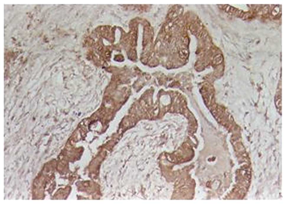
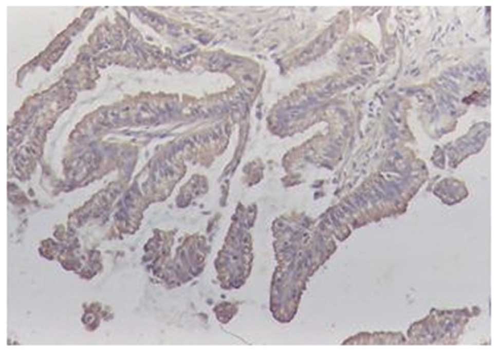
The number of cases staining positive for ODC and
GAD65 were 65 and 59, respectively, among the 108 patients with
gallbladder adenocarcinoma, 13 and 14, respectively, among the 46
peritumoral tissues, 3 and 4, respectively, among the 15 patients
with adenomatous polyps and 4 and 6, respectively, among the 35
patients with chronic cholecystitis (Figs. 1–4). The number of positive stainings of
ODC and GAD65 in the gallbladder adenocarcinoma tissues were
significantly higher than those in the peritumoral tissues
(P<0.01), adenomatous polyps (P<0.01 for ODC; P<0.05 for
GAD65) and chronic cholecystitis (P<0.01). Moreover, mild,
moderate or severe atypical proliferation was observed in all cases
expressing ODC or GAD65.
Correlation of ODC and GAD65 expression
with clinical and pathological features of gallbladder
adenocarcinoma
The expression levels of ODC and GAD65 in cases with
adenoma canceration or well-differentiated carcinoma or without
lymph node metastasis or surrounding invasion were significantly
lower than those in cases with poorly differentiated carcinoma,
lymph node metastasis or surrounding invasion. No significant
correlation was observed between the expression levels of ODC and
GAD65 and the clinicopathological features of patients with
gallbladder adenocarcinoma, including gender, age, tumor size and
gallstones (Table I).
 | Table ICorrelation between gallbladder cancer
and expression of ODC and GAD65. |
Table I
Correlation between gallbladder cancer
and expression of ODC and GAD65.
| | ODC | GAD65 |
|---|
| |
|
|
|---|
| Clinicopathological
features | Total | Positive (%) | P-value | Positive (%) | P-value |
|---|
| Gender |
| Male | 31 | 15 (48.4) | >0.05 | 14 (45.2) | >0.05 |
| Female | 77 | 50 (64.9) | | 45 (58.4) | |
| Age |
| ≤45 | 24 | 15 (62.5) | >0.05 | 12 (50.0) | >0.05 |
| >45 | 84 | 50 (59.5) | | 47 (56.0) | |
| Pathological
classification |
| Adenoma
canceration | 9 | 3 (33.3) | <0.05 | 2 (22.2) | <0.01 |
|
Well-differentiated | 29 | 13 (44.8) | | 11 (37.9) | |
| Moderately
differentiated | 29 | 18 (62.1) | | 16 (55.2) | |
| Poorly
differentiated | 30 | 25 (83.3) | | 24 (80.0) | |
| Mucous
adenocarcinoma | 11 | 6 (54.5) | | 6 (54.5) | |
| Tumor diameter |
| <2 cm | 31 | 15 (48.4) | >0.05 | 13 (41.9) | >0.05 |
| ≥2 cm | 77 | 50 (64.9) | | 46 (59.7) | |
| Lymph node
metastasis |
| No | 49 | 21 (42.9) | <0.01 | 19 (38.8) | <0.01 |
| Yes | 59 | 44 (74.6) | | 40 (67.8) | |
| Surrounding
invasion |
| No | 49 | 22 (44.9) | <0.01 | 20 (40.8) | <0.01 |
| Yes | 59 | 43 (72.9) | | 39 (66.1) | |
| Gallstones |
| No | 50 | 26 (52.0) | >0.05 | 23 (46.0) | >0.05 |
| Yes | 58 | 39 (67.2) | | 36 (62.1) | |
Correlation between disease-specific
overall survival and expression of ODC and GAD65
Following surgical resection, only 67 of the 108
patients were successfully followed up via telephone or mail
surveys, among which 20 cases had survived ≥1 year and 47 cases had
succumbed to the disease <1 year postoperatively, with a mean
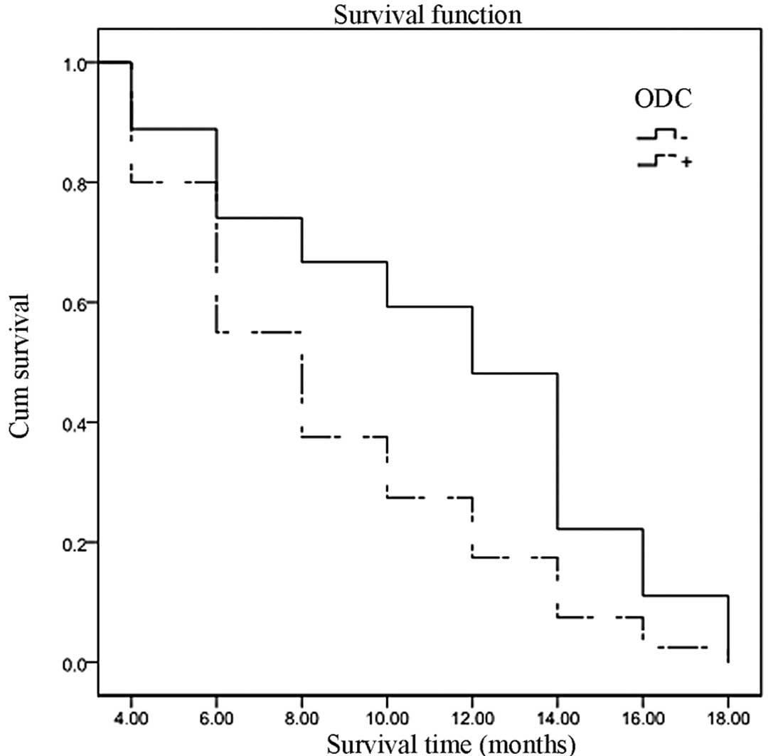
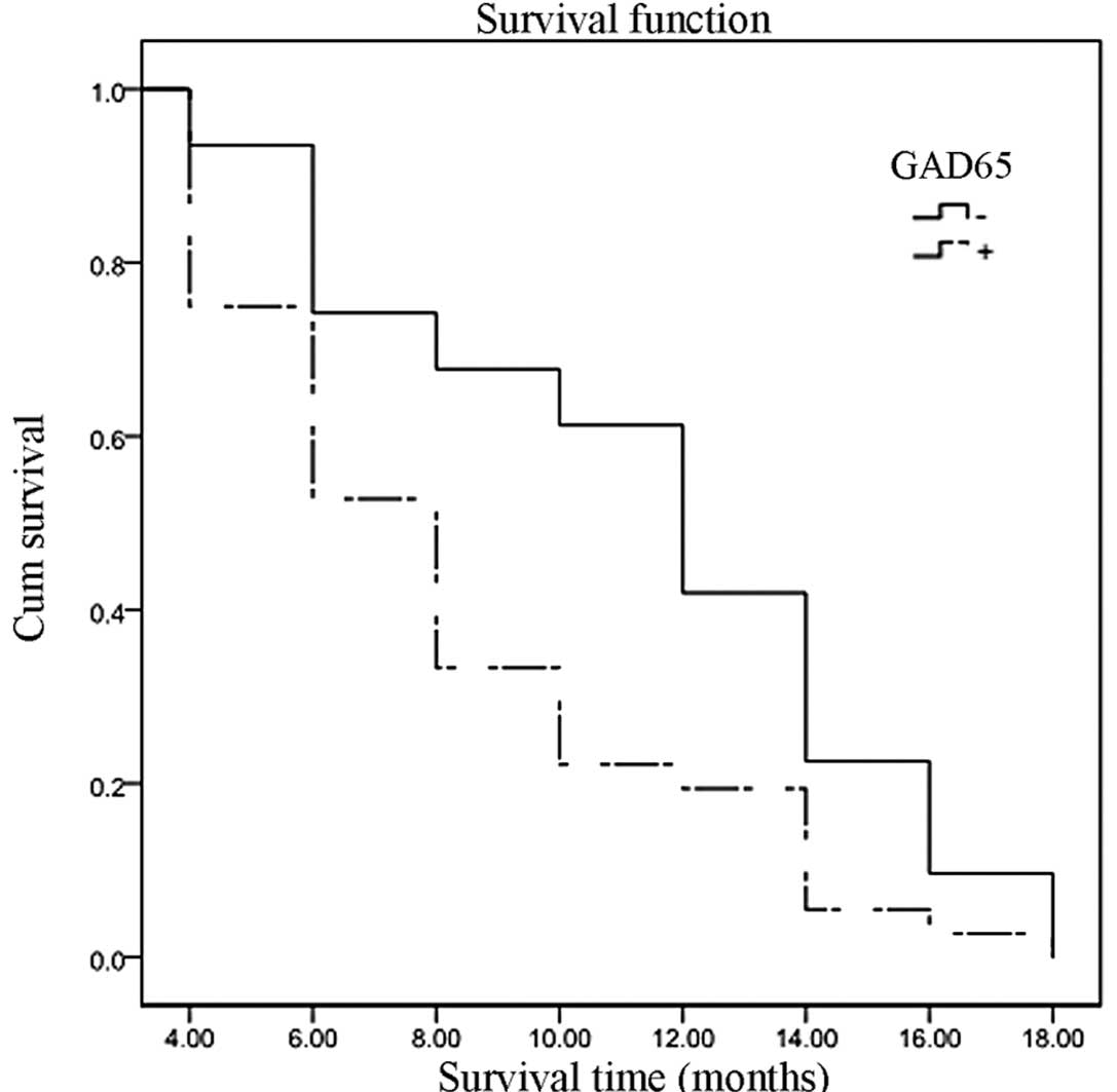
survival time of 9.7±4.4 months. Of the 67 surviving patients, 40
had positive immunohistochemical staining of ODC and 36 had
positive staining of GAD65. The relevance of the patients’ survival
to the positive expression of ODC and GAD65 was examined by
univariate Kaplan-Meier survival analysis. Overall survival was
inversely associated with positive or increased expression of ODC
(Fig. 5; P=0.009) and GAD65
(Fig. 6; P=0.006). The relevance
of overall survival to other clinicopathological characteristics
was also assessed by univariate analysis, which revealed that the
overall survival was associated with tumor pathological type
(P<0.01), tumor diameter (P<0.01), lymph node metastasis
(P<0.01) and surrounding tissue invasion (P<0.01). All
factors that showed significant association in the univariate
Kaplan-Meier analysis were subsequently subjected to multivariate
Cox regression survival analysis, which indicated that tumor
maximum diameter ≥2 cm, lymph node metastasis and surrounding
tissue invasion followed by ODC or GAD65 positive expression were
the most significant predictors of short overall survival (Table II).
| Table IIMultivariate Cox regression analysis
of overall survival in 67 patients with surgical resection of
gallbladder carcinoma. |
Table II
Multivariate Cox regression analysis
of overall survival in 67 patients with surgical resection of
gallbladder carcinoma.
| | | | | | 95% CI for
Exp(B) |
|---|
| | | | | |
|
|---|
| Factor | Category | B | SE(B) | Exp(B) | P | Inferior | Superior |
|---|
| Pathology type | Adenoma
canceration/well-/moderately/poorly differentiated/mucous
carcinoma | 0.208 | 0.123 | 1.231 | 0.091 | 0.967 | 1.567 |
| Tumor diameter | <2.0 cm/≥2.0
cm | 0.617 | 0.274 | 1.853 | 0.025 | 1.082 | 3.173 |
| Lymph node
metastasis | No/Yes | 0.561 | 0.275 | 1.752 | 0.041 | 1.022 | 3.002 |
| Surrounding tissue
invasion | No/Yes | 0.634 | 0.266 | 1.886 | 0.017 | 1.120 | 3.175 |
| ODC | −/+ | 0.568 | 0.285 | 1.765 | 0.046 | 1.011 | 3.084 |
| GAD65 | −/+ | 0.585 | 0.269 | 1.795 | 0.030 | 1.059 | 3.042 |
Discussion
ODC, a key enzyme in polyamine metabolism, is
observed in numerous types of cells from humans and animals. It is
a dimer consisting of two 50-kDa subunits. The ODC-related
sequences, including ODC1 and ODC2, are present on human
chromosomes 2p25 and 7q31 respectively. ODC1 is an important
functional gene which exists universally in organisms, whereas ODC2
is a pseudogene which exists only in the human body and is without
a clear function. The degradation of ODC depends mainly on ODC
antizyme (OAZ) which functions not by suppressing the synthesis of
ODC but by formulation of an ODC-antizyme complex. The latter is
generated to promote the degradation of ODC by the 26S proteasome
and polyamine synthesis is subsequently decreased. Polyamines have
been shown to be closely associated with cell growth,
proliferation, division, differentiation and malignant
transformation. As the first rate-limiting enzyme in polyamine
synthesis, ODC has a significant effect on polyamine levels. ODC is
overexpressed in cancerous tissues from a number of malignancies,
including gastric cancer, pancreatic cancer and colorectal cancer
(5,6,14),
and the inhibition of ODC may be a therapeutic strategy for
malignancies, including breast cancer (15). It has been reported that the
overexpression of ODC may lead to increased polyamine levels and
thus induce the overproliferation and malignant transformation of
cells (4,16–19).
It contributes to the development, progression, metastasis and
surrounding invasion of malignant tumors, and serves as a useful
biological marker for certain precancerous lesions of digestive
malignancies (4–8,16,17,20).
A previous study demonstrated that Helicobacter pylori
upregulates the expression of ODC, resulting in gastric precursor
lesions and cancer (21).
Carcinomas with highly expressed ODC are more progressive and
poorly differentiated, with increased metastasis, surrounding
invasion and poor prognosis. Hence, ODC may be a novel target for
use in the development of new anticancer drugs and as an indicator
for the efficacy of treatments (4–8,16,17,20).
Tissue-specific markers are useful for the
identification of tumor type in advanced cancers of unknown origin.
Jaraj et al investigated the expression of GAD67 (GAD1) in
the prostate and found that GAD67 expression was significantly
higher in malignant and benign prostatic tissue than in
non-prostatic control tissues. It was suggested that GAD67 may
serve as a highly prostate-specific tissue biomarker (22). However, whether GAD 65 (GAD2) plays
a similar role in malignancies of the gallbladder remains unknown.
In our study, we identified that GAD65 expression levels were
significantly higher in gallbladder malignancy than in benign
tissue.
Previous studies have shown that GABA stimulates the
growth of a variety of types of cancer (23,24).
The key enzyme for GABA synthesis is GAD which appears in two
isoforms, GAD65 and GAD67, named according to their molecular
weights (25). According to a
study by Moon et al, the proliferation of HT-29 colon cancer
cells was significantly inhibited when GAD67 expression was
repressed (26). GAD65 is
overexpressed in the cancerous tissues of numerous malignancies,
including colon cancer, breast cancer and gastric cancer, and is
closely associated with the clinicopathology of the diseases.
Increased levels of GAD65 expression represent a more malignant,
progressive and invasive nature of the lesion (11–13,27).
The administration of a GABA receptor antagonist inhibits the
growth and development of numerous types of cells, including
malignant cells (9,10).
In the present study, the positive staining of ODC
and GAD65 observed in gallbladder adenocarcinoma was significantly
higher than in peritumoral tissues, adenomatous polyps and chronic
cholecystitis. Furthermore, each case with expression of ODC and
GAD65 was accompanied by mild, moderate or severe atypical
proliferation of the gallbladder mucosal epithelium. As revealed in
the present study, the expression levels of ODC and GAD65 in cases
with adenoma canceration or well-differentiated carcinoma or
without lymph node metastasis or surrounding invasion were
significantly lower than those in cases with poorly differentiated
carcinoma, lymph node metastasis or surrounding invasion. The
expression levels of ODC and GAD65 in cases surviving >one year
were significantly lower than those in cases succumbing to the
disease within one year. Cox regression analysis revealed that the
expression levels of ODC and GAD65 were ideal prognostic markers in
gallbladder carcinoma. These results are in accordance with
findings reported from previous studies. This suggests that ODC and
GAD65 are closely correlated with the development, progression,
metastasis and surrounding invasion of gallbladder adenocarcinoma
and may be important biological markers for the early screening of
gallbladder carcinoma in benign lesions and for the prognosis of
gallbladder malignancy.
Acknowledgements
The authors would like to thank Professor Zhulin
Yang for designing the study and examining the specimens.
References
|
1
|
Ito H, Matros E, Brooks DC, Osteen RT,
Zinner MJ, Swanson RS, Ashley SW and Whang EE: Treatment outcomes
associated with surgery for gallbladder cancer: a 20-year
experience. J Gastrointest Surg. 8:183–190. 2004.PubMed/NCBI
|
|
2
|
Randi G, Franceschi S and La Vecchia C:
Gallbladder cancer worldwide: geographical distribution and risk
factors. Int J Cancer. 118:1591–1602. 2006. View Article : Google Scholar : PubMed/NCBI
|
|
3
|
Shukla PJ and Barreto SG: Gallbladder
cancer: we need to do better! Ann Surg Oncol. 16:2084–2085.
2009.
|
|
4
|
Gerner EW and Meyskens FL Jr: Polyamines
and cancer: old molecules, new understanding. Nat Rev Cancer.
4:781–792. 2004. View
Article : Google Scholar : PubMed/NCBI
|
|
5
|
Miao XP, Li JS, Li HY, Zeng SP, Zhao Y and
Zeng JZ: Expression of ornithine decarboxylase in precancerous and
cancerous gastric lesions. World J Gastroenterol. 13:2867–2871.
2007.PubMed/NCBI
|
|
6
|
Subhi AL, Tang B, Balsara BR, et al: Loss
of methylthioadenosine phosphorylase and elevated ornithine
decarboxylase is common in pancreatic cancer. Clin Cancer Res.
10:7290–7296. 2004. View Article : Google Scholar : PubMed/NCBI
|
|
7
|
Love RR, Astrow SH, Cheeks AM and
Havighurst TC: Ornithine decarboxylase (ODC) as a prognostic factor
in operable breast cancer. Breast Cancer Res Treat. 79:329–334.
2003. View Article : Google Scholar : PubMed/NCBI
|
|
8
|
Young L, Salomon R, Au W, Allan C, Russell
P and Dong Q: Ornithine decarboxylase (ODC) expression pattern in
human prostate tissues and ODC transgenic mice. J Histochem
Cytochem. 54:223–229. 2006. View Article : Google Scholar : PubMed/NCBI
|
|
9
|
Matsuba T, Yano M, Abiru N, Takino H,
Akazawa S, Nagataki S and Yasukawa K: Expression of recombinant
human glutamic acid decarboxylase (GAD) in myeloma cells and
enzyme-linked immunosorbent assay (ELISA) for autoantibodies to
GAD. J Biochem. 121:20–24. 1997. View Article : Google Scholar : PubMed/NCBI
|
|
10
|
Pinal CS, Cortessis V and Tobin AJ:
Multiple elements regulate GAD65 transcription. Dev Neurosci.
19:465–475. 1997. View Article : Google Scholar : PubMed/NCBI
|
|
11
|
Matuszek M, Jesipowicz M and Kleinrok Z:
GABA content and GAD activity in gastric cancer. Med Sci Monit.
7:377–381. 2001.PubMed/NCBI
|
|
12
|
Opolski A, Mazurkiewicz M, Wietrzyk J,
Kleinrok Z and Radzikowski C: The role of GABA-ergic system in
human mammary gland pathology and in growth of transplantable
murine mammary cancer. J Exp Clin Cancer Res. 19:383–390.
2000.PubMed/NCBI
|
|
13
|
Kleinrok Z, Matuszek M, Jesipowicz J,
Matuszek B, Opolski A and Radzikowski C: GABA content and GAD
activity in colon tumors taken from patients with colon cancer or
from xenografted human colon cancer cells growing as s. c tumors in
athymic nu/nu mice. J Physiol Pharmacol. 49:303–310.
1998.PubMed/NCBI
|
|
14
|
Kumar KN, Raja SB, Vidhya N and Devaraj
SN: Ellagic acid modulates antioxidant status, ornithine
decarboxylase expression, and aberrant crypt foci progression in
1,2-dimethylhydrazine-instigated colon preneoplastic lesions in
rats. J Agric Food Chem. 60:3665–3672. 2012. View Article : Google Scholar
|
|
15
|
Arisan ED, Obakan P, Coker A and
Palavan-Unsal N: Inhibition of ornithine decarboxylase alters the
roscovitine-induced mitochondrial-mediated apoptosis in MCF-7
breast cancer cells. Mol Med Rep. 5:1323–1329. 2012.
|
|
16
|
Manni A, Washington S, Griffith JW, et al:
Influence of polyamines on in vitro and in vivo features of
aggressive and metastatic behavior by human breast cancer cells.
Clin Exp Metastasis. 19:95–105. 2002. View Article : Google Scholar : PubMed/NCBI
|
|
17
|
Kubo S, Tamori A, Tanaka H, et al:
Polyamine metabolism and recurrence after resection for
hepatocellular carcinoma. Hepatogastroenterology. 51:208–210.
2004.PubMed/NCBI
|
|
18
|
Smirnova OA, Isaguliants MG, Hyvonen MT,
et al: Chemically induced oxidative stress increases polyamine
levels by activating the transcription of ornithine decarboxylase
and spermidine/spermine-N(1)-acetyltransferase in human hepatoma
HUH7 cells. Biochimie. 94:1876–1883. 2012. View Article : Google Scholar
|
|
19
|
Barry EL, Mott LA, Sandler RS, Ahnen DJ
and Baron JA: Variants downstream of the ornithine decarboxylase
gene influence risk of colorectal adenoma and aspirin
chemoprevention. Cancer Prev Res (Philadelphia). 4:2072–2082. 2011.
View Article : Google Scholar : PubMed/NCBI
|
|
20
|
Wolf C, Brüss M, Hänisch B, Göthert M, von
Kügelgen I and Molderings GJ: Molecular basis for the
antiproliferative effect of agmatine in tumor cells of colonic,
hepatic, and neuronal origin. Mol Pharmacol. 71:276–283. 2007.
View Article : Google Scholar : PubMed/NCBI
|
|
21
|
Xu X, Liu Z, Fang M, et al: CagA induces
ornithine decarboxylase upregulation via Src/MEK/ERK/c-Myc pathway:
implication for progression of gastric diseases Helicobacter
pylori. Exp Biol Med (Maywood). 237:435–441. 2012. View Article : Google Scholar : PubMed/NCBI
|
|
22
|
Jaraj SJ, Augsten M, Häggarth L, Wester K,
Pontén F, Ostman A and Egevad L: GAD1 is a biomarker for benign and
malignant prostatic tissue. Scand J Urol Nephrol. 45:39–45. 2011.
View Article : Google Scholar : PubMed/NCBI
|
|
23
|
Takehara A, Hosokawa M, Eguchi H, Ohigashi
H, Ishikawa O, Nakamura Y and Nakagawa H: Gamma-aminobutyric acid
(GABA) stimulates pancreatic cancer growth through overexpressing
GABAA receptor pi subunit. Cancer Res. 67:9704–9712. 2007.
View Article : Google Scholar
|
|
24
|
Maemura K, Shiraishi N, Sakagami K, et al:
Proliferative effects of gamma-aminobutyric acid on the gastric
cancer cell line are associated with extracellular signal-regulated
kinase 1/2 activation. J Gastroenterol Hepatol. 24:688–696. 2009.
View Article : Google Scholar
|
|
25
|
Walls AB, Nilsen LH, Eyjolfsson EM, et al:
GAD65 is essential for synthesis of GABA destined for tonic
inhibition regulating epileptiform activity. J Neurochem.
115:1398–1408. 2010. View Article : Google Scholar : PubMed/NCBI
|
|
26
|
Moon MS, Cho EW, Byun HS, Jung IL and Kim
IG: GAD 67KD antisense in colon cancer cells inhibits cell growth
and sensitizes to butyrate and pH reduction and
H2O2 and gamma-radiation. Arch Biochem
Biophys. 430:229–236. 2004. View Article : Google Scholar : PubMed/NCBI
|
|
27
|
Mazurkiewicz M, Opolski A, Wietrzyk J,
Radzikowski C and Kleinrok Z: GABA level and GAD activity in human
and mouse normal and neoplastic mammary gland. J Exp Clin Cancer
Res. 18:247–253. 1999.PubMed/NCBI
|