Introduction
Prostate cancer (PCa) is one of the leading causes
of cancer-related mortality worldwide. Early-stage PCa is
androgen-dependent and may be treated effectively with androgen
ablation therapy, radiation and/or surgery. However, all the
patients eventually progress to an androgen-independent state,
referred to as castration-resistant PCa, resulting in metastasis or
death (1). At this stage, there are
no effective treatment options available for PCa (2). Therefore, there is an urgent requirement
for the development of novel treatment methods for PCa (3).
Ultrasound has been proven to be a useful
therapeutic and diagnostic method, which is safe, non-invasive and
cost-effective (4). Ultrasound has
been widely used in cancer therapy and has been shown to mediate
apoptosis through thermal, cavitation, sonoporation and
sonochemical effects; however, the underlying mechanisms remain
unclear (5). Furthermore, a number of
experiments indicated that the effects of ultrasound may be
enhanced by microbubbles (6,7).
Autophagy maintains homeostasis in normal cells and
is triggered as a protective response to stressful factors,
including nutrient deprivation, oxidative stress and infection. It
was previously demonstrated that disorders in autophagy may lead to
cancer; this may also be used as a type of anticancer therapy by
promoting autophagic cell death in tumors (8). However, since the role of autophagy in
cell survival and death remains unclear, further studies are
required to determine its function (9). The aim of the present study was to
determine whether ultrasound is able to modify autophagy in PCa
cells.
Materials and methods
Cell lines
The human DU-145 PCa cell line was obtained from the
Cell Bank of the Chinese Academy of Sciences (Shanghai, China) and
used in all the experiments. The cells were incubated at 37°C in 5%
CO2 and Dulbecco's modified Eagle's medium supplemented
with 10% fetal bovine serum (Gibco-BRL, Grand Island, NY, USA) was
used as the culture medium, which was replaced every second
day.
Ultrasound apparatus and
microbubbles
The experiment was performed using FS-450 ultrasonic
processing (Shanghai Institute of Ultrasound in Medicine, Shanghai,
China). In all the procedures, the probe frequency was fixed at 21
kHz, the intensity was 4.6 mW/cm2 and the duty cycle was
20% (i.e., 2 sec ‘on’ and 8 sec ‘off’ time), with a total exposure
time of 5 min. The SonoVue™ microbubble echo-contrast agent (Bracco
SpA, Milan, Italy) was reconstituted in 5 ml phosphate-buffered
saline, 200 µl of which was used in the experimental group
(10). The cells were divided into
two groups, namely the control group (CON; no treatment) and the
group administered ultrasound combined with microbubbles (US + MB).
Each test was repeated three times.
Measurement of cell proliferation by
the MTT assay
Following treatment, the cells in each group were
grown to 80% confluence in 96-well plates for 24 h and their
viability was assessed with the MTT assay (Wellscan MK3; Ani
Labsystems Ltd. OY, Vantaa, Finland). According to the
manufacturer's instructions, MTT reagent (50 µl) was incubated with
the cells for 4 h at 37°C. Subsequently, the MTT reagent was
removed and 150 µl dimethylsulfoxide was added to each well and
agitated for 15 min. The optical density was measured at a
wavelength of 492 nm using a microculture plate reader (BioTek,
Winooski, VT, USA). The result was calculated as follows: Cell
viability (%) = (AbsorbanceUS +
MB/AbsorbanceCON) × 100. Cell viability was
calculated based on the average percentage.
Transmission electron microscopy
Ultra-thin specimens of DU-145 cells from each group
were prepared according to the conventional method (11) and were observed and photographed using
transmission electron microscopy (Hitachi S-4800; Hitachi
High-Technologies Corporation, Tokyo, Japan) at a magnification of
×24,500. Autophagosomes were identified by the characteristic
structure of a double- or multi-lamellar smooth membrane completely
surrounding compressed mitochondria, or as membrane-bound
electron-dense material.
Reverse transcription-polymerase chain
reaction (RT-PCR) analysis
After having grown to ~85% confluence, the cells
were harvested for RNA isolation using TRIzol® reagent (Invitrogen
Life Technologies, Carlsbad, CA, USA), according to the
manufacturer's instructions. Following purification, RNA was
subjected to quantitative PCR analysis using an iQ5™ multicolor
detection system (Bio-Rad, Hercules, CA, USA); GAPDH expression
levels were used as control. The results of the relative expression
levels were calculated using the relative quantitative
2−ΔΔ cycle threshold method. The primer sequences are
presented in Table I.
 | Table I.Oligonucleotide sequences for reverse
transcription polymerase chain reaction. |
Table I.
Oligonucleotide sequences for reverse
transcription polymerase chain reaction.
| Gene | Primer sequence
(5′-3′) |
|---|
| BECN-1 |
|
|
Forward |
ATGGAGGGGTCTAAGGCGTC |
|
Reverse |
TGGGCTGTGGTAAGTAATGGA |
| GAPDH |
|
|
Forward |
AATGGATTTGGACGCATTGGT |
|
Reverse |
TTTGCACTGGTACGTGTTGAT |
Western blot analysis
After having grown to 80% confluence, the cells were
harvested using lysis buffer (Beyotime Institute of Biotechnology,
Shanghai, China) and protein samples were collected and quantified
by running on SDS-PAGE gel (Beyotime Institute of Biotechnology)
and transferring onto a nitrocellulose membrane (Sigma-Aldrich, St.
Louis, MO, USA). Protein samples were probed with the indicated
primary antibody (dilution, 1:500; catalog no., sc-11427; Santa
Cruz Biotechnology, Inc., Santa Cruz, CA, USA) overnight at 4°C and
then incubated with secondary antibody (anti-mouse IgG; dilution,
1:5,000; Beyotime Institute of Biotechnology) for 1 h at 25°C;
β-actin was used as control. Protein was visualized using an
enhanced chemiluminescence system and band intensity was quantified
using Image J software (National Institutes of Health, Bethesda,
MD, USA).
Statistical analysis
Independent Student's t-tests were performed
using SPSS 13.0 software (SPSS Inc., Chicago, IL, USA). P<0.05
was considered to indicate statistically significant differences
between the CON and US + MB groups. All the experiments were
performed at least 3 times. The results of the statistical analyses
are presented as means ± standard deviation.
Results
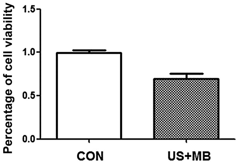
Measurement of cell proliferation
The MTT assay was used to evaluate cell viability.
The percentage of cell viability was 99.2±7.5% in the CON and
69.3±14.7% in the US + MB group (Fig.
1). Therefore, DU-145 cell growth was inhibited by ultrasound
combined with microbubbles.
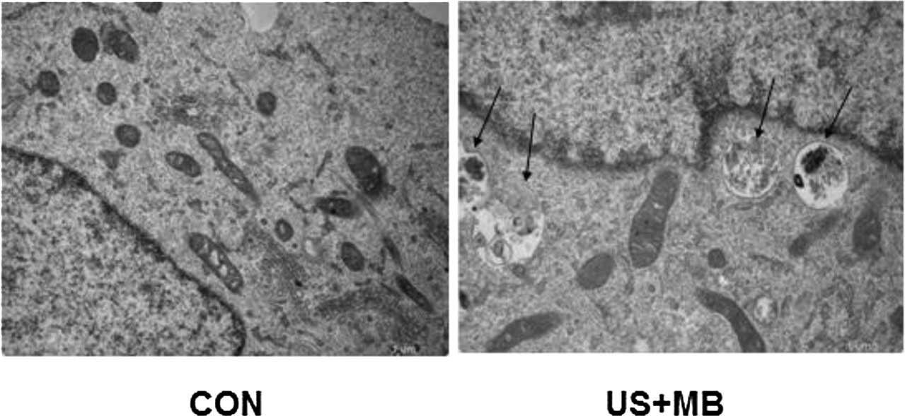
Analysis of autophagosomes by
transmission electron microscopy
Autophagosomes contain organelles or other
decomposed residues and have a characteristic double- or
multi-membrane structure. The number of autophagosomes under 10
different vision fields at a magnification of ×13,500 was
calculated in a blinded manner. Compared with CON, the number of
autophagosomes was significantly increased in the US + MB group
(0.2±0.42 vs. 2.6±2.12, respectively; P<0.05) (Fig. 2).
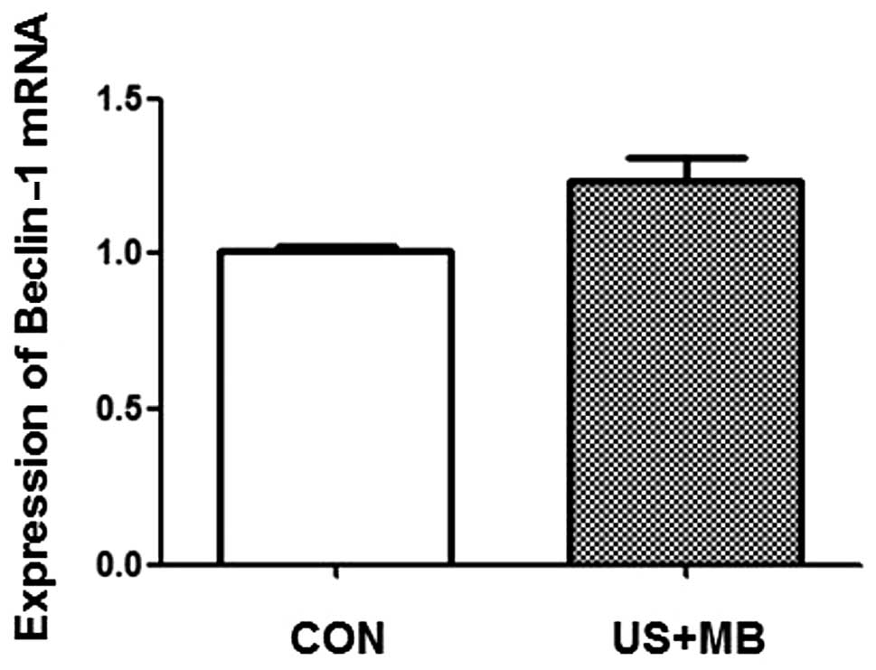
Beclin-1 induces autophagy in DU-145
cells following ultrasound irradiation
Compared with the CON group, the mRNA level of the
autophagy-related gene Beclin-1 was significantly increased in the
DU-145 cells of the US + MB group (1.01±0.03 vs. 1.23±0.08,
respectively; P<0.05; Fig. 3).
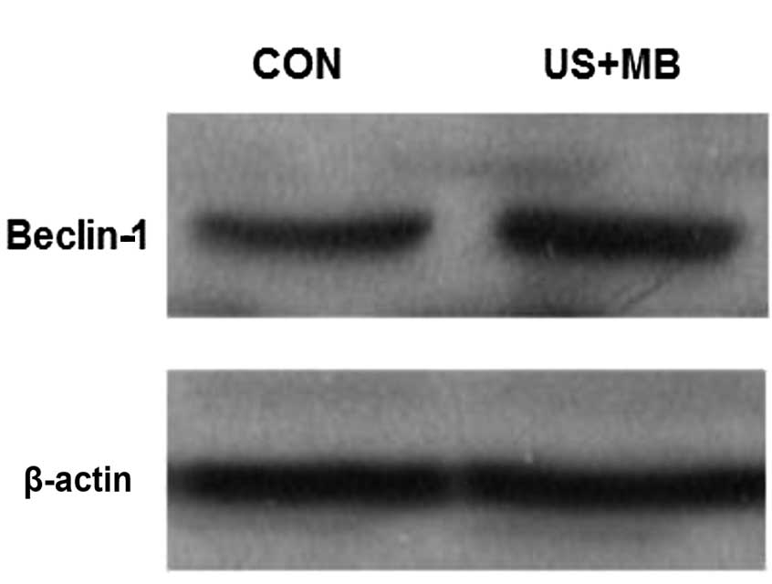
Western blot analysis of the Beclin-1
protein
The Beclin-1 protein expresion was assessed by
western blotting and was found to be higher in the DU-145 cells of
the US +MB group compared with the CON group (Fig. 4). Thus, ultrasound combined with
microbubbles increased the expression level of the Beclin-1
protein.
Discussion
Autophagy, a highly conserved mechanism present in
almost all species, constitutes an important part of the
degradation/recirculation system, which is widely encountered from
simple unicellular organisms and plants to mammalian cells
(12–14). Autophagy is the main channel for the
degradation of intact organelles and macromolecular protein and
autophagy disorders are closely associated with the occurrence,
development and outcome of cancer. Autophagosome formation is
associated with autophagy-related genes (12–14). In
the present study, it was demonstrated that there were more
autophagosomes in the DU-145 cells of the US + MB group compared
with the CON group (P<0.05). The autophagosomes of the DU-145
cells in the US + MB group exhibited typical characteristics, with
double- or multi-membrane structures containing organelles or other
decomposed residues (Fig. 2),
indicating that ultrasound combined with microbubbles may induce
autophagy in PCa cells.
Autophagy is regulated by a series of
autophagy-related genes and changes in autophagy may occur in
various human diseases. Liang et al (15) made the landmark discovery that cancer
is genetically linked to impaired autophagy and that Beclin-1 is a
phylogenetically conserved protein essential for autophagy.
Beclin-1 monoallelic deletion on chromosome locus 17q21 occurs in
40–75% of human PCa, ovarian and breast cancer cases (16). Beclin-1 in mammals is homologous with
autophagy-related gene 6 in yeast. Increased expression of Beclin-1
in mammalian cells may stimulate autophagy. Beclin-1-mediated
autophagy maintains cellular homeostasis by adjusting the level of
recycling and reuse of macromolecular materials. The Beclin-1 gene
is considered to be a type of tumor suppressor gene. As
demonstrated by RT-PCR and western blot analysis (Figs. 3 and 4)
in the present study, DU-145 cells treated with ultrasound with
microbubbles exhibited an increase in Beclin-1 expression, which
favored autophagy (Fig. 2)
An increasing number of studies on the biological
effect of ultrasound have indicated that the effects of ultrasound
reach far beyond its classic role in diagnosis (17–22).
Ultrasound therapy may be divided into different types according to
different frequencies. The mechanisms of conventional low-frequency
ultrasound in the treatment of tumors include the cytotoxic,
apoptosis-promoting and sensitization effects of chemotherapy. At
present, the majority of studies investigate the effect of
low-frequency ultrasound on the biological behavior of tumor cells
at the genetic level. Tabuchi et al (23) observed that 193 genes were
downregulated and 201 were upregulated in tumor cells following
irradiation by low-frequency ultrasound. These genes are associated
with cell growth, proliferation, apoptosis, movement, polymorphism
and death (24).
Low-frequency ultrasound has certain advantages in
disease treatment. However, studies (25–28) on
low-frequency ultrasound therapy are at an early stage and certain
areas require further investigation: i) The mechanisms underlying
the role of ultrasound in the treatment of certain diseases remain
unclear; ii) the optimal parameters are under debate; iii) it has
not been definitively established how to combine ultrasound with
other methods of treatment; iv) the development of low-frequency
ultrasound instruments cannot yet meet the personalized
requirements of disease treatment; v) ultrasound cannot be
concomitantly used for diagnosis and treatment; vi) the majority of
the studies are currently at the laboratory stage and ultrasound
has not yet been widely applied in clinical practice. Further
studies may lead to low-frequency ultrasound becoming a highly
efficient, convenient and widely used treatment method in the
clinical setting.
In conclusion, the present study demonstrated that
ultrasound combined with microbubbles induced autophagy through the
upregulation of Beclin-1 in DU-145 PCa cells. Additionally,
ultrasound combined with microbubbles significantly suppressed
DU-145 cell growth. Based on these results, ultrasound combined
with microbubbles shows considerable promise as a treatment for
androgen-independent PCa. However, the underlying mechanisms
require further investigation.
Acknowledgements
This study was supported by grants from the National
Natural Science Foundation of China (nos. 81271597 and 81401421)
and the Major Infrastructure Projects of Shanghai Science and
Technology (no. 10JC1412600).
References
|
1
|
Chaturvedi S and Garcia JA: Novel agents
in the management of castration resistant prostate cancer. J
Carcinog. 13:52014. View Article : Google Scholar : PubMed/NCBI
|
|
2
|
Shin SW, Kim SY and Park JW: Autophagy
inhibition enhances ursolic acid-induced apoptosis in PC3 cells.
Biochim Biophys Acta. 1823:451–457. 2012. View Article : Google Scholar : PubMed/NCBI
|
|
3
|
Jácome-Pita F, Sánchez-Salas R, Barret E,
Amaruch N, Gonzalez-Enguita C and Cathelineau X: Focal therapy in
prostate cancer: the current situation. Ecancermedicalscience.
8:4352014.PubMed/NCBI
|
|
4
|
Franco de Oliveira R, DA Pires Oliveira
and Soares CP: Effect of low-intensity pulsed ultrasound on l929
fibroblasts. Arch Med Sci. 7:224–229. 2011. View Article : Google Scholar : PubMed/NCBI
|
|
5
|
Feng Y, Tian Z and Wan M: Bioeffects of
low-intensity ultrasound in vitro: apoptosis, protein profile
alteration and potential molecular mechanism. J Ultrasound Med.
29:963–974. 2010.PubMed/NCBI
|
|
6
|
Wang Y, Bai WK, Shen E and Hu B:
Sonoporation by low-frequency and low-power ultrasound enhances
chemotherapeutic efficacy in prostate cancer cells in vitro. Oncol
Lett. 6:495–498. 2013.PubMed/NCBI
|
|
7
|
Yang SL, Tang KQ, Bai WK, Shen E, Zhao YW,
Lin YD, Nan SL and Bing H: Effects of low-frequency ultrasound
combined with microbubbles on benign prostate hyperplasia. Can Urol
Assoc J. 7:E681–E686. 2013. View
Article : Google Scholar : PubMed/NCBI
|
|
8
|
Law BY, Chan WK, Xu SW, Wang JR, Bai LP,
Liu L and Wong VK: Natural small-molecule enhancers of autophagy
induce autophagic cell death in apoptosis-defective cells. Sci Rep.
4:55102014. View Article : Google Scholar : PubMed/NCBI
|
|
9
|
Wei MF, Chen MW, Chen KC, Lou PJ, Lin SY,
Hung SC, Hsiao M, Yao CJ and Shieh MJ: Autophagy promotes
resistance to photodynamic therapy-induced apoptosis selectively in
colorectal cancer stem-like cells. Autophagy. 10:1179–1192. 2014.
View Article : Google Scholar : PubMed/NCBI
|
|
10
|
Bai WK, Yang SL, Shen E, et al: Treatment
of PC-3 cells with ultrasound combined with microbubbles induces
distinct alterations in the expression of Bcl-2 and Bax. Chin Sci
Bull. 58:3535–3540. 2013. View Article : Google Scholar
|
|
11
|
Fan X, Wang J, Hou J, et al: Berberine
alleviates ox-LDL induced inflammatory factors by up-regulation of
autophagy via AMPK/mTOR signaling pathway. J Transl Med. 13:922015.
View Article : Google Scholar : PubMed/NCBI
|
|
12
|
Yorimitsu T and Klionsky DJ: Autophagy:
Molecular machinery for self-eating. Cell Death Differ. 12 (Suppl
2):1542–1552. 2005. View Article : Google Scholar : PubMed/NCBI
|
|
13
|
Xie Z, Nair U and Klionsky DJ: Atg8
controls phagophore expansion during autophagosome formation. Mol
Biol Cell. 19:3290–3298. 2008. View Article : Google Scholar : PubMed/NCBI
|
|
14
|
Shintani T and Klionsky DJ: Autophagy in
health and disease: Adouble-edged sword. Science. 306:990–995.
2004. View Article : Google Scholar : PubMed/NCBI
|
|
15
|
Liang XH, Jackson S, Seaman M, Brown K,
Kempkes B, Hibshoosh H and Levine B: Induction of autophagy and
inhibition of tumorigenesis by Beclin-1. Nature. 402:672–676. 1999.
View Article : Google Scholar : PubMed/NCBI
|
|
16
|
Aita VM, Liang XH, Murty VV, Pincus DL, Yu
W, Cayanis E, Kalachikov S, Gilliam TC and Levine B: Cloning and
genomic organization of Beclin-1, a candidate tumor suppressor gene
on chromosome 17q21. Genomics. 59:59–65. 1999. View Article : Google Scholar : PubMed/NCBI
|
|
17
|
Tsivgoulis G, Eggers J, Ribo M, et al:
Safety and efficacy of ultrasound-enhanced thrombolysis: a
comprehensive review and meta-analysis of randomized and
nonrandomized studies. Stroke. 41:280–287. 2010. View Article : Google Scholar : PubMed/NCBI
|
|
18
|
Hensel K, Mienkina MP and Schmitz G:
Analysis of ultrasound fields in cell culture wells for in vitro
ultrasound therapy experiments. Ultrasound Med Biol. 37:2105–2115.
2011. View Article : Google Scholar : PubMed/NCBI
|
|
19
|
Rapoport NY, Nam KH, Gao Z and Kennedy A:
Application of ultrasound for targeted nanotherapy of malignant
tumors. Acoust Phys. 55:594–601. 2009. View Article : Google Scholar : PubMed/NCBI
|
|
20
|
Kawai N and Iino M: Molecular damage to
membrane proteins induced by ultrasound. Ultrasound Med Biol.
29:609–614. 2003. View Article : Google Scholar : PubMed/NCBI
|
|
21
|
Marentis TC, Kusler B, Yaralioglu GG, et
al: Microfluidic sonicator for real-time disruption of eukaryotic
cells and bacterial spores for DNA analysis. Ultrasound Med Biol.
31:1265–1277. 2005. View Article : Google Scholar : PubMed/NCBI
|
|
22
|
Nyborg WL, Leighton TG, Miller D, et al:
Nonthermal issues: cavitation - its nature, detection and
measurement. Ultrasound Med Biol. 6:11–16. 1998.
|
|
23
|
Tabuchi Y, Takasaki I, Zhao QL, et al:
Genetic networks responsive to low-intensity pulsed ultrasound in
human lymphoma U937 cells. Cancer Lett. 270:286–294. 2008.
View Article : Google Scholar : PubMed/NCBI
|
|
24
|
Xu WP, Shen E, Bai WK, Wang Y and Hu B:
Enhanced antitumor effects of low-frequency ultrasound and
microbubbles in combination with simvastatin by downregulating
caveolin-1 in prostatic DU145 cells. Oncol Lett. 7:2142–2148.
2014.PubMed/NCBI
|
|
25
|
Maruani A, Boucaud A, Perrodeau E, et al:
Low-frequency ultrasound sonophoresis to increase the efficiency of
topical steroids: a pilot randomized study of humans. Int J Pharm.
395:84–90. 2010. View Article : Google Scholar : PubMed/NCBI
|
|
26
|
Liu HL, Chen WS, Chen JS, et al:
Cavitation-enhanced ultrasound thermal therapy by combined low- and
high-frequency ultrasound exposure. Ultrasound Med Biol.
32:759–767. 2006. View Article : Google Scholar : PubMed/NCBI
|
|
27
|
Ren ST, Liao YR, Kang XN, et al: The
antitumor effect of a new docetaxel-loaded microbubble combined
with low-frequency ultrasound in vitro: Preparation and parameter
analysis. Pharm Res. 30:1574–1585. 2013. View Article : Google Scholar : PubMed/NCBI
|
|
28
|
Yang SL, Tang KQ, Bai WK, et al: Combined
low-frequency ultrasound and microbubble contrast agent for the
treatment of benign prostatic hyperplasia. J Endourol.
27:1020–1026. 2013. View Article : Google Scholar : PubMed/NCBI
|