Introduction
Tongue squamous cell carcinoma (TSCC) is the most
common malignant tumor in the oral and maxillofacial region
(1). Compared with other oral
malignant tumors, TSCC exhibits features of an increased degree of
malignancy, more rapid proliferation and more marked invasion.
Owing to the tongue's rich blood supply and frequent movement, TSCC
often exhibits early cervical lymph node metastasis and increased
transfer rates (2). Currently, the
most effective treatment of TSCC is surgical resection. However,
excessive surgical excision often affects oral function and
postoperative quality of life in patients with TSCC (3). Following adjuvant treatment
radiotherapy, chemotherapy or immunotherapy, the survival rate of
patients with TSCC has markedly improved, but it remains
unsatisfactory. Among the treatment options, chemotherapy has an
important role in the comprehensive treatment of TSCC. As it
exhibits side effects, including liver and kidney toxicity,
inhibition of the hematopoietic system and damage to the body's
immune system, its clinical application is limited to a certain
extent (4). Therefore, the search for
novel drugs with low toxicity and high efficacy is vital in the
comprehensive treatment of TSCC. Traditional Chinese medicine
primarily consists of the use of natural plants and minerals, which
exhibit characteristics of low side effects and being rich in
resources (5). Therefore, traditional
Chinese medicine is a focus of research for antitumor drugs.
Psoralea corylifolia is an annual herb widely
distributed in China. The fruit of P. corylifolia has been
used in traditional Chinese medicine, exhibiting diuretic,
analeptic and laxative effects. Pharmacology studies have
demonstrated that P. corylifolia exhibits a number of
biological activities including enhancement of immunity,
antibacterial effects and treatment of vitiligo. Isobavachalcone
(2′,4′,4-trihydroxy-3′-[3′-methylbut-3′-ethyl] chalcone or IBC) is
a natural chalcone compound isolated from P. corylifolia.
IBC exhibits anti-inflammatory, antifungal, antioxidant and
antimicrobial pharmacological activities. Previously, it has been
reported that IBC is able to promote tumor cell apoptosis without
exhibiting toxic effects towards normal cells, including human
neuroblastoma, ovarian cancer and gastric cancer cells (6–9). However,
only a limited number of studies have been performed to investigate
the effects of IBC on human TSCC.
Furthermore, the antimicrobial activities of IBC may
prevent infection during chemotherapy for patients with cancer
(10). These results suggested that
IBC as a newly discovered anticancer drug exhibited characteristics
of low toxicity and high efficacy. IBC has the potential for
clinical application to decrease the side effects of
chemotherapy.
Therefore, the aim of the present study was to
investigate the effect of various concentrations of IBC on human
TSCC Tca8113 cells, including inhibition of cell proliferation,
induction of apoptosis, and the ability to inhibit migration and
invasion. In addition, changes in the expression of various
proteins including the apoptosis-related protein kinase B (Akt),
extracellular-signal-regulated kinase (ERK), Bcl-2-associated X
protein (Bax), B-cell lymphoma 2 (Bcl-2) and caspase-3, and the
invasion-related matrix metalloproteinase (MMP)-2 and MMP-9 were
determined. These results provide a theoretical basis for the
application of IBC in the clinical treatment of TSCC.
Materials and methods
Cell culture and treatment
Human TSCC Tca8113 cells were provided by Shanghai
Institute of Biochemistry and Cell Biology (Shanghai, China).
Tca8113 cells were cultured in RPMI 1640 medium (Gibco; Thermo
Fisher Scientific, Inc., Waltham, MA, USA) supplemented with 10%
fetal bovine serum (Gibco; Thermo Fisher Scientific, Inc.) and 100
U/ml penicllin/streptomycin (Nanjing KeyGen Biotech Co. Ltd.,
Nanjing, China), and maintained at 37°C in a humidified atmosphere
containing 5% CO2. The medium was changed three
times/week. IBC was purchased from Shanghai Tauto Biotech Co., Ltd.
(Shanghai, China). IBC was dissolved in dimethylsulfoxide (DMSO) as
a 100-mM stock solution to give a final concentration of 0.1% in
the culture medium.
MTT analysis
Tca8113 cells (5,000 cells/well) were seeded in
96-well cell culture plates and incubated overnight. Various
concentrations (10, 20, 40 and 80 µM) of IBC were added, and cells
were incubated for a further 24 or 48 h. The control groups
included a blank control, treated with RPMI 1640, and a negative
control, treated with DMSO. The cells were incubated with 20 µl 5
mg/ml MTT solution (Sigma-Aldrich; Merck KGaA, Darmstadt, Germany)
for a further 4 h at 37°C in a humidified atmosphere containing 5%
CO2. Following lysis of the cells and dissolution of the
formazan product in 150 µl DMSO, the optical density (OD) was
measured at 490 nm using a spectrophotometer (Bio-Rad Laboratories,
Inc., Hercules, CA, USA). The proliferation inhibition rate was
calculated using the following formula: Proliferation inhibition
rate=1-(experimental sample OD-blank control sample OD)/(negative
control sample OD-blank control sample OD) ×100%. All experiments
were performed in triplicate and mean values were plotted to create
cell proliferation inhibition curves.
Morphological staining
Tca8113 cells were cultured in 6-well culture plates
at a density of 5×10⁵ cells/well. Tca8113 cells were
treated with 40 µM of IBC and incubated for 48 h; control cells
received no treatment. Following treatment, the cells were
collected and centrifuged at 300 × g for 5 min at room temperature.
The cell smears were stained with Wright-Giemsa solution
(Sigma-Aldrich; Merck KGaA) for 15 min, and washed with running
water. Alterations in cellular morphology were observed using light
microscopy.
Annexin V and propidium iodide (PI)
staining
Tca8113 cells were cultured in 6-well culture plates
at a density of 5×10⁵ cells/well. Tca8113 cells were
incubated for 48 h with various concentrations (0, 20 and 40 µM) of
IBC. The cells were collected, and centrifuged at 200 × g for 5 min
at room temperature. The cells were washed with PBS and suspended
in binding buffer (Nanjing KeyGen Biotech Co. Ltd.). Cells were
incubated with 5 µl annexin V-fluorescein isothiocyanate and 5 µl
PI (both Nanjing KeyGen Biotech Co. Ltd., Nanjing, China) for 15
min in the dark at room temperature. The percentage of apoptotic
cells was evaluated using the BD FACSVerse flow cytometer (BD
Biosciences, San Jose, CA, USA).
Wound healing analysis
Tca8113 cells were cultured in 6-well culture plates
at a density of 5×10⁵ cells/well, and incubated in RPMI
1640 medium supplemented with 10% fetal bovine serum and 100 U/ml
penicillin/streptomycin. A central denuded wound was made in a
confluent cell monolayer using a 200 µl pipette tip. Subsequently,
cells were cultured in serum-free RPMI 1640 containing 0, 20 or 40
µM IBC. Wound closure was observed, and images were captured under
an inverted microscope 0 and 24 h after treatment. Relative cell
migration was calculated using the formula: Relative cell
migration=(mean distance of original wound-mean distance of 24 h
wound)/mean distance of original wound ×100%.
Transwell invasion analysis
Transwell invasion analysis was determined using a
Transwell permeable support system (BD Biosciences, San Jose, CA,
USA) containing 24-well Transwell chambers (8 µm pore size
polycarbonate membranes). The upper chamber was coated with 30 µl
Matrigel (BD Biosciences, San Jose, CA, USA). Tca8113 cells and
various concentrations (0, 20 and 40 µM) of IBC were seeded into
the upper chambers at densities of 5×10⁴ cells/well in
serum-free RPMI 1640. RPMI 1640 containing 600 µl 10% fetal bovine
serum was added to the lower chamber as a chemoattractant.
Following incubation at 37°C for 24 h, the cells on the lower
surface of the polycarbonate membranes were fixed with 4%
formaldehyde and stained with 0.5% crystal violet. The invading
cells were counted visually in five random fields at ×400
magnification using light microscopy.
Western blot analysis
Tca8113 cells were cultured in 6-well culture plates
at a density of 5×10⁵ cells/well. Following treatment
with IBC (0, 20 and 40 µM) for 48 h, Tca8113 cells were washed with
ice-cold PBS and protein was extracted with lysis buffer containing
50 mmol/l Tris-HCl, pH 7.4, 150 mmol/l NaCl, 1% Triton X-100, 1
mmol/l EDTA, 0.1% SDS and 2 mmol/l PMSF. The protein concentrations
in the supernatant were estimated using a BCA Protein Assay kit
(Beyotime Institute of Biotechnology, Haimen, Jiangsu, China) using
bovine serum albumin as a standard. Samples of 20 µl protein were
separated by SDS-PAGE (12% gel) and then transferred onto
polyvinylidene fluoride membranes. Following incubation for 1 h in
blocking buffer (5% non-fat dry milk in PBS containing Tween-20),
the membranes were incubated with the primary antibodies overnight
at 4°C. Primary antibodies were as follows: Akt (dilution, 1:200;
cat. no., sc-8312); p-Akt (dilution, 1:200; cat. no., sc-135650);
ERK (dilution, 1:200; cat. no., sc-94); p-ERK (dilution, 1:200;
cat. no., sc-16982); Bax (dilution, 1:300; cat. no., sc-493); Bcl-2
(dilution, 1:300; cat. no., sc-492); caspase-3 (dilution, 1:200;
cat. no., sc-7184); MMP-2 (dilution, 1:200; cat. no., sc-10736);
MMP-9 (dilution, 1:200; cat. no., sc-10737); β-actin (dilution,
1:1,000; cat. no., sc-1616); all antibodies were from Santa Cruz
Biotechnology (Dallas, TX, USA). The membranes were then treated
with horseradish peroxidase-conjugated secondary antibodies,
including chicken anti-mouse (cat. no., sc-2954) and chicken
anti-rabbit (cat. no., sc-2955; both dilution, 1:800; Santa Cruz
Biotechnology. Inc.) and chemiluminescence substrate (ECL plus,
Beyotime Institute of Biotechnology, Haimen, Jiangsu, China). The
digital images were captured and analyzed densitometrically using
ImageJ 2X software (National Institutes of Health, Bethesda, MD,
USA).
Statistical analysis
Statistical analysis was carried out using SPSS for
Windows (version 16.0; SPSS, Inc., Chicago, IL, USA). Results were
expressed as the mean ± standard deviation. Multi-group comparisons
of the means were carried out by one-way analysis of variance test,
and the post hoc analysis was performed using the
Student-Newman-Keuls test. P<0.05 was considered to indicate a
statistically significant difference. Three independent experiments
were performed for all assays.
Results
IBC inhibits the proliferation of
Tca8113 cells
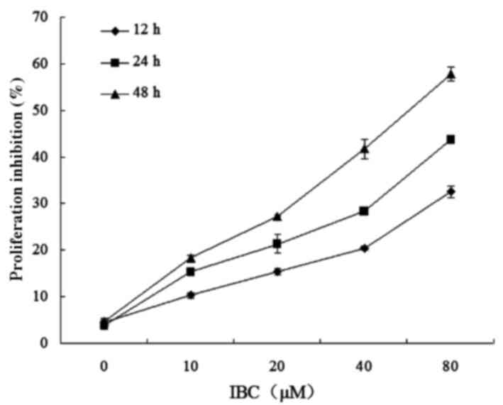
Tca8113 cells were treated with between 0 and 80 µM
IBC for 12, 24 and 48 h. An MTT assay demonstrated that the
half-maximal inhibitory concentrations of IBC at 12, 24 and 48 h
were 285.13±8.97, 132.40±7.76 and 58.56±5.93 µΜ, respectively
(P<0.05; Fig. 1).
IBC induces the apoptotic morphology
of Tca8113 cells
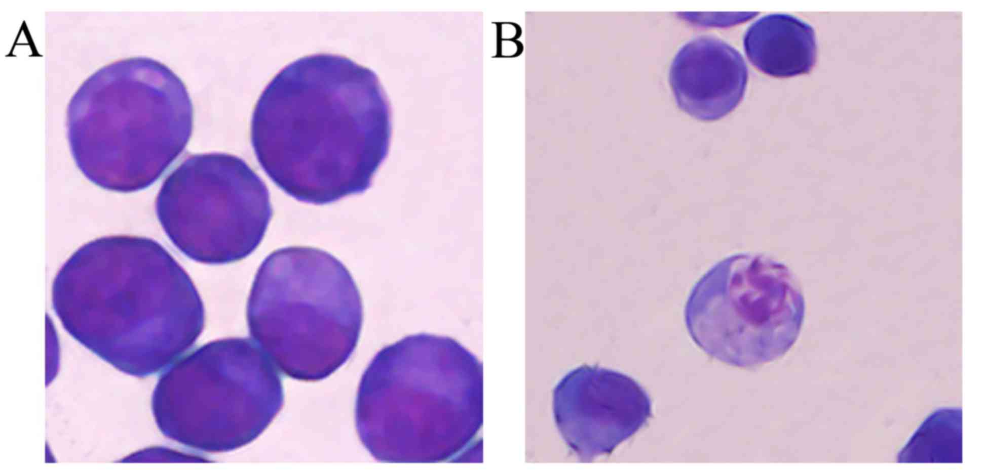
Tca8113 cells were treated with 40 µM IBC for 48 h
to observe the alterations in cell morphology. IBC treatment
resulted in cell shrinkage and nuclear DNA fragmentation in Tca8113
cells (Fig. 2), suggesting that IBC
induced apoptosis in Tca8113 cells.
IBC induces the apoptosis of Tca8113
cells
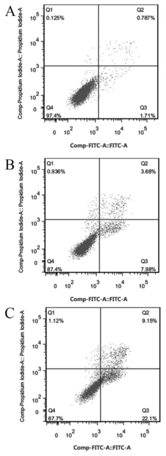
Tca8113 cells were cultured in 20 and 40 µM IBC for
48 h. The pro-apoptotic rates were 8.21±2.32 and 22.45±1.18%,
respectively, which were significantly increased compared with the
untreated control (1.69±0.65%) (P<0.05; Fig. 3). These results suggested that IBC
treatment induced apoptosis in Tca8113 cells. The cell apoptosis
rate was positively associated with drug concentration.
IBC inhibits the migration and
invasion of Tca8113 cells
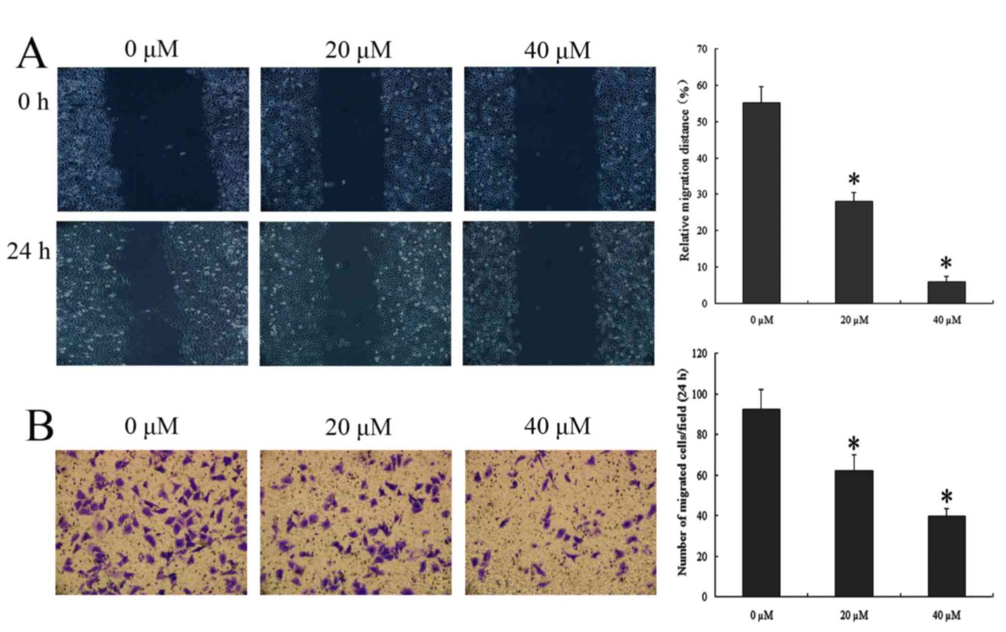
To determine whether IBC treatment has an effect on
wound repair, wound healing was compared between Tca8113 cells with
and without IBC. To eliminate the effect of cell apoptosis on the
results, an IBC treatment time of 24 h was selected rather than 48
h. The results of the wound healing and Transwell invasion analysis
demonstrated the effect of IBC on the migration and invasion of
Tca8113 cells respectively. The migration and invasion ability of
20 and 40 µΜ IBC-treated Tca8113 cells were significantly decreased
compared with the untreated control (P<0.05; Fig. 4). The results indicated that IBC
caused marked decreases in cell migration and invasion, indicating
that IBC serves a vital role in cell migration.
Effect of IBC on the expression of
Tca8113 cell apoptosis-related proteins
IBC treatment at 20 and 40 µΜ IBC resulted primarily
in significant downregulation of the anti-apoptotic protein Bcl-2,
and, in contrast, significant upregulation of the apoptotic protein
Bax (P<0.05; Fig. 5). Members of
the caspase protein family are known to induce apoptosis in cancer
cells. As presented in Fig. 5, IBC
treatment with 20 and 40 µΜ IBC increased caspase-3 activity in
Tca8113 cells. These results suggested that IBC treatment led to a
concentration-dependent decrease in Bcl-2 expression, and a
concentration-dependent increase in Bax and caspase-3 expression in
Tca8113 cells.
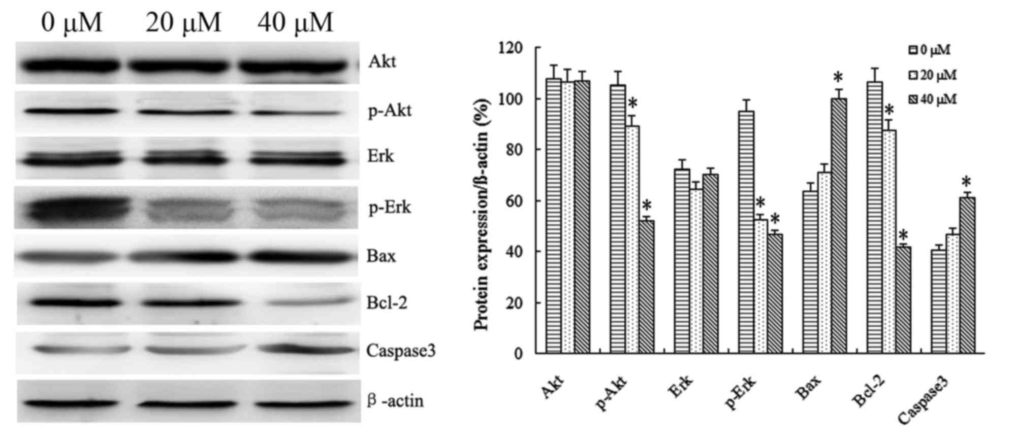 | Figure 5.IBC regulates the protein expression
of Akt, p-Akt, ERK, p-ERK, Bax, Bcl-2 and caspase-3 in Tca8113
cells in a concentration-dependent manner. At 48 h after incubation
with 0, 20 or 40 µM IBC, the expression levels of all proteins were
analyzed by western blotting. β-actin served as a loading control.
Results are expressed as the mean ± standard deviation of three
independent experiments. *P<0.05 vs. control. IBC,
isobavachalcone; Akt, protein kinase B; p, phospho; ERK,
extracellular-signal-regulated kinase; Bax, Bcl-2-associated X
protein; Bcl-2, B-cell lymphoma 2. |
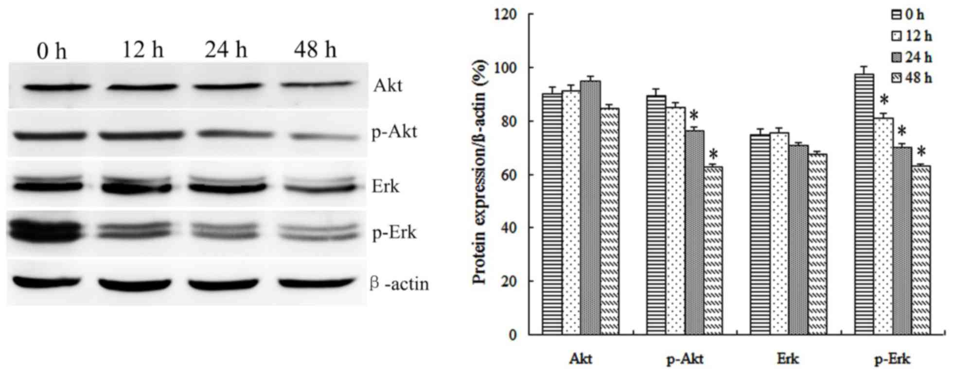
A number of protein kinases (including Akt or ERK)
are involved in cell proliferation and transformation. Therefore,
the effect of IBC on activities of Akt and ERK in Tca8113 cells was
investigated. It was identified that 20 and 40 µM IBC treatment for
48 h significantly decreased the protein levels of phospho (p)-Akt
and p-ERK compared with the untreated control in Tca8113 cells. In
order to observe the effect of IBC on protein expression in Tca8113
cells following various incubation times, Tca8113 cells were
cultured in 40 µM IBC for 12, 24 and 48 h. It was identified that
IBC treatment for 48 h significantly decreased the protein
expression levels of p-Akt and p-ERK compared with the untreated
control in Tca8113 cells (P<0.05; Fig.
6). Total Akt and ERK levels were not affected by the
concentration or duration of IBC treatment. These results suggested
the ability of IBC to inactivate Akt and ERK in Tca8113 cells.
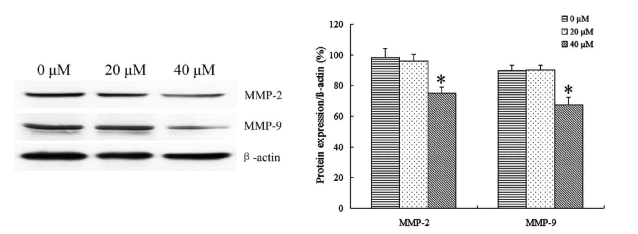
Effect of IBC on the expression of
Tca8113 cell invasion-related proteins
In order to identify the effect of IBC on the
expression of Tca8113 cell invasion-related proteins, Tca8113 cells
were treated with 20 and 40 µM IBC for 48 h to observe the protein
expression of MMP-2 and MMP-9 by Western blot analysis. It was
identified that IBC significantly decreased MMP-2 and MMP-9 protein
expression in Tca8113 cells compared with the control (P<0.05;
Fig. 7). These results indicated that
IBC may regulate the protein expression of MMP-2 and MMP-9 at the
transcriptional level in TSCC cells.
Discussion
Malignant tumors are one of the most serious threats
to human health. In comparison with normal cells, malignant tumor
cells are able to divide and proliferate endlessly. Malignant tumor
cells are able to damage the body's histological barrier by various
means, invade normal tissue, metastasize, and spread via the
bloodstream and lymphatic system (11). Current clinical treatments for TSCC
are primarily surgery, radiotherapy, chemotherapy and
immunotherapy. Surgery, which is the preferred method of TSCC
treatment, is limited by tumor invasion, metastasis and a number of
other factors. Chemotherapy, owing to its poor specificity, would
also inevitably kill normal tissue cells and destroy the human
immune system. The clinical application of chemotherapy drugs has
become a ‘double-edged sword’. Seeking novel ways to treat TSCC and
the development of effective antitumor drugs has become important
in modern medicine, particularly in the field of clinical
medicine.
P. corylifolia is a legume member of the
angiosperms. It has been demonstrated previously that a variety of
active ingredients extracted from P. corylifolia are able to
serve a antitumor role through various mechanisms (12). IBC, an active ingredient from P.
corylifolia, was initially extracted by Bhalla et al
(13) in 1968. It has been
demonstrated that IBC exhibited antibacterial, antifungal,
anti-inflammatory, antimicrobial and antitumor activities (14). In the development of a novel
anticancer drug with low toxicity and cost, IBC is a promising
candidate for clinical application. Previous in vitro
experiments confirmed that, without damaging normal tissue cells,
IBC inhibited the proliferation and induced apoptosis in a variety
of tumor cells including OVCAR-8, PC3, A549 and MCF-7 cells
(6–9).
However, little is known about the association between IBC and
human TSCC Tca8113 cells.
In the present study, the effect of IBC on cell
proliferation and apoptosis of TSCC Tca8113 cells in vitro
was determined using an MTT assay, Wright-Giemsa staining and flow
cytometric analysis. Using an MTT assay, IBC demonstrated
significant inhibition of cell proliferation in a marked
concentration- and time-dependent manner. Tca8113 cells treated
with 40 µM IBC for 48 h demonstrated typical apoptotic morphology
of nuclear fragmentation and apoptotic bodies. Furthermore, flow
cytometric analysis identified pro-apoptotic rates of 8.21±2.32 and
22.45±1.18% for Tca8113 cells treated with 20 and 40 µΜ IBC for 48
h, respectively, which were significantly increased compared with
the untreated control. These results indicated that IBC inhibited
the proliferation of Tca8113 cells and induced apoptosis in a
concentration- and time-dependent manner.
The primary characteristics of malignant tumors is
that tumor cells exhibit unrestricted proliferation and are able to
circumvent apoptosis. Members of the Bcl-2 family encode
anti-apoptotic proteins and pro-apoptotic proteins (15). Bcl-2 and Bax are, respectively,
important anti-apoptotic and pro-apoptotic genes in the Bcl-2 gene
family. The expression of Bcl-2 protein is generally low in normal
cells, but abnormally increased in tumor cells. The prognoses of
cancer patients are markedly associated with Bcl-2 protein
expression (16). Zhang et al
(17) observed the expression of
Bcl-2 protein in TSCC tissues using immunohistochemistry. They
identified that, compared with the adjacent tissues, Bcl-2 protein
expression was significantly increased in TSCC tissues. Increased
expression of Bcl-2 was also markedly associated with platinum drug
resistance in TSCC cells. Overexpression of Bcl-2 protein may
inhibit apoptosis induced by growth factor deficiency, and caused
by chemotherapy and radiotherapy (18). Therefore, it hypothesized that there
is an association between expression levels of Bcl-2 protein in
TSCC and tumor cell apoptosis. Decreasing the level of Bcl-2
protein expression may improve the drug resistance and low
radiosensitivity of tumor cells, thereby enhancing the curative
effect of chemotherapy and radiotherapy for patients with tongue
cancer, and increasing the range of options for clinical treatment
of TSCC. In the present study, using western blot analysis, the
expression of Bcl-2 and Bax protein was observed in Tca8113 cells
cultured with various concentrations of IBC for 48 h. The results
demonstrated that Bcl-2 protein expression was significantly
decreased in association with an increase in the concentration of
IBC. However, Bax protein expression was significantly increased in
association with an increase in IBC concentration.
The serine/threonine protein kinase Akt, an
important target of phosphoinositide 3-kinase/Akt signal
transduction pathway, may exhibit abnormal activation in a variety
of malignant tumors including gastric, cervical and tongue cancer.
Akt is able to phosphorylate and regulate a number of proteins
which were associated with cell metabolism, apoptosis,
proliferation and differentiation, thereby inhibiting the apoptosis
of tumor cells and promoting the proliferation of tumor cells. Akt
protein expression may be regulated to alter the balance between
cell proliferation and apoptosis, to achieve the purpose of
inhibiting tumor growth (19). In
addition, p-Akt may phosphorylate Bcl-2-associated death promotor
protein to inhibit its ability to bind to the B-cell lymphoma extra
large protein, restoring the apoptosis resistance of Bcl-2 protein
(20). ERK is a special type of cell
signaling protein, whose function is to conduct the signal from the
cell surface to the nucleus (21).
Abnormal activation of the ERK signaling pathway has been
demonstrated in a number of types of tumor cell, including TSCC
cells, which has marked importance for the development of tumors.
Our previous study demonstrated that IBC dose-dependently inhibited
the activation of the Akt and ERK signaling pathways in gastric
carcinoma MGC803 cells (22). These
results are consistent with those of the present study that, under
various concentrations or with various incubation times, IBC
inhibited p-Akt and p-ERK protein expression. These results
indicated that IBC may inhibit the Akt and ERK signaling pathways
to promote the apoptosis of TSCC.
The present study observed the effects of IBC on
Tca8113 cell migratory ability using wound healing analysis. It was
identified that in association with the increase in drug
concentration, the migratory ability of Tca8113 cells gradually
increased. However, tumor invasion and metastasis is a complex
process. Tumor cells metastasize following the degradation of
extracellular matrix (ECM) and entry into the blood circulation or
lymphatic system. In the present study, the effects of IBC on the
Tca8113 cell invasive ability was observed using Transwell invasion
analysis. Transwell chambers were coated with Matrigel, which was
used to simulate ECM. It was demonstrated that Tca8113 cells are
able to penetrate the Matrigel into the lower chamber in the
control group. At increasing concentrations of IBC, Tca8113 cells
gradually lost the ability to penetrate the Matrigel. These results
indicated that IBC inhibited the migratory and invasive ability of
TSCC cells in a concentration-dependent manner. In order to invade
and spread to the surrounding normal tissue, tumor cells secrete
MMPs, a group of zinc-dependent endopeptidases that are able to
degrade almost all extracellular matrix components (23). Aparna et al (24) identified that the high expression
level of MMP-2 and MMP-9 proteins was a risk factor of local
recurrence of patients with TSCC. Furthermore, MMP-9 was also
identified to be associated with distant organ metastasis and
survival of cancer patients. MMPs, therefore, were identified as
key proteins of cancer cell invasion and metastasis. The increased
expression level of MMP-2 and MMP-9 proteins represents to a
certain extent the increased invasion of tongue cancer cells. In
the present study, western blot analysis indicated that IBC
significantly decreased the expression of MMP-2 and MMP-9 proteins
in the cytoplasm of Tca8113 cells. It was suggested that IBC
inhibited the invasion of Tca8113 cells by downregulating MMP-2 and
MMP-9 protein expression. Previous studies have demonstrated that
abnormal activation of ERK/p-ERK is able to activate a series of
cytoplasmic proteins to promote the invasion and metastasis of
cancer cells (25,26). In addition, the Akt signaling pathway
also served a role in the expression of MMP-2 and MMP-9 proteins
(27). The results of the present
study indicated that IBC may significantly decrease p-Akt and p-ERK
protein expression in Tca8113 cells. Therefore, the inhibition of
abnormal activation of the Akt and ERK signaling pathways may be
one of the mechanisms of IBC inhibiting the invasiveness of Tca8113
cells.
The results of the present study confirmed that IBC
inhibited Tca8113 cell proliferation, induced Tca8113 apoptosis,
and exhibited an antitumor effect in a concentration- and
time-dependent manner. IBC inhibited the migration and invasion of
Tca8113 cells by downregulating MMP-2 and MMP-9 protein expression.
Furthermore, the Akt and ERK signaling pathways may be one of the
mechanisms by which IBC inhibits the invasiveness of tongue cancer
cells. The present study indicated that IBC may downregulate the
apoptosis-related protein Bcl-2, upregulate the expression of Bax
protein, and dephosphorylate Akt and ERK proteins, in a
concentration- and time-dependent manner, which may be one of the
mechanisms of promoting tumor cell apoptosis. In addition, the
decrease in Bcl-2 protein expression suggests that IBC may be used
as a radiotherapy and chemotherapy sensitizer of TSCC in a clinical
setting. The results of the present study provide an experimental
basis for the clinical application of IBC in the treatment of
tongue cancer and other malignancies.
Acknowledgements
The present study was supported by The First
Affiliated Hospital of Liaoning Medical University (Jinzhou,
China). The authors would like to thank Professor Zhi-tu Zhu for
valuable support.
References
|
1
|
Clump DA, Bauman JE and Ferris RL: Cancer
of the oropharynx. Surg Oncol Clin N Am. 24:509–520. 2015.
View Article : Google Scholar : PubMed/NCBI
|
|
2
|
Guo XH, Wang JY, Gao Y, Gao M, Yu GY,
Xiang RL, Li L, Yang NY, Cong X, Xu XY, et al: Decreased
adiponectin level is associated with aggressive phenotype of tongue
squamous cell carcinoma. Cancer Sci. 104:206–213. 2013. View Article : Google Scholar : PubMed/NCBI
|
|
3
|
Dzebo S, Mahmutovic J and Erkocevic H:
Quality of life of patients with oral cavity cancer. Mater
Sociomed. 29:30–34. 2017. View Article : Google Scholar : PubMed/NCBI
|
|
4
|
Chan KK, Glenny AM, Weldon JC, Furness S,
Worthington HV and Wakeford H: Interventions for the treatment of
oral and oropharyngeal cancers: Targeted therapy and immunotherapy.
Cochrane Database Syst Rev. 1:CD0103412015.
|
|
5
|
Dashtdar M, Dashtdar MR, Dashtdar B, Kardi
K and Shirazi MK: The concept of wind in traditional chinese
medicine. J Pharmacopuncture. 19:293–302. 2016. View Article : Google Scholar : PubMed/NCBI
|
|
6
|
Jing H, Zhou X, Dong X, Cao J, Zhu H, Lou
J, Hu Y, He Q and Yang B: Abrogation of Akt signaling by
Isobavachalcone contributes to its anti-proliferative effects
towards human cancer cells. Cancer Lett. 294:167–177. 2010.
View Article : Google Scholar : PubMed/NCBI
|
|
7
|
Nishimura R, Tabata K, Arakawa M, Ito Y,
Kimura Y, Akihisa T, Nagai H, Sakuma A, Kohno H and Suzuki T:
Isobavachalcone, a chalcone constituent of Angelica keiskei,
induces apoptosis in neuroblastoma. Biol Pharm Bull. 30:1878–1883.
2007. View Article : Google Scholar : PubMed/NCBI
|
|
8
|
Szliszka E, Jaworska D, Ksek M, Czuba ZP
and Król W: Targeting death receptor TRAIL-R2 by chalcones for
TRAIL-induced apoptosis in cancer cells. Int J Mol Sci.
13:15343–15359. 2012. View Article : Google Scholar : PubMed/NCBI
|
|
9
|
Szliszka E, Czuba ZP, Mazur B, Sedek L,
Paradysz A and Krol W: Chalcones enhance TRAIL-induced apoptosis in
prostate cancer cells. Int J Mol Sci. 11:1–13. 2009. View Article : Google Scholar : PubMed/NCBI
|
|
10
|
Dzoyem JP, Hamamoto H, Ngameni B, Ngadjui
BT and Sekimizu K: Antimicrobial action mechanism of flavonoids
from Dorstenia species. Drug Discov Ther. 7:66–72. 2013.PubMed/NCBI
|
|
11
|
Jin X, Zhu Z and Shi Y: Metastasis
mechanism and gene/protein expression in gastric cancer with
distant organs metastasis. Bull Cancer. 101:E1–E12. 2014.PubMed/NCBI
|
|
12
|
Akihisa T, Tokuda H, Hasegawa D, Ukiya M,
Kimura Y, Enjo F, Suzuki T and Nishino H: Chalcones and other
compounds from the exudates of Angelica keiskei and their cancer
chemopreventive effects. J Nat Prod. 69:38–42. 2006. View Article : Google Scholar : PubMed/NCBI
|
|
13
|
Bhalla VX, Nayak UR and Dev S: Some new
fiavonoids from Psoralea corylifolia. Tetrahedron Lett.
20:2401–2406. 1968. View Article : Google Scholar
|
|
14
|
Nowakowska Z: A review of anti-infective
and anti-inflammatory chalcones. Eur J Med Chem. 42:125–137. 2007.
View Article : Google Scholar : PubMed/NCBI
|
|
15
|
Green DR and Reed JC: Mitochondria and
apoptosis. Science. 281:1309–1312. 1998. View Article : Google Scholar : PubMed/NCBI
|
|
16
|
Scott N, Hale A, Deakin M, Hand P, Adab
FA, Hall C, Williams GT and Elder JB: A histopathological
assessment of the response of rectal adenocarcinoma to combination
chemo-radiotherapy: Relationship to apoptotic activity, p53 and
bcl-2 expression. Eur J Surg Oncol. 24:169–173. 1998. View Article : Google Scholar : PubMed/NCBI
|
|
17
|
Zhang B, Liu M, Tang HK, Ma HB, Wang C,
Chen X and Huang HZ: The expression and significance of MRP1, LRP,
TOPOIIβ, and BCL2 in tongue squamous cell carcinoma. J Oral Pathol
Med. 41:141–148. 2012. View Article : Google Scholar : PubMed/NCBI
|
|
18
|
Meterissian SH, Kontogiannea M, Po J,
Jensen G and Ferdinand B: Apoptosis induced in human colorectal
carcinoma by anti-Fas antibody. Ann Surg Oncol. 4:169–175. 1997.
View Article : Google Scholar : PubMed/NCBI
|
|
19
|
Wang Y, Jiang XY, Liu L and Jiang HQ:
Phosphatidylinositol 3-kinase/Akt pathway regulates hepatic
stellate cell apoptosis. World J Gastroenterol. 14:5186–5191. 2008.
View Article : Google Scholar : PubMed/NCBI
|
|
20
|
Chong ZZ and Maises K: Targeting WNT
protein kinase B, and mitochondrial membrane integrity to foster
cellular survival in the nervous system. Histol Histopathol.
19:495–504. 2004.PubMed/NCBI
|
|
21
|
Aranda F, Vacchelli E, Eggermont A, Galon
J, Fridman WH, Zitvogel L, Kroemer G and Galluzzi L: Trial Watch:
Immunostimulatory monoclonal antibodies in cancer therapy.
Oncoimmunology. 3:e272972014. View Article : Google Scholar : PubMed/NCBI
|
|
22
|
Jin X and Shi YI: Isobavachalcone induces
the apoptosis of gastric cancer cells via inhibition of the Akt and
Erk pathways. Exp Ther Med. 11:403–408. 2016.PubMed/NCBI
|
|
23
|
Rao JS: Molecular mechanisms of glioma
invasiveness: The role of proteases. Nat Rev Cancer. 3:489–501.
2003. View
Article : Google Scholar : PubMed/NCBI
|
|
24
|
Aparna M, Rao L, Kunhikatta V and
Radhakrishnan R: The role of MMP-2 and MMP-9 as prognostic markers
in the early stages of tongue squamous cell carcinoma. J Oral
Pathol Med. 44:345–352. 2015. View Article : Google Scholar : PubMed/NCBI
|
|
25
|
Yin B, Liu Z, Wang Y, Wang X, Liu W, Yu P,
Duan X, Liu C, Chen Y, Zhang Y, et al: RON and c-Met facilitate
metastasis through the ERK signaling pathway in prostate cancer
cells. Oncol Rep. 37:3209–3218. 2017.PubMed/NCBI
|
|
26
|
Wang D, Wang D, Wang N, Long Z and Ren X:
Long non-coding RNA BANCR promotes endometrial cancer cell
proliferation and invasion by regulating MMP2 and MMP1 via ERK/MAPK
signaling pathway. Cell Physiol Biochem. 40:644–656. 2016.
View Article : Google Scholar : PubMed/NCBI
|
|
27
|
Cheng CY, Hsieh HL, Hsiao LD and Yang CM:
PI3-K/Akt/JNK/NF-κB is essential for MMP-9 expression and outgrowth
in human limbal epithelial cells on intact amniotic membrane. Stem
Cell Res. 9:9–23. 2012. View Article : Google Scholar : PubMed/NCBI
|