Introduction
As one of the three common major malignant
gynecological tumors, ovarian cancer does greater harm to women's
health than cervical cancer and endometrial cancer (1) due to the difficult early diagnosis and
low diagnosis rate. Clinical statistics show that (2) the pathogenic site of ovarian cancer is
mainly located deep in the pelvis. So it is hard to detect the site
of the lesion. Moreover, ovarian cancer has no significant clinical
symptoms, and the lack of effective detection greatly contributed
to the mortality associated with ovarian cancer in China that has
an increasing trend in recent years (3) albeit the 5-year survival rate is
maintained between 20 and 30% (2).
So, the study on pathogenesis of ovarian cancer has become an
important direction for treatment and diagnosis of ovarian cancer.
In the 1950s, the nerve growth factor (NGF) was found for the first
time, and its important role (4) not
only in the nerve cell proliferation and function, but also in
non-nerve cells was established. Study results showed that
(5) the level of expression of NGF
and its receptor had significant correlation with the
proliferation, differentiation and migration of tumor cells in
esophageal cancer and colon cancer (6). The detection of NGF in lesion tissue of
colon cancer and normal colon tissue show that the quantity of
expression of NGF and its receptor in colon cancer tissue is 57.3
times of that in normal colon tissue. Although NGF and its
receptors cannot be used as the marker of malignant tumor (7), its abnormal expression in malignant
tumor can significantly promote the abnormal proliferation of
malignant tumor and cancer cells. Besides, it was reported that
(8) NGF and its receptors can promote
the spread of malignant melanoma, acute leukemia and pancreatic
cancer cells in the body through blood circulation, in both
paracrine and autocrine manner. But there is little study at
present on the NGF and its receptors, onset and diagnosis of
ovarian cancer. Herein, we studied the effect of NGF and its
receptors on the onset and diagnosis of ovarian cancer for the
first time to provide certain theoretical and experimental basis
for the early diagnosis and treatment of ovarian cancer.
Patients and methods
Patient information
The study was conducted on 35 patients with ovarian
tumor admitted in the First Affiliated Hospital of Nanchang
University from July 2014 to July 2015. They were selected as the
observation group (21 patients with benign ovarian tumor, with
average age of 47.4±5.4 years and 14 patients with malignant
ovarian tumor with an average age of 43.6±6.2 years) and 21 healthy
women, selected as control group (their average age was 45.2±3.8
years). This study was approved by the Ethics Committee of the
First Affiliated Hospital of Nanchang University. Signed written
informed consents were obtained from all the participants before
the study.
Main reagents
RNA extraction kit (Takara Bio, Dalian, China),
total zooblast protein extraction kit (Axygen, Beijing, China),
Rabbitpolyclonal NGF antibody (dilution, 1:500; cat. no. ab6199),
rabbit monoclonal TrkA antibody (dilution, 1:500; cat. no.
ab76291), rabbit polyclonal p75NTR antibody (dilution, 1:500; cat.
no. ab8874) and secondary goat anti-rabbit (HRP) IgG antibody
(dilution, 1:2,000; cat. no. ab6721) were all purchased from Abcam
(Cambridge, MA, USA), immunohistochemical kit (Axygen).
Main instruments
Fluorescent microscope (ABM China Co., Ltd.),
protein electrophoresis apparatus (Liuyi, Beijing, China),
fluorescent microscope (Olympus Corp., Tokyo, Japan),
multifunctional microplate reader (Bio-Rad, Berkeley, CA, USA).
Methods
Fluorescent quantitative PCR
RNA extraction
In the experimental process, RNA in tissue samples
of different study subjects was extracted using Takara RNA
extraction kit. Tissue sample (0.5 g) was taken accurately and
lysis buffer was added using a pipette. The RNA Plus was added for
cell lysis, and centrifuged for 10 min at 800 × g at 4°C. The
supernatant was added into the isometric chloroform, and the liquid
was violently shaken, mixed and centrifuged for 5 min at 800 × g.
The supernatant was then added into the isometric isoamyl alcohol,
placed at room temperature for 5 min, and centrifuged for 15 min at
800 × g at 4°C. The supernatant was abandoned, washed using 75%
absolute ethyl alcohol twice, placed at room temperature for 20
min. De-RNA enzyme sterile water was added after ethyl alcohol was
volatilized; RNA concentration was detected and RNA integrity was
verified (9).
Fluorescent quantitation
The total RNA extracted was used as the template,
reversed to obtain cDNA, and then fluorescent quantitation was
carried out in accordance with the specifications. The total volume
was 10 µl. Primers are shown in Table
I.
 | Table I.Fluorescent quantitative PCR
primers. |
Table I.
Fluorescent quantitative PCR
primers.
| Primer | Primer sequence |
|---|
| NGF-F |
CGTAGCTAGGATCGATGACTAG |
| NGF-R |
TAGCTAGGCGATAGACTGCGATGC |
| TrkA-F |
AGCTAGCGGCCTCGTAGCTAGC |
| TrkA-R |
CGTAGCGGCTAGATCGATCGC |
| p75NTR-F |
CGTCGTGCTGACAGCTAGCGCTC |
| p75NTR-R |
CGTAGTCGATCGATCGACCGTCG |
| GAPDH-F |
CGTCGTCGGGACAGCTAGCTGAG |
| GAPDH-R |
CGTAGTCGACAGCTGATCGCAGCTG |
ELISA
The total protein extracted in the experiment was
used as in study, and the total protein content in different
samples was detected through Coomassie brilliant blue staining.
There was 2.5 µg total protein in the process of ELISA (Keygen,
Nanjing, China). Sample (100 µl) was placed into a 96-well plate,
35 µl detection liquid A was added, and incubated for 1 h at room
temperature; detection liquid B was added, and incubated for 30 min
at 37°C. The stop solution was added finally, and 475 nm absorbance
was detected (10).
Western blotting
We used the zooblast protein extraction kit (Axygen)
to extract the total protein in the sample (the concrete operation
should be in accordance with the specification), and with
improvements (11). Different study
samples (0.5 mg) were taken accurately; after the total protein was
extracted, the quantification was performed through Coomassie
brilliant blue staining. The primary antibody in the experiment was
used diluted 1:1,000.
Immunohistochemistry
The regular antibody incubation and staining for
tissue samples in nerve cells and its receptor were conducted
through the streptavidin peroxidase method. The standard
immunohistochemical evaluation was as follows: The sample with
membrane staining <10% or that was negative after staining was
considered as negative (12); the
sample with only membrane staining or membrane staining >10% was
considered as positive.
Data processing
The results were analyzed, and the data are
presented as mean ± standard deviation; SPSS 10.0 (SPSS, Inc.,
Chicago, IL, USA) was used for analysis; P<0.05 was considered
to indicate a statistically significant difference.
Results
Expression quantity of mRNA in nerve
cells and its receptor in the patients with ovarian cancer and
healthy people
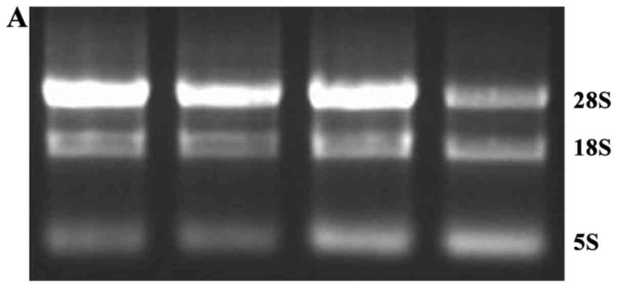
The total RNA extracted from different study samples
was assessed (Fig. 1A).
Electrophoretogram showed that the RNA extracted was of good
quality and no significant degradation was observed. Fluorescent
quantitative PCR was used to detect the difference of expression
quantity of NGF and its receptor in different samples, and the
results are shown in Fig. 1,
indicating that compared with healthy women, the expression
quantity of mRNA in NGF, TrkA and p75NTR gene in patients with
ovarian cancer was significantly increased, and there was
significant difference in the quantity of expression of mRNA in
NGF, TrkA and p75NTR gene and healthy ovary (P<0.05).
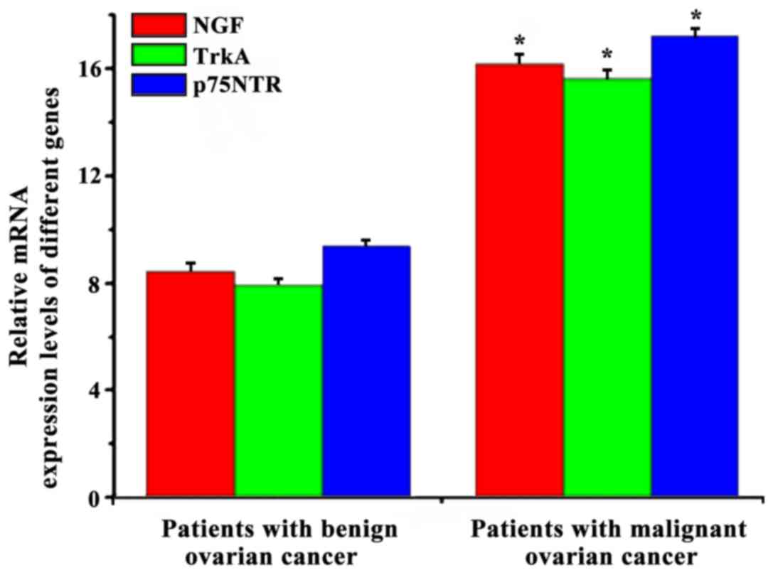
Quantity of expression of mRNA in
nerve cells and its receptor in patients with benign ovarian tumor
and patients with malignant ovarian tumor
RNA extracted from the lesion tissue sample of
patients with ovarian cancer in the observation group was studied.
The difference in quantity of expression of mRNA in nerve cells and
its receptor in patients with benign ovarian tumor and patients
with malignant ovarian cancer was detected (Fig. 2). The quantity of expression of mRNA
in GF, TrkA and p75NTR gene in patients with benign ovarian cancer
was significantly lower than that in patients with malignant
ovarian cancer. This suggested correlation between the quantity of
expression of mRNA in GF, TrkA and p75NTR gene and the severity of
ovarian cancer.
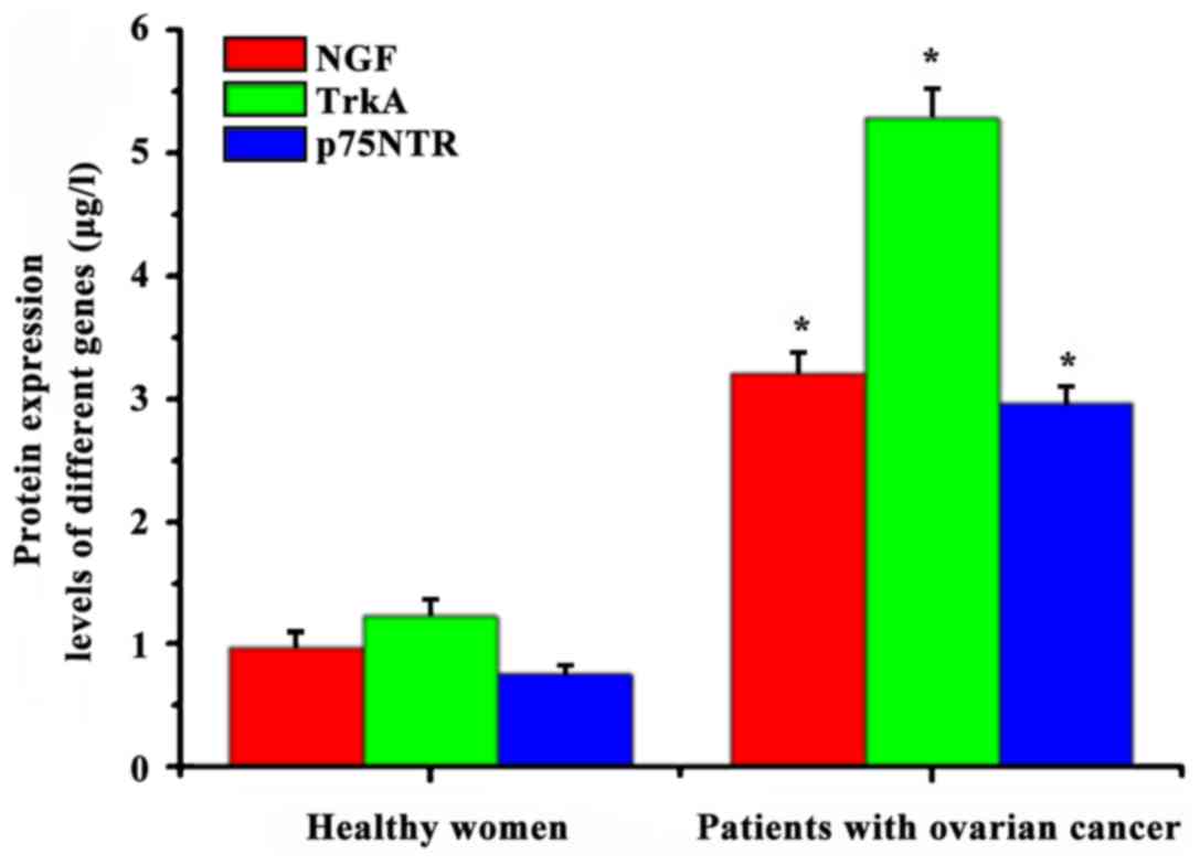
Protein expression quantity of nerve
cells and its receptor in the patients with ovarian cancer and
healthy women
The quantity of protein expression in NGF and its
receptor in different samples was detected using ELISA, and the
results are shown in Fig. 3. The
quantity of expression of NGF in healthy women (0.98±0.12 µg/l) was
significantly lower than that in lesion tissue in patients with
ovarian cancer (3.21±0.16 µg/l), and there were significant
differences between them (P<0.05). The quantity of expression of
NGF receptor, TrkA and p75NTR, in healthy women (1.23±0.14 and
0.76±0.07 µg/l, respectively) was significantly lower than that in
lesion tissue in patients with ovarian cancer (5.28±0.25 and
2.97±0.13 µg/l, respectively), and there were significant
differences (P<0.05).
The result was consistent with that of fluorescent
quantitative PCR. This shows that the content of NGF, TrkA and
p75NTR genes is different in healthy women and patients with
ovarian cancer, which may be associated with ovarian cancer.
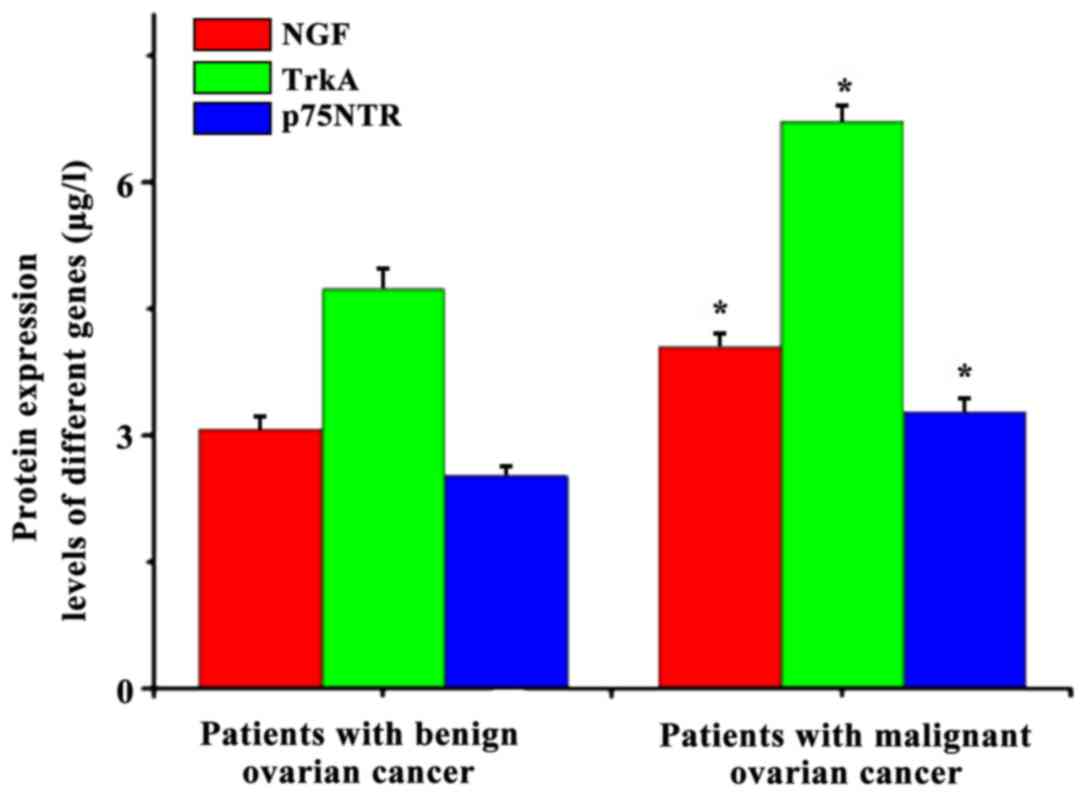
The quantity of protein expression of
nerve cells and its receptors in women with benign ovarian tumor
and patients with malignant ovarian tumor
The protein extracted from the lesion tissue samples
of patients with ovarian cancer in observation group was used, and
the difference of protein expression quantity of NGF and its
receptor in the patients with benign ovarian tumor and patients
with malignant ovarian tumor was detected, and the results are
shown in Fig. 4. The quantity of
protein expression of GF, TrkA and p75NTR gene in patients with
benign ovarian tumor (3.08±0.14, 4.73±0.25 and 2.53±0.11 µg/l,
respectively) was significantly lower than that in patients with
malignant ovarian tumor (4.06±0.16, 6.73±0.18 and 3.29±0.15 µg/l,
respectively). This suggests that there is a certain correlation
between the quantity of protein expression of GF, TrkA and p75NTR
gene and the severity of ovarian cancer.
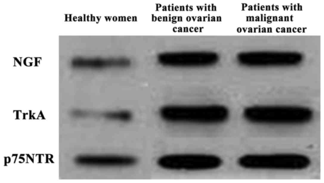
Detection of protein expression
quantity of nerve cells and its receptor in different samples using
western blot analysis
The total protein extracted from the healthy women,
patients with benign ovarian tumor and patients with malignant
ovarian tumor was assessed, and the difference of protein
expression quantity of nerve cells and its receptor in the samples
was detected using western blot analysis, and the results are shown
in Fig. 5. The quantity of protein
expression of NGF, TrkA and p75NTR gene in patients with benign
ovarian tumor and patients with malignant ovarian tumor was
increased significantly compared with that in healthy women; and
there were significant differences, which was consistent with the
results of ELISA. It suggests that there is a certain correlation
between the NGF and its receptor and the onset of ovarian
cancer.
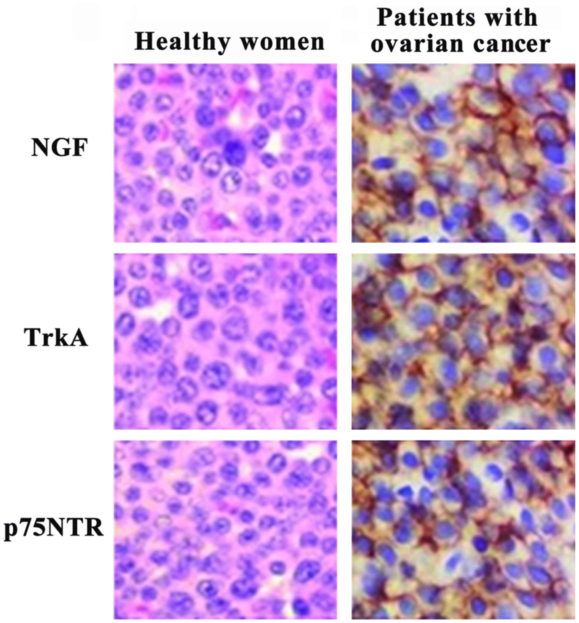
Detection of the quantity of protein
expression of nerve cells and its receptor in samples using
immunohistochemistry
The distribution and expression of protein in NGF
and its receptor in the samples (Fig.
6) were detected using immunohistochemical method. Compared
with healthy ovarian tissue, the quantity of expression of NGF and
its receptor in patients with ovarian cancer was higher, and mainly
located on the cell membrane surface. The result of positive cell
count showed that (Table II) the
number of positive cells in NGF, TrkA and p75NTR in normal ovarian
tissue (8.75, 6.5 and 4.5%) was significantly lower than that in
lesion tissue in patients with ovarian cancer (91.25, 93.5 and
95.5%, respectively). The result was consistent with that of
fluorescent quantitative PCR and ELISA, suggesting that the NGF and
its receptors are related to the onset of ovarian cancer.
| Table II.Positive cell count in NGF and its
receptor in different tissue samples. |
Table II.
Positive cell count in NGF and its
receptor in different tissue samples.
|
| Healthy women | Patients with ovarian
cancer |
|---|
|
|
|
|
|---|
| Gene | Total cell count
(pcs) | Positive cell count
(pcs) | Positive cell rate
(%) | Positive cell count
(pcs) | Positive cell rate
(%) |
|---|
| NGF | 400 | 35 | 8.75 | 365 |
91.25 |
| TrkA | 400 | 26 | 6.5 | 374 | 93.5 |
| p75NTR | 400 | 18 | 4.5 | 382 | 95.5 |
Discussion
The morbidity and mortality of ovarian cancer, as
the main gynecological disease endangering women's health at
present, has showed an increasing trend year by year in women in
China. But the early diagnosis of ovarian cancer is difficult and
there is no special drug treatment. So no significant improvement
has been made in the treatment of patients with ovarian cancer in
China in recent years. The study results show that (13) the onset of ovarian cancer is
influenced not only by environmental factors, but also by genetic
factors. For example, it is believed in most studies that the onset
of ovarian cancer is related to many other factors, such as the
body immune dysfunction, endocrine dyscrasia and diet. Studies have
shown that (14) the NGF, as a kind
of polypeptide hormone, mainly aims to promote the nerve cell
proliferation and the transmission of substance and signal between
nerve cells. The abnormal increase of quantity of expression of NGF
in many tumors and cancers have been shown (15). For example, some studies have shown
that (16) the expression quantity of
NGF in colon cancer is 28 times of that in normal colon tissue. The
statistical result of patients' rehabilitation states after colon
cancer surgery shows that the NGF and its receptor can affect the
peripheral tumor cell proliferation and migration process in
autocrine and paracrine manner suggesting that the NGF and its
receptor may be associated with the late recovery of the tumor.
Data showed that (17) the NGF
receptors mainly include TrkA and p75NTR, among which NGF can be
combined with TrkA in the early stage of tumor, thus promoting the
abnormal increase of expression quantity of TrkA in tumor cells.
Data showed that TrkA is considered to be the proto-oncogene
expression product in human body, and further studies found that
(18) TrkA can be involved in the
expression of vascular endothelial growth factor; thus promoting
the formation of tumor vessels. With the deepening of the study on
NGF and TrkA, it has been found that the NGF receptor p75NTR can
interact with TrkA in some cancers, such as cervical cancer,
promoting the tumor cell proliferation and migration process. Only
by combining with p75NTR can TrkA play its function. But in
endometrial carcinoma, the individual TrkA can also play the role
of NGF receptor. Therefore, no final conclusion has yet been
reached on the relationship between NGF and its receptors (19). In our study, it was found that mRNA
and protein content in NGF, TrkA and p75NTR genes in ovarian cancer
tissue were significantly increased compared with those in normal
ovarian tissue, and there were also differences in the expression
quantity of NGF, TrkA and p75NTR between the benign ovarian tumor
and malignant ovarian tumor. This suggests that NGF, TrkA and
p75NTR play important roles in the progression of ovarian cancer,
so they can act as the markers and diagnostic criteria of detecting
ovarian cancer to some extent.
Acknowledgements
This study was supported by the Natural Science
Foundation of Jiangxi Province (project no. 20132BAB205107).
References
|
1
|
Li L, Ji J, Wang JB, Niyazi M, Qiao YL and
Boffetta P: Attributable causes of breast cancer and ovarian cancer
in China: Reproductive factors, oral contraceptives and hormone
replacement therapy. Chin J Cancer Res. 24:9–17. 2012. View Article : Google Scholar : PubMed/NCBI
|
|
2
|
Gao J, Gao G, Zhang Y and Wang F:
Proteomic analysis of human epithelial ovarian cancer xenografts in
immunodeficient mice exposed to chronic psychological stress. Sci
China Life Sci. 54:112–120. 2011. View Article : Google Scholar : PubMed/NCBI
|
|
3
|
Itamochi H: Targeted therapies in
epithelial ovarian cancer: Molecular mechanisms of action. World J
Biol Chem. 1:209–220. 2010. View Article : Google Scholar : PubMed/NCBI
|
|
4
|
Bilici A, Ustaalioglu BB, Seker M,
Canpolat N, Tekinsoy B, Salepci T and Gumus M: Clinical value of
FDG PET/CT in the diagnosis of suspected recurrent ovarian cancer:
Is there an impact of FDG PET/CT on patient management? Eur J Nucl
Med Mol Imaging. 37:1259–1269. 2010. View Article : Google Scholar : PubMed/NCBI
|
|
5
|
Theveneau E, Marchant L, Kuriyama S, Gull
M, Moepps B, Parsons M and Mayor R: Collective chemotaxis requires
contact-dependent cell polarity. Dev Cell. 19:39–53. 2010.
View Article : Google Scholar : PubMed/NCBI
|
|
6
|
Emoto S, Ishigami H, Yamashita H,
Yamaguchi H, Kaisaki S and Kitayama J: Clinical significance of
CA125 and CA72-4 in gastric cancer with peritoneal dissemination.
Gastric Cancer. 15:154–161. 2012. View Article : Google Scholar : PubMed/NCBI
|
|
7
|
Wang Y, Xu RC, Zhang XL, Niu XL, Qu Y, Li
LZ and Meng XY: Interleukin-8 secretion by ovarian cancer cells
increases anchorage-independent growth, proliferation, angiogenic
potential, adhesion and invasion. Cytokine. 59:145–155. 2012.
View Article : Google Scholar : PubMed/NCBI
|
|
8
|
Barton DL, Burger K, Novotny PJ, Fitch TR,
Kohli S, Soori G, Wilwerding MB, Sloan JA, Kottschade LA, Rowland
KM Jr, et al: The use of Ginkgo biloba for the prevention of
chemotherapy-related cognitive dysfunction in women receiving
adjuvant treatment for breast cancer, N00C9. Support Care Cancer.
21:1185–1192. 2013. View Article : Google Scholar : PubMed/NCBI
|
|
9
|
Guerrero AT, Zarpelon AC, Vieira SM, Pinto
LG, Ferreira SH, Cunha FQ, Verri WA Jr and Cunha TM: The role of
PAF/PAFR signaling in zymosan-induced articular inflammatory
hyperalgesia. Naunyn Schmiedebergs Arch Pharmacol. 386:51–59. 2013.
View Article : Google Scholar : PubMed/NCBI
|
|
10
|
Liu C, Liu H, Wang X and Xinbo S: Clinical
significance and expression of PAF and TNF-alpha in seminal plasma
of leukocytospermic patients. Mediators Inflamm. 2012:6397352012.
View Article : Google Scholar : PubMed/NCBI
|
|
11
|
Rota M, Pasquali E, Scotti L, Pelucchi C,
Tramacere I, Islami F, Negri E, Boffetta P, Bellocco R, Corrao G,
et al: Alcohol drinking and epithelial ovarian cancer risk. a
systematic review and meta-analysis. Gynecol Oncol. 125:758–763.
2012. View Article : Google Scholar : PubMed/NCBI
|
|
12
|
Kim HA, Kim KJ, Seo KH, Lee HK and Im SY:
PTEN/MAPK pathways play a key role in platelet-activating
factor-induced experimental pulmonary tumor metastasis. FEBS Lett.
586:4296–4302. 2012. View Article : Google Scholar : PubMed/NCBI
|
|
13
|
Arai Y, Totoki Y, Hosoda F, Shirota T,
Hama N, Nakamura H, Ojima H, Furuta K, Shimada K, Okusaka T, et al:
Fibroblast growth factor receptor 2 tyrosine kinase fusions define
a unique molecular subtype of cholangiocarcinoma. Hepatology.
59:1427–1434. 2014. View Article : Google Scholar : PubMed/NCBI
|
|
14
|
Bilici A, Seker M, Ustaalioglu BB, Kefeli
U, Yildirim E, Yavuzer D, Aydin FM, Salepci T, Oncel M and Gumus M:
Prognostic significance of perineural invasion in patients with
gastric cancer who underwent curative resection. Ann Surg Oncol.
17:2037–2044. 2010. View Article : Google Scholar : PubMed/NCBI
|
|
15
|
Kemp SW, Webb AA, Dhaliwal S, Syed S,
Walsh SK and Midha R: Dose and duration of nerve growth factor
(NGF) administration determine the extent of behavioral recovery
following peripheral nerve injury in the rat. Exp Neurol.
229:460–470. 2011. View Article : Google Scholar : PubMed/NCBI
|
|
16
|
Rogers ML, Bailey S, Matusica D, Nicholson
I, Muyderman H, Pagadala PC, Neet KE, Zola H, Macardle P and Rush
RA: ProNGF mediates death of Natural Killer cells through
activation of the p75NTR-sortilin complex. J Neuroimmunol.
226:93–103. 2010. View Article : Google Scholar : PubMed/NCBI
|
|
17
|
Teng KK, Felice S, Kim T and Hempstead BL:
Understanding proneurotrophin actions: Recent advances and
challenges. Dev Neurobiol. 70:350–359. 2010.PubMed/NCBI
|
|
18
|
Li C, Ma H, Wang Y, Cao Z, Graves-Deal R,
Powell AE, Starchenko A, Ayers GD, Washington MK, Kamath V, et al:
Excess PLAC8 promotes an unconventional ERK2-dependent EMT in colon
cancer. J Clin Invest. 124:2172–2187. 2014. View Article : Google Scholar : PubMed/NCBI
|
|
19
|
Zhu Y, Li Y, Haraguchi S, Yu M, Ohira M,
Ozaki T, Nakagawa A, Ushijima T, Isogai E, Koseki H, et al:
Dependence receptor UNC5D mediates nerve growth factor
depletion-induced neuroblastoma regression. J Clin Invest.
123:2935–2947. 2013. View
Article : Google Scholar : PubMed/NCBI
|