Introduction
Thyroid cancer is the most common tumour of the
endocrine system, and its incidence rate has dramatically increased
over the past several decades (1).
Roughly 300,000 new cases with a median age at diagnosis of 50
years and nearly 40,000 deaths are estimated to occur each year
worldwide (2). Depending on
pathological type, thyroid cancer can be classified into four
types: papillary, follicular, medullary and anaplastic thyroid
cancer (3). Papillary thyroid
carcinoma (PTC) is the most common type of thyroid cancer,
accounting for approximately 90% of all thyroid cancer cases
(1). Majority of PTC patients have a
good prognosis after surgical resection in combination with
radioiodine and levothyroxine treatment (4). However, PTC patients with large primary
tumour, extrathyroidal invasion, lymph node metastasis, advanced
tumour-node-metastasis stage or recurrences typically have a poor
prognosis (5). Therefore, the
molecular mechanisms underlying the formation and progression of
PTC must be elucidated to improve the diagnosis, therapy and
prevention of this disease.
MicroRNAs (miRNAs) belong to a large family of
endogenous, single-strand and short non-coding RNAs with a length
of approximately 22 nucleotides (6).
MiRNAs negatively regulate gene expression through imperfect base
pairing with the 3′-untranslated regions (3′-UTRs) of their target
messenger RNA (mRNA) and result in mRNA destabilisation and/or
translational inhibition (7). MiRNAs
have been implicated in various biological processes, such as cell
growth, cell cycle regulation, differentiation, apoptosis,
development and metastasis (8,9).
Dysregulated miRNAs have been observed in various types of human
cancers, such as miR-126 in thyroid cancer (10), miR-122 in gastric cancer (11), miR-660 in breast cancer (12) and miR-335 in bladder cancer (13). Thus, miRNAs may function as important
regulators in tumourigenesis and tumour development (14,15).
MiRNAs can function as either tumour suppressors or oncogenes in
different human cancers depending on the characteristics of their
target genes (16). Therefore, miRNAs
may serve as therapeutic targets for cancer treatments (17).
MiR-139 has been recently reported to be aberrantly
expressed in several types of cancer (18–21).
However, the expression levels, biological functions and associated
molecular mechanism of miR-139 in PTC have not been clearly
elucidated. In the present study, we measured miR-139 expression in
PTC tissues and cell lines. We also investigated the regulatory
roles of miR-139 in PTC cells. Moreover, we explored the underlying
molecular mechanism of its actions in PTC cells.
Materials and methods
Tissue samples and cell lines
This study was approved by the Medical Ethics
Committee of The Seventh People's Hospital of Shanghai University
of Traditional Chinese Medicine. All patients included in this
research were required to offer written informed consent.
Forty-three paired PTC tissues and adjacent normal tissues were
obtained from patients (age range, 35–67 years; median age, 48;
eighteen males and twenty-five females) who underwent surgical
resection in The Seventh People's Hospital of Shanghai University
of Traditional Chinese Medicine between February 2014 and December
2015. All tissue samples were snap-frozen in liquid nitrogen
immediately after surgery and stored at −80°C until RNA
extraction.
The human PTC cell lines (TPC-1, HTH83, and BCPAP)
were acquired from Cell Bank of the Chinese Academy of Sciences
(Shanghai, China). Normal human thyroid cell line (Nthy-ori 3–1)
was obtained from European Collection of Authenticated Cell
Cultures (ECACC; Salisbury, UK). All of the cell lines were grown
in Dulbecco's modified Eagle's medium (DMEM; Gibco, Grand Island,
NY) supplemented with 10% fetal bovine serum (FBS; Gibco, Grand
Island, NY), 100 units of penicillin/ml and 100 ng of
streptomycin/ml (Gibco, Grand Island, NY) at 37°C in humidified air
with 5% CO2.
Transfection assay
MiR-139 mimics and corresponding miRNA negative
control (miR-NC) were chemically synthesized and purified by
Guangzhou RiboBio Co., Ltd (Guangzhou, China). Overexpression FN1
plasmid (pcDNA3.1-FN1) and blank vector (pcDNA3.1) were obtained
from GeneCopoeia (Guangzhou, China). Cells were seeded into 6-well
plates at a density of 8×10⁵ cells per well and
maintained in DMEM medium without antibiotics. When the cell
density reached 60–70%, transfection was performed using
Lipofectamine 2000 (Invitrogen, Carlsbad, CA, USA) according to the
manufacturer's protocol. Tranfected cells were incubated at 37°C
with 5% CO2 for 6 h and the medium was replaced by DMEM
with 10% FBS.
RNA preparation and reverse
transcription-quantitative polymerase chain reaction (RT-qPCR)
According to the manufacturer's instructions, total
RNA was isolated from the tissue specimens or cells using TRIzol
(Invitrogen, Carlsbad, CA, USA) and stored at −80°C. To determine
miR-139 expression levels, cDNA was generated by reverse
transcription using a TaqMan MicroRNA Reverse Transcription Kit
(Applied Biosystems, Carlsbad, CA, USA). Quantification PCR (qPCR)
was performed with TaqMan MicroRNA PCR Kit (Applied Biosystems,
Carlsbad, CA, USA) on an ABI Prism 7900 Sequence Detection System
(Applied Biosystems, Carlsbad, CA, USA). To quantify FN1 mRNA
expression, cDNA was synthesized with PrimeScript RT Reagent kit
(Takara Biotechnology Co., Ltd., Dalian, China) and qPCR was
conducted with SYBR Premix Ex Taq™ (Takara Biotechnology Co., Ltd.,
Dalian, China). U6 and GAPDH were used to normalize the level of
miR-139 and FN1 mRNA expression, respectively. The data were
analyzed using the 2−ΔΔCq method (22). The primers used were as followed:
miR-139: 5′-GCTCTACAGTGCACGTGTC-3′, 5′-GTGCAGGGTCCGAGGT-3′. U6:
5′-CTCGCTTCGGCAGCACA-3′, 5′-AACGCTTCACGAATTTGCGT-3′. FN1:
5′-CAGTGGGAGACCTCGAGAAG-3′, 5′-TCCCTCGGAACATCAGAAAC-3′. GAPDH:
5′-GCTGGCGCTGAGTACGTCGTGGAGT-3′,
5′-CACAGTCTTCTGGGTGGCAGTGATGG-3′.
Cell Counting kit-8 (CCK-8) assay
Cells were seeded into 96-well plates at
3×10³ cells per well. After incubation overnight, cell
transfection was performed and then incubated at 37°C in humidified
air with 5% CO2. Cell proliferation was examined at 0,
24, 48, and 72 h after transfection. Briefly, 10 µl of CCK-8
reagent (Dojindo, Kumamoto, Japan) was added into each well and
incubated at 37°C for another 2 h. Finally, the optical density
(OD) was detected at a wavelength of 450 nm using the ELISA plate
reader (Model 550; Bio-Rad Laboratories, Hercules, CA, USA). At
least three independent experiments were performed.
Transwell invasion assay
Transwell invasion assay was performed to assess
cell invasion capacity by using Matrigel-coated Transwell chambers
(Millipore, Billerica, MA, USA). A total of 1×10⁵
transfected cells in 100 µl of FBS-free DMEM medium were placed in
the upper chambers. DMEM with 10% FBS was added into the lower
chamber as chemoattractant. After 24 h of incubation, the upper
surface of the membrane was wiped with a cotton tip. Subsequently,
invasive cells were fixed with methanol, stained with 0.5% crystal
violet, washed with PBS and photographed under an inverted
microscope at 200× magnification (X71; Olympus, Tokyo, Japan). The
number of invasive cells was counted at five randomly selected
fields.
Bioinformatics analysis
To predict the potential targets of miR-139,
bioinformatics analysis was performed with TargetScan (http://www.targetscan.org) and miRanda (http://www.microrna.org/microrna/getExprForm.do).
Luciferase reporter assay
The pMIR-FN1-3′-UTR-wild-type (Wt) and
pMIR-FN1-3′-UTR-mutant (Mut) containing the putative binding site
of miR-139 were synthesized and confirmed by GenePharma, Co., Ltd.
(Shanghai, China). Cells were seeded in 24-well plates and cultured
until the cell density reached 80–90% confluence. Subsequently,
cells were transfected with either the pMIR-FN1-3′-UTR-Wt or the
pMIR-FN1-3′-UTR-Mut reporter vector, together with miR-139 mimics
or miR-NC using Lipofectamine 2000. After incubation 48 h, the
activities of firefly and Renilla luciferases were determined in
transfected cells using the dual-luciferase reporter assay system
(Promega, Madison, WI) according to the manufacturer's
recommendations. Renilla-luciferase activity was assayed for
normalization.
Western blot analysis
Total protein was isolated form tissue samples or
cells with RIPA lysis buffer (Beyotime Biotechnology, Jiangsu,
China) containing 1% protease inhibitors (Pierce, Rockford, IL,
USA). The concentration of total protein was examined by Bradford
assay (Biorad Laboratories, Hercules, CA, USA). Equal amounts of
protein samples (about 30 µg) were resolved by 10% sodium dodecyl
sulfate-polyacrylamide gel electrophoresis and transferred to PVDF
membrane (Millipore, Billerica, MA, USA). After blocking with 5%
non-fat milk in Tris Buffered saline with Tween (TBST), the
membranes were incubated with primary antibodies overnight at 4°C.
The primary antibodies used in this study include rabbit anti-human
polyclonal FN1 (15613–1-AP; 1:200 dilution; Proteintech, USA) and
mouse anti-human monoclonal GAPDH antibody (sc-47724; 1:1,000
dilution; Santa Cruz Biotechnology, CA, USA). The membranes were
then washed with TBST and incubated with corresponding
HRP-conjugated secondary antibodies (sc-2204 and sc-2005; 1:5,000
dilution; Santa Cruz Biotechnology, Santa Cruz, CA, USA) at room
temperature for 2 h. Band signals were visualized using an enhanced
chemiluminescence kit (Pierce, Minneapolis, MN, USA), and analyzed
with Quantity One software version 4.6.2 (Bio-Rad Laboratories,
Hercules, CA, USA). GAPDH was used as an internal control.
Statistical analysis
The statistical analyses were performed using the
SPSS 17.0 software (SPSS, Inc., Chicago, IL, USA). All data were
presented as mean ± SEM, and differences between groups were
analyzed using two-tailed student's t-test or a one-way ANOVA.
P<0.05 was considered to indicate a statistically significant
difference.
Results
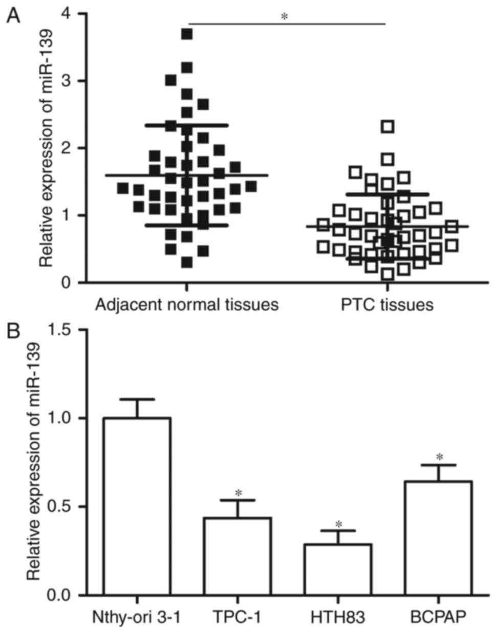
MiR-139 is frequently down-regulated
in PTC tissues and cell lines
RT-qPCR was performed in 43 pairs of PTC tissues and
adjacent normal tissues to determine miR-139 expression levels in
PTC. Results showed that miR-139 was down-regulated in PTC tissues
compared with adjacent normal tissues (Fig. 1A, P<0.05). MiR-139 expression was
further detected in PTC cell lines (TPC-1, HTH83 and BCPAP) and
normal human thyroid cell line (Nthy-ori 3-1) through RT-qPCR. The
expression level of miR-139 was significantly reduced in the PTC
cell lines compared with Nthy-ori 3-1 (Fig. 1B, P<0.05). These results suggest
that low miR-139 levels correlate with PTC development.
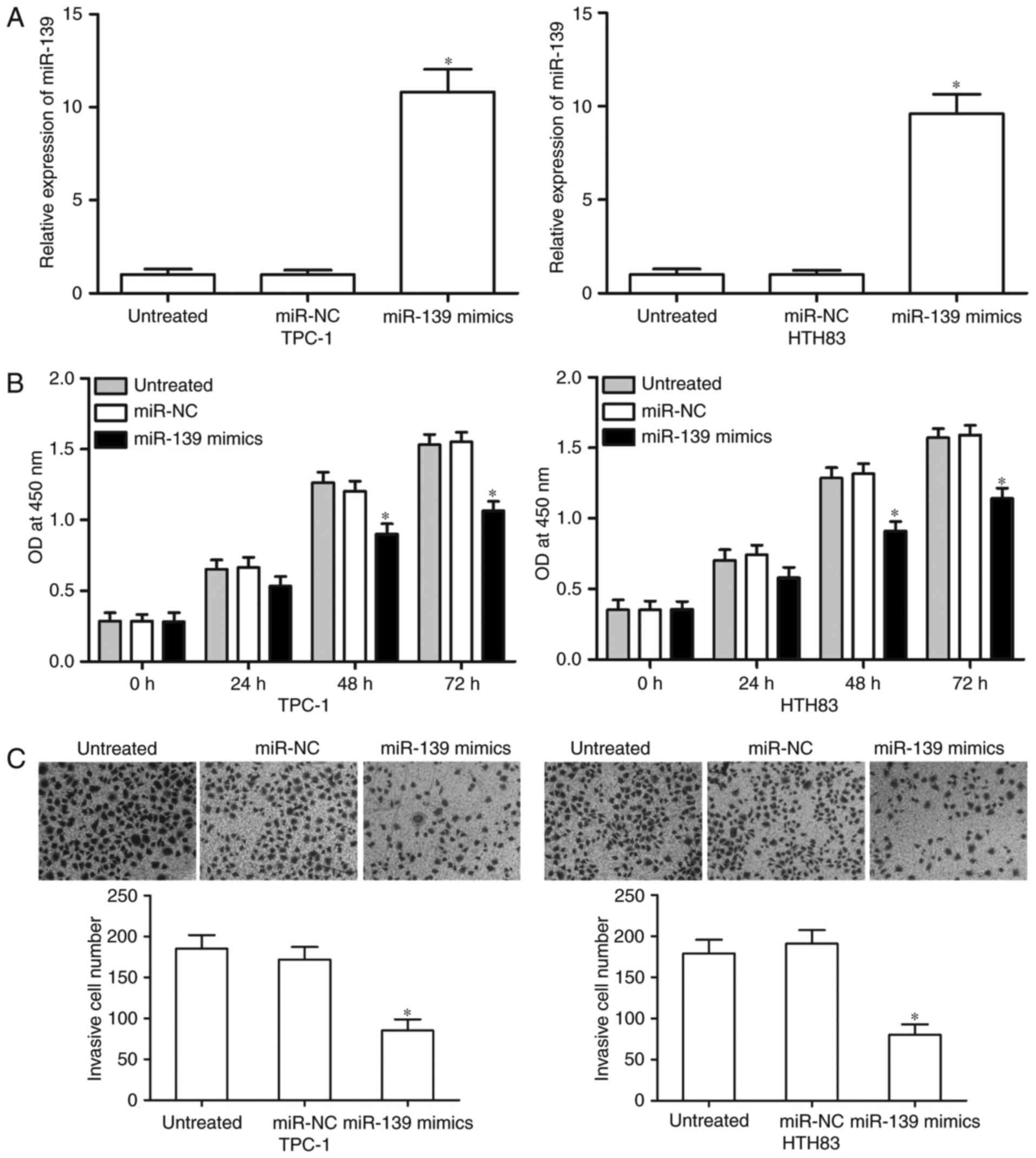
MiR-139 overexpression inhibits the
proliferation and invasion of PTC cells
We up-regulated miR-139 expression in TPC-1 and
HTH83 cells through miR-139 mimics transfection to determine the
functional roles of miR-139 in PTC. RT-qPCR analysis demonstrated
that miR-139 evidently increased in the TPC-1 and HTH83 cells
transfected with the miR-139 mimics compared with that in the cells
transfected with miR-NC and untreated cells (Fig. 2A, P<0.05). We then performed CCK-8
assay to examine cell proliferation. Our results revealed that
miR-139 up-regulation obviously suppressed the proliferation of
TPC-1 and HTH83 cells (Fig. 2B,
P<0.05). Transwell invasion assays were utilized to examine the
invasion capacities of TPC-1 and HTH83 cells after transfection
with the miR-139 mimics, miR-NC or untreated cells. According to
the CCK-8 assay, we revealed miR-139 did not significantly affected
the cell proliferation at 24 h incubation time. Hence, we believed
that the anti-proliferative role of miR-139 did not be responsible
for part of the observed effect on PTC cell invasion. Fig. 2C shows that the number of invasive
cells significantly decreased in the miR-139 overexpression group
compared with the miR-NC and untreated groups in TPC-1 and HTH83
cells (P<0.05). These results suggest that miR-139 serves
tumour-suppressive functions in PTC cells.
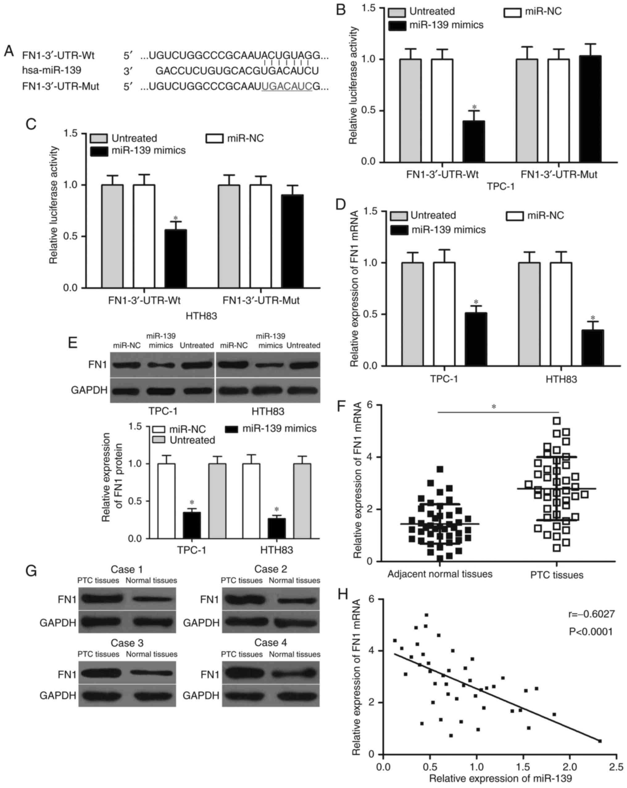
FN1 is a direct target of miR-139 in
PTC
The potential targets of miR-139 were predicted
through bioinformatics analysis to investigate the mechanism by
which miR-139 inhibits PTC cell proliferation and invasion. FN1
plays oncogenic roles in PTC (23)
and thus was selected for further confirmation (Fig. 3A). Luciferase reporter assays were
carried out to explore whether miR-139 targets FN1 by binding to
its 3′-UTR. TPC-1 and HTH83 cells were co-transfected with the
pMIR-FN1-3′-UTR-Wt or pMIR-FN1-3′-UTR-Mut reporter vector and
miR-139 mimics or miR-NC. The results showed that luciferase
activities significantly decreased in the TPC-1 and HTH83 cells
transfected with the wild-type FN1 reporter vector (Fig. 3B and C, P<0.05) but not in the
cells with the mutant reporter vector. In addition, RT-qPCR and
Western blot analyses indicated that miR-139 overexpression in
TPC-1 and HTH83 cells decreased the mRNA and protein expression of
FN1 (Fig. 3D and E, P<0.05). These
results suggest that miR-139 directly targets FN1 by binding to its
3′-UTR region in PTC cells.
We subsequently examined the mRNA and protein
expression levels of FN1 in PTC tissues and adjacent normal tissues
through RT-qPCR and Western blot analyses. Results showed that FN1
expression was significantly up-regulated at both mRNA (Fig. 3F, P<0.05) and protein (Fig. 3G, P<0.05) levels in PTC tissues
compared with adjacent normal tissues. Moreover, Spearman's
correlation analysis demonstrated that the expression levels of
miR-139 were inversely correlated with FN1 mRNA in PTC tissues
(Fig. 3H, r=−0.6027, P<0.001).
Overall, these results indicate that FN1 is a direct target of
miR-139 in PTC.
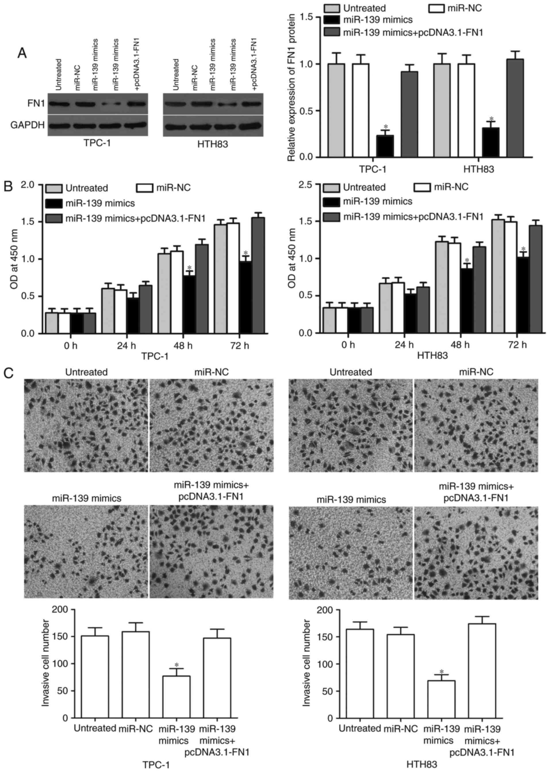
Restoration of FN1 reverses the
effects of miR-139 in PTC cells
We carried out rescue experiments in TPC-1 and HTH83
cells co-transfected with the miR-139 mimics with or without
pcDNA3.1-FN1 to test whether miR-139 exerts tumour-suppressive
functions in PTC cells via the down-regulation of FN1. Western blot
analysis determined that FN1 expression was restored in the miR-139
mimics combined with the FN1 overexpression plasmid (Fig. 4A, P<0.05). In addition, the
restoration expression of FN1 can rescue the inhibitory effects of
miR-139 overexpression on the proliferation (Fig. 4B, P<0.05) and invasion (Fig. 4C, P<0.05) of TPC-1 and HTH83 cells.
In summary, rescue experiments suggest that miR-139 inhibits PTC
cell proliferation and invasion through regulation of FN1.
Luciferase reporter assay, RT-qPCR and Western blotting analysis
also indicated that miR-139 could directly targeted the 3′UTR of
FN1 and decreased FN1 expression at both mRNA and protein level. We
think this is the mechanism underlying the tumor-suppressing roles
of miR-139 in PTC.
Discussion
Aberrantly expressed miRNAs are involved in PTC
formation and progression by regulating their target genes. Thus,
miRNAs may be investigated as molecular biomarkers for the
prediction and prognosis of PTC and as novel therapeutic targets
for PTC patients (24,25). The present study demonstrated that
miR-139 expression was significantly down-regulated in PTC tissues,
and a similarly reduced expression was confirmed in PTC cell lines.
Increased miR-139 expression suppressed the proliferation and
invasion of PTC cells in vitro. Furthermore, FN1 was
validated as a novel direct target of miR-139 in PTC. The
tumour-suppressive roles of miR-139 overexpression on PTC cells
were rescued by ectopic FN1 expression. Overall, the present study
demonstrates that miR-139 expression is down-regulated in PTC and
suggests that this reduced expression impedes PTC tumourigenesis
and tumour progression by inhibiting FN1 expression.
MiR-139 has been observed to be abnormally expressed
in various human cancers. For instance, miR-139 was found
underexpressed in tumour tissues and cell lines in colorectal
cancer (26). Low miR-139 expression
was obviously correlated with disease progression and metastasis
(26). Guo et al (27) found that the downregulation of miR-139
was associated with advanced stage and low overall survival of
patients with colorectal cancer. Liu et al (28) reported that miR-139 was lowly
expressed in esophageal squamous-cell carcinoma. Low miR-139
expression was also associated with lymph node metastases of
esophageal squamous-cell carcinoma (28). Wong et al reported that the
decreased expression of miR-139 in hepatocellular carcinoma was
correlated with venous invasion, microsatellite formation, absence
of tumour encapsulation and reduced differentiation (29). MiR-139 downregulation was also
observed in breast cancer (18),
glioma (19), tongue squamous-cell
carcinoma (20), oral cancer
(21) and non-small-cell lung cancer
(30). These findings suggested that
the aberrant downregulation of miR-139 might represent a prognostic
marker of human cancer.
Accumulated studies reported that miR-139 functions
as a regulator in various cancers through its targeting. Shen et
al found that miR-139 overexpression decreased cell invasion
and metastasis of colorectal cancer in vitro and in
vivo by targeting IGF-1R, AMFR and NOTCH1 (26,31). In
addition, upregulation of miR-139 reduced colorectal cancer cell
proliferation via downregulation of PAP1B (27). MiR-139 inhibited cell invasion and
enhanced cell-cycle arrest in the G0/G1 phase via blockade of NR5A2
in esophageal squamous-cell carcinoma (28). In hepatocellular carcinoma, enforced
expression of miR-139 repressed tumour cell migration and invasion
in vitro, as well as the incidence and severity of lung
metastasis from orthotopic liver tumours in vivo by negative
regulation of ROCK2 (29). Gu et
al revealed that restoration of miR-139 suppressed
hepatocellular carcinoma cell proliferation and invasion by
directly targeting the WNT/TCF-4 pathway (32). In breast cancer, ectopic miR-139
expression suppressed proliferation, migration and invasion;
induced apoptosis and cell-cycle arrest; and improved
chemosensitivity to docetaxel by downregulation of Notch1 (18). In glioma, miR-139 re-expression
inhibited cell proliferation and invasion both in vitro and
in vivo by directly targeting IGF-1 R, AMY-1 and PGC-1β
(19). These studies suggested that
miR-139 may act as tumour suppressor in various human cancers and
can be investigated as a therapeutic target for cancer
treatments.
FN1, a member of the FN family, is up-regulated in
multiple types of human cancer, including anal cancer (33), breast cancer (34), head and neck squamous cell carcinoma
(35) and ovarian cancer (36). Functional experiments indicated that
FN1 is involved in several biological processes, such as cell
proliferation, embryogenesis, wound healing, platelet aggregation
host defence, blood coagulation, EMT and metastasis (37–39).
Previous studies reported that FN1 plays important roles in
tumourigenesis and tumour development. For example, FN1 expression
increases in cisplatin-resistant lung cancer. The down-regulation
of FN1 suppresses lung cancer cell migration, induces apoptosis and
increases cisplatin sensitivity (40). In PTC, FN1 is highly expressed in
tumour tissues compared with non-tumour tissues. The knockdown of
FN1 represses PTC cell proliferation, adhesion and metastasis in
vitro (23). These findings
suggest that FN1 targeting in PTC provides a novel strategy to
treat PTC patients.
In conclusion, the study is the first to demonstrate
that miR-139 expression is down-regulated in PTC tissues and cell
lines. In addition, miR-139 inhibits the proliferation and invasion
of PTC cells by directly targeting FN1. These results provide novel
insights into the molecular mechanism underlying PTC progression
and suggest that miR-139 can potentially serve as an anti-tumour
agent in PTC treatment.
In thyroid carcinoma, the dysregulation of thyroid
hormone is critical symptom and FN1 may be involved in the
production of thyroid hormone. In this study, we did not explore
the effect of miR-139 on thyroid hormone. In the following
experiments, we will examine the effect of miR-139 on thyroid
hormone. In addition, we can not analyze the association between
miR-139 and clinical parameters of patients with PTC using TCGA,
and this issue is a limitation of the present study. In the
following experiments, we will collect more PTC tissues and
explored the association between miR-139 and clinical parameters of
patients with PTC.
Acknowledgements
The present study was supported by grants from the
Key Disciplines Group Construction Project of Pudong Health Burea
of Shanghai (grant no. PWZxq2014-12), the Natural Science
Foundation of China (grant no. 81571718), the Shanghai Sailing
Program (grant no. 16YF1408800) and the Shanghai Science and
Technology Committee Foundation (grant no. 14DZ1940605).
References
|
1
|
Liebner DA and Shah MH: Thyroid cancer:
Pathogenesis and targeted therapy. Ther Adv Endocrinol Metab.
2:173–195. 2011. View Article : Google Scholar : PubMed/NCBI
|
|
2
|
Sondermann A, Andreghetto FM, Moulatlet
AC, da Silva Victor E, de Castro MG, Nunes FD, Brandão LG and
Severino P: MiR-9 and miR-21 as prognostic biomarkers for
recurrence in papillary thyroid cancer. Clin Exp Metastasis.
32:521–530. 2015. View Article : Google Scholar : PubMed/NCBI
|
|
3
|
Gonzalez-Gonzalez R, Bologna-Molina R,
Carreon-Burciaga RG, Gómezpalacio-Gastelum M, Molina-Frechero N and
Salazar-Rodríguez S: Papillary thyroid carcinoma: Differential
diagnosis and prognostic values of its different variants: Review
of the literature. ISRN Oncol. 2011:9159252011.PubMed/NCBI
|
|
4
|
Shi X, Liu R, Basolo F, Giannini R, Shen
X, Teng D, Guan H, Shan Z, Teng W, Musholt TJ, et al: Differential
clinicopathological risk and prognosis of major papillary thyroid
cancer variants. J Clin Endocrinol Metab. 101:264–274. 2016.
View Article : Google Scholar : PubMed/NCBI
|
|
5
|
Voutilainen PE, Multanen MM, Leppäniemi
AK, Haglund CH, Haapiainen RK and Franssila KO: Prognosis after
lymph node recurrence in papillary thyroid carcinoma depends on
age. Thyroid. 11:953–957. 2001. View Article : Google Scholar : PubMed/NCBI
|
|
6
|
Bartel DP: MicroRNAs: Genomics,
biogenesis, mechanism, and function. Cell. 116:281–297. 2004.
View Article : Google Scholar : PubMed/NCBI
|
|
7
|
He H, Jazdzewski K, Li W, Liyanarachchi S,
Nagy R, Volinia S, Calin GA, Liu CG, Franssila K, Suster S, et al:
The role of microRNA genes in papillary thyroid carcinoma. Proc
Natl Acad Sci USA. 102:pp. 19075–19080. 2005, View Article : Google Scholar : PubMed/NCBI
|
|
8
|
Maroney PA, Yu Y and Nilsen TW: MicroRNAs,
mRNAs, and translation. Cold Spring Harb Symp Quant Biol.
71:531–535. 2006. View Article : Google Scholar : PubMed/NCBI
|
|
9
|
Guo H, Ingolia NT, Weissman JS and Bartel
DP: Mammalian microRNAs predominantly act to decrease target mRNA
levels. Nature. 466:835–840. 2010. View Article : Google Scholar : PubMed/NCBI
|
|
10
|
Qian Y and Wang X, Lv Z, Guo C, Yang Y,
Zhang J and Wang X: MicroRNA-126 is downregulated in thyroid cancer
cells, and regulates proliferation, migration and invasion by
targeting CXCR4. Mol Med Rep. 14:453–459. 2016. View Article : Google Scholar : PubMed/NCBI
|
|
11
|
Rao M, Zhu Y, Zhou Y, Cong X and Feng L:
MicroRNA-122 inhibits proliferation and invasion in gastric cancer
by targeting CREB1. Am J Cancer Res. 7:323–333. 2017.PubMed/NCBI
|
|
12
|
Shen Y, Ye YF, Ruan LW, Bao L, Wu MW and
Zhou Y: Inhibition of miR-660-5p expression suppresses tumor
development and metastasis in human breast cancer. Genet Mol Res.
16:2017. View Article : Google Scholar
|
|
13
|
Wu D, Niu X, Pan H, Zhou Y, Qu P and Zhou
J: MicroRNA-335 is downregulated in bladder cancer and inhibits
cell growth, migration and invasion via targeting ROCK1. Mol Med
Rep. 13:4379–4385. 2016. View Article : Google Scholar : PubMed/NCBI
|
|
14
|
Esquela-Kerscher A and Slack FJ:
Oncomirs-microRNAs with a role in cancer. Nat Rev Cancer.
6:259–269. 2006. View
Article : Google Scholar : PubMed/NCBI
|
|
15
|
Kent OA and Mendell JT: A small piece in
the cancer puzzle: microRNAs as tumor suppressors and oncogenes.
Oncogene. 25:6188–6196. 2006. View Article : Google Scholar : PubMed/NCBI
|
|
16
|
Jones KB, Salah Z, Del Mare S, Galasso M,
Gaudio E, Nuovo GJ, Lovat F, LeBlanc K, Palatini J, Randall RL, et
al: miRNA signatures associate with pathogenesis and progression of
osteosarcoma. Cancer Res. 72:1865–1877. 2012. View Article : Google Scholar : PubMed/NCBI
|
|
17
|
Kong YW, Ferland-McCollough D, Jackson TJ
and Bushell M: microRNAs in cancer management. Lancet Oncol.
13:e249–e258. 2012. View Article : Google Scholar : PubMed/NCBI
|
|
18
|
Zhang HD, Sun DW, Mao L, Zhang J, Jiang
LH, Li J, Wu Y, Ji H, Chen W, Wang J, et al: MiR-139-5p inhibits
the biological function of breast cancer cells by targeting Notch1
and mediates chemosensitivity to docetaxel. Biochem Biophys Res
Commun. 465:702–713. 2015. View Article : Google Scholar : PubMed/NCBI
|
|
19
|
Wang H, Yan X, Ji LY, Ji XT, Wang P, Guo
SW and Li SZ: miR-139 functions as an antioncomir to repress glioma
progression through targeting IGF-1 R, AMY-1, and PGC-1β. Technol
Cancer Res Treat. 16:497–511. 2017. View Article : Google Scholar : PubMed/NCBI
|
|
20
|
Duz MB, Karatas OF, Guzel E, Turgut NF,
Yilmaz M, Creighton CJ and Ozen M: Identification of miR-139-5p as
a saliva biomarker for tongue squamous cell carcinoma: A pilot
study. Cell Oncol (Dordr). 39:187–193. 2016. View Article : Google Scholar : PubMed/NCBI
|
|
21
|
Ren Y, Zhu H, Chi C, Yang F and Xu X:
MiRNA-139 regulates oral cancer Tca8113 cells apoptosis through Akt
signaling pathway. Int J Clin Exp Pathol. 8:4588–4594.
2015.PubMed/NCBI
|
|
22
|
Livak KJ and Schmittgen TD: Analysis of
relative gene expression data using real-time quantitative PCR and
the 2(-Delta Delta C(T)) method. Methods. 25:402–408. 2001.
View Article : Google Scholar : PubMed/NCBI
|
|
23
|
Sponziello M, Rosignolo F, Celano M,
Maggisano V, Pecce V, De Rose RF, Lombardo GE, Durante C, Filetti
S, Damante G, et al: Fibronectin-1 expression is increased in
aggressive thyroid cancer and favors the migration and invasion of
cancer cells. Mol Cell Endocrinol. 431:123–132. 2016. View Article : Google Scholar : PubMed/NCBI
|
|
24
|
Han Aragon P, Weng CH, Khawaja HT,
Nagarajan N, Schneider EB, Umbricht CB, Witwer KW and Zeiger MA:
MicroRNA expression and association with clinicopathologic features
in papillary thyroid cancer: A systematic review. Thyroid.
25:1322–1329. 2015. View Article : Google Scholar : PubMed/NCBI
|
|
25
|
Hua K, Jin J, Zhang H, Zhao B, Wu C, Xu H
and Fang L: MicroRNA-7 inhibits proliferation, migration and
invasion of thyroid papillary cancer cells via targeting CKS2. Int
J Oncol. 49:1531–1540. 2016. View Article : Google Scholar : PubMed/NCBI
|
|
26
|
Shen K, Liang Q, Xu K, Cui D, Jiang L, Yin
P, Lu Y, Li Q and Liu J: MiR-139 inhibits invasion and metastasis
of colorectal cancer by targeting the type I insulin-like growth
factor receptor. Biochem Pharmacol. 84:320–330. 2012. View Article : Google Scholar : PubMed/NCBI
|
|
27
|
Guo H, Hu X, Ge S, Qian G and Zhang J:
Regulation of RAP1B by miR-139 suppresses human colorectal
carcinoma cell proliferation. Int J Biochem Cell Biol.
44:1465–1472. 2012. View Article : Google Scholar : PubMed/NCBI
|
|
28
|
Liu R, Yang M, Meng Y, Liao J, Sheng J, Pu
Y, Yin L and Kim SJ: Tumor-suppressive function of miR-139-5p in
esophageal squamous cell carcinoma. PLoS One. 8:e770682013.
View Article : Google Scholar : PubMed/NCBI
|
|
29
|
Wong CC, Wong CM, Tung EK, Au SL, Lee JM,
Poon RT, Man K and Ng IO: The microRNA miR-139 suppresses
metastasis and progression of hepatocellular carcinoma by
down-regulating Rho-kinase 2. Gastroenterology. 140:322–331. 2011.
View Article : Google Scholar : PubMed/NCBI
|
|
30
|
Xu W, Hang M, Yuan CY, Wu FL, Chen SB and
Xue K: MicroRNA-139-5p inhibits cell proliferation and invasion by
targeting insulin-like growth factor 1 receptor in human non-small
cell lung cancer. Int J Clin Exp Pathol. 8:3864–3870.
2015.PubMed/NCBI
|
|
31
|
Song M, Yin Y, Zhang J, Zhang B, Bian Z,
Quan C, Zhou L, Hu Y, Wang Q, Ni S, et al: MiR-139-5p inhibits
migration and invasion of colorectal cancer by downregulating AMFR
and NOTCH1. Protein Cell. 5:851–861. 2014. View Article : Google Scholar : PubMed/NCBI
|
|
32
|
Gu W, Li X and Wang J: miR-139 regulates
the proliferation and invasion of hepatocellular carcinoma through
the WNT/TCF-4 pathway. Oncol Rep. 31:397–404. 2014. View Article : Google Scholar : PubMed/NCBI
|
|
33
|
Waalkes S, Atschekzei F, Kramer MW,
Hennenlotter J, Vetter G, Becker JU, Stenzl A, Merseburger AS,
Schrader AJ, Kuczyk MA and Serth J: Fibronectin 1 mRNA expression
correlates with advanced disease in renal cancer. BMC Cancer.
10:5032010. View Article : Google Scholar : PubMed/NCBI
|
|
34
|
Ruiz-Garcia E, Scott V, Machavoine C,
Bidart JM, Lacroix L, Delaloge S and Andre F: Gene expression
profiling identifies Fibronectin 1 and CXCL9 as candidate
biomarkers for breast cancer screening. Br J Cancer. 102:462–468.
2010. View Article : Google Scholar : PubMed/NCBI
|
|
35
|
Jerhammar F, Ceder R, Garvin S, Grénman R,
Grafström RC and Roberg K: Fibronectin 1 is a potential biomarker
for radioresistance in head and neck squamous cell carcinoma.
Cancer Biol Ther. 10:1244–1251. 2010. View Article : Google Scholar : PubMed/NCBI
|
|
36
|
Helleman J, Jansen MP, Span PN, van
Staveren IL, Massuger LF, Meijer-van Gelder ME, Sweep FC, Ewing PC,
van der Burg ME, Stoter G, et al: Molecular profiling of platinum
resistant ovarian cancer. Int J Cancer. 118:1963–1971. 2006.
View Article : Google Scholar : PubMed/NCBI
|
|
37
|
Nadamuni M, Piras R, Mazbar S, Higgins JP
and Kambham N: Fibronectin glomerulopathy: An unusual cause of
adult-onset nephrotic syndrome. Am J Kidney Dis. 60:839–842. 2012.
View Article : Google Scholar : PubMed/NCBI
|
|
38
|
Baydar Ertoy D, Kutlugun AA, Bresin E and
Piras R: A case of familial glomerulopathy with fibronectin
deposits caused by the Y973C mutation in fibronectin. Am J Kidney
Dis. 61:514–518. 2013. View Article : Google Scholar : PubMed/NCBI
|
|
39
|
Park J and Schwarzbauer JE: Mammary
epithelial cell interactions with fibronectin stimulate
epithelial-mesenchymal transition. Oncogene. 33:1649–1657. 2014.
View Article : Google Scholar : PubMed/NCBI
|
|
40
|
Gao W, Liu Y, Qin R, Liu D and Feng Q:
Silence of fibronectin 1 increases cisplatin sensitivity of
non-small cell lung cancer cell line. Biochem Biophys Res Commun.
476:35–41. 2016. View Article : Google Scholar : PubMed/NCBI
|