Introduction
Colon cancer is a common malignant tumor of the
digestive tract, and the morbidity and mortality rates are high in
China; recent environmental and lifestyle changes have increased
the incidence rate of colorectal cancer in China (1). The 5-year survival rate for patients
with primary colon cancer is 80–90%; however, the 5-year survival
rate for patients with metastatic colon cancer is 5–15% (2). Therefore, it is important to investigate
and clarify the molecular pathological mechanism of colon cancer
metastasis and identify new therapeutic targets for the prevention
and treatment of colon cancer (3).
MicroRNAs (miRNAs/miRs) are single stranded
non-coding endogenous RNA molecules of ~22 nucleotides. miRNAs can
bind with mRNA from a target gene at the 3′-untranslated region
(3′-UTR), leading to post-transcriptional silencing, which
regulates the expression of the target gene (4). It is established that the abnormal
expression of miRNAs, including miR-511-3p (5) and miR-135b (6), serves an important function in the
occurrence and development of colon cancer.
miR-383 is located in chromosome 8p22. Previous
studies have demonstrated that miR-383 is associated with
medulloblastoma (7), glioma (8) and hepatocellular carcinoma (9), and may serve important function(s) in
these types of tumor. Previous studies have demonstrated that
majority of miR-383's target genes in numerous tumors (7–9); however,
target genes of miR-383 in colon cancer remain unclear. A
proliferating-inducing ligand (APRIL) is a member of the tumor
necrosis factor (TNF) super-family. Initially, it was discovered as
a cytokine that was able to stimulate tumor cell growth, induce
tumor cell proliferation and modulate tumor cell apoptosis
(10). APRIL is predominantly
expressed in tumor tissues in colon cancer, pancreatic cancer,
gastric cancer and bladder cancer (11,12). The
expression of APRIL promotes tumor growth and metastasis in
colorectal cancer cells (13,14), and APRIL knockdown suppresses
migration and invasion of colon caner cells (15). Cyclooxygenase-2 (COX-2) is markedly
expressed in tumor tissues in colon cancer and breast cancer
(16,17). Overexpression of COX-2 is associated
with the aggressive and invasive potential of tumor cells (18) and is positively associated with the
tumor-node-metastasis (TNM) staging and lymph node metastasis in
colon cancer (16). Myeloid cell
leukemia-1 (MCL-1) is an anti-apoptotic protein of the B cell
lymphoma-2 (BCL-2) family (19).
Over-expression of MCL-1 is associated with the inhibition of
apoptosis in colon cancer cells and the promotion of drug
resistance in colon cancer liver metastasis (20,21).
In the present study, reverse
transcription-quantitative polymerase chain reaction (RT-qPCR) was
used to determine expression levels of miR-383 in colon cancer and
adjacent normal tissues. The associations between miR-383
expression and the clinicopathological characteristics of colon
cancer were analyzed. A miR-383 mimic was used to overexpress
miR-383 in colon cancer HT-29 and LoVo cell lines, and the effect
of miR-383 on colorectal cancer cell proliferation, migration and
invasion was analyzed. Western blot analysis assessed the effect of
miR-383 upregulation on the protein expression levels of previously
identified miR-383 target genes, including A proliferating-inducing
ligand (APRIL), myeloid cell leukemia-1 (MCL-1) and
cyclooxygenase-2 (COX-2) (9,22,23). The
aim of the present study is to provide a theoretical basis for the
future exploration of the molecular mechanism(s) of miR-383, and
the identification of novel therapeutic targets in colon
cancer.
Materials and methods
Patients
A total of 64 pairs of matched colon tumor and
adjacent non-tumor mucosal tissues (>5 cm from the edge of the
tumor) were obtained from patients with colon cancer at Zhejiang
Provincial People's Hospital (Zhejiang, China) between March 2014
and January 2015.
In total, 30 patients were <60 years, 34 patients
were ≥60 years (range, 34–86 years), 36 patients were male and 28
patients were female. Overall, 26 patients had a left colon tumor
and 38 patients had a right colon tumor. Furthermore, 45 patients
presented with adenocarcinoma and 19 with mucinous carcinoma cases.
A total of 55 patients were at tumor-node-metastasis stages I, II
or III, and 9 were at TNM stage IV (24).
Written informed consent was obtained from all
patients prior to surgery for the use of their tissues in the
present study. The use of all specimens was approved by the Ethics
Committee of Zhejiang Provincial People's Hospital (Zhejiang,
China). No patients received radiotherapy or chemotherapy prior to
surgery. Following resection, tissues were immediately frozen in
liquid nitrogen and stored at −80°C until further use.
Cell lines
LoVo and HT-29 colon cancer cell lines were
purchased from the Chinese Academy of Sciences, Shanghai Institute
of Life Sciences Cell Bank (Shanghai, China). Cells were cultured
in RPMI-1640 medium (Gibco; Thermo Fisher Scientific, Inc.,
Waltham, MA, USA), supplemented with 10% fetal bovine serum (FBS,
Dingguo Biotechnology Co., Ltd., Beijing, China) and incubated at
37°C in a 5% CO2 incubator with saturated humidity.
Reagents
TRIzol reagent was purchased from Invitrogen (Thermo
Fisher Scientific, Inc.). The PrimeScript One Step miRNA cDNA
Synthesis kit and the SYBR premix Ex Taq™ II qPCR reagents were
purchased from Takara Bio, Inc. (Otsu, Japan). An rno-miR-383 mimic
kit (cat. no., miR1003114-1-2) and a corresponding negative control
(cat no., miR01201-1-2) were procured from Guangzhou Ruibo
Biological Technology Co., Ltd. (Guangzhou, China). A Transwell
chamber migration assay kit was obtained from BD Biosciences
(Franklin Lakes, NJ, USA). A Boyden chamber invasion assay kit was
purchased from EMD Millipore (Billerica, MA, USA). β-actin and
primary antibodies for western blot against β-actin (ab8227), APRIL
(ab64967), MCL-1 (ab28147) and COX-2 (ab102005) were obtained from
Abcam (Cambridge, UK). Horseradish peroxidase-conjugated-Goat
Anti-Rabbit immunoglobulin (Ig)G (cat. no. ab6721) used as a
secondary antibody was obtained from Abcam.
Total RNA extraction and reverse
transcription
Total RNA was extracted using RNA Extraction kit
(Takara Bio. Inc., Otsu, Japan), according to the manufacturer's
protocol. Total RNA (1 µg) was reverse transcribed to cDNA by a
reverse transcription reaction, using PrimeScript One Step miRNA
cDNA Synthesis kit (Takara Bio. Inc.), with the following reaction
conditions: 37°C for 60 min, 85°C for 5 sec.
RT-qPCR assay for miR-383
Primer sequences were as follows: miR-383 forwards,
5′-CACGAAAGATCAGAAGGTGATTG-3′; U6B forwards,
5′-ACGCAAATTCGTGAAGCGTT-3′. The reverse primer was the universal
primer provided by the reverse transcription kit. The qPCR reaction
system contained 10 µl SYBR premix, 0.4 µl Rox, 0.5 µl reverse
primer, 1 µl template and 7.6 µl distilled H2O. Reaction
conditions were as follows: 94°C for 4 min; then 40 cycles of 94°C,
5 sec, 55°C, 20 sec and 72°C, 20 sec; melting curve program: 95°C,
1 min, 55°C, 30 sec and 95°C, 30 sec, 1 cycle. The expression of
miR-383 was calculated relative to U6B with the 2−ΔΔCq
method (25).
Transfection of the HT-29 and LoVo
cell lines with an miR-383 mimic
HT-29 and LoVo cells in the logarithmic growth phase
were seeded onto 6-well plates at a density of
2×10⁵/well. The cells were transfected according to the
protocol of the rno-miR-383 kit (Guangzhou Ruibo Biological
Technology Co., Ltd.) using Lipofectamine® 2000
(Invitrogen; Thermo Fisher Scientific, Inc.). Cells transfected
with the rno-miR-383 mimic were defined as the experimental group
and the cells transfected with the mimic negative control were
defined as the control group. At 24 h post-transfection, the
efficiency was assessed using RT-qPCR.
MTT assay for cell proliferation
rno-miR-383-transfected HT-29 and LoVo cells were
added to 96-well plates at a concentration of 2×10³
cells/well in sextuplicate, and incubated at 37°C in a 5%
CO2 incubator with saturated humidity. Following 24, 48
or 72 h incubation, 20 µl MTT reagent (5 mg/ml) was added to each
well and incubated for a further 4 h. Dimethyl sulfoxide (150 µl)
was added to dissolve the reaction products, and the absorbance (A)
was measured at 570 and 630 nm using a microplate reader. The
relative cell proliferation rate was calculated as follows:
Relative proliferation rate (%)=[(A570-A630)
of study group/(A570-A630) of control group]
× 100.
Colony formation assay
A total of 200 cells from the experimental and
control groups were cultured on a 6-well plate, and media was
replenished every 5 days. At 2 weeks, the colonies were fixed with
4% poly methanol for 15 min at room temperature, and stained with
10% Giemsa stain for 20 min at room temperature.
Migration assay
Migration assays were performed using a 24-well
Transwell chamber kit. In the upper chamber, 200 µl of cell
suspension containing 4×10⁴ experimental or control
cells was added to serum-free Dulbecco's modified Eagle's medium
(DMEM) culture medium; the lower chamber contained DMEM with 10%
FBS. Following 24 h of incubation, cells remaining on the upper
surface of the membrane were removed with a cotton swab, whereas
adhered cells on the lower membrane surface were fixed in 4% poly
methanol for 15 min at room temperature and stained with 10% Giemsa
stain for 20 min at room temperature. Subsequently, five fields of
view were selected at random and observed under a microscope
(brightfield; magnification, ×200). The number of cells was counted
and the mean was calculated.
Invasion assay
A Matrigel invasion chamber kit was used. A total of
300 µl serum-free DMEM culture medium was added to the upper
chamber, including 5×10⁵ experimental or control cells.
DMEM with 10% FBS was added to the lower chamber. Following
incubation for 48 h, the cells were fixed in 4% poly methanol for
15 min at room temperature and stained with 10% Giemsa stain for 20
min at room temperature. Subsequently, five fields of view were
selected at random and observed under a microscope (brightfield,
magnification, ×200). The number of cells was counted and the
median was calculated.
Western blotting
Western blotting was employed to examine the protein
expression of APRIL, MCL-1 and COX-2, target genes of miR-383.
Total protein was extracted from cells with TRIzol, according to
the manufacturer's protocol. Use BCA protein assay kit (Thermo
Fisher Scientific, Inc., Waltham, MA, USA) to quantify protein
concentration. Total protein (30 µg per lane) was resolved by 15%
SDS-PAGE gel electrophoresis for 2 h and transferred to a
polyvinylidene fluoride membrane. The membrane was washed once in
TBST (20 mM Tris, 150 mM NaCl, containing 0.05% Tween-20, pH 7.4),
for 5 min. The membrane was blocked by incubation for 1 h in 1% BSA
(bovine serum albumin) at room temperature. Following 3 washes in
TBST for 10 min each, the membrane was probed overnight at 4°C with
APRIL, MCL-1 or COX-2 antibodies (dilution, 1:2,000), and β-actin
(dilution, 1:5,000) as a loading control. The membrane was washed 3
times in TBST for 10 min each. Membranes were then incubated with
the enzyme-labeled secondary antibody (Goat Anti-Rabbit IgG (HRP),
dilution, 1:5,000) at room temperature for 1 h prior to detection
with enhanced chemiluminscence (Beyotime Institute of
Biotechnology, Shanghai, China) and X-ray film.
Statistical analysis
Statistical analysis was performed using SPSS 18.0
software (SPSS, Inc., Chicago, IL, USA). Measurement data are
presented as the mean ± standard deviation. Paired measurement data
were analyzed with a paired samples t-test; categorical data were
compared using a χ2 or Fisher's exact test. P<0.05
was considered to indicate a statistically significant
difference.
Results
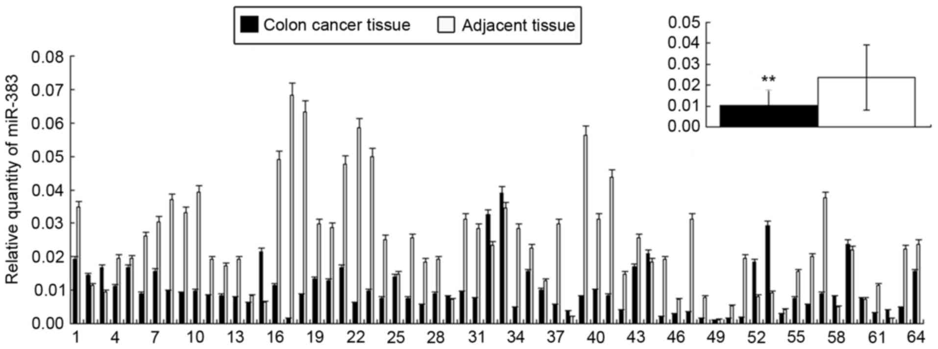
miR-383 expression is decreased in
colon cancer tissue
The relative expression of miR-383 in colon cancer
tissues was
1.04x10⁻²±7.54x10⁻³,
significantly lower than in adjacent tissues
(2.57x10⁻²±1.42x10⁻³; Fig. 1; P<0.01). In addition, the
expression of miR-383 was negatively associated with the
pathological type and TNM staging of patients with colon cancer
(P<0.05), whereas it was not associated with patient sex or age,
or tumor location and differentiation (Table I).
 | Table I.Association of clinical features with
the relative expression of miR-383, as calculated with the
χ2 test. |
Table I.
Association of clinical features with
the relative expression of miR-383, as calculated with the
χ2 test.
| Clinical feature | n | Relative expression
of miR-383, mean ± standard deviation | P-value |
|---|
| Sex |
|
| 0.374 |
|
Male | 36 |
1.07×10⁻²±8.03×10⁻³ |
|
|
Female | 28 |
1.01×10⁻²±6.98×10⁻³ |
|
| Age, years |
|
| 0.215 |
|
<60 | 30 |
9.58×10⁻²±8.75×10⁻³ |
|
|
≥60 | 34 |
1.11×10⁻²±6.32×10⁻³ |
|
| Tumor location |
|
| 0.458 |
| Left
hemicolon | 26 |
1.03×10⁻²±8.17×10⁻³ |
|
| Right
hemicolon | 38 |
1.05×10⁻³±4.41×10⁻³ |
|
| Pathological
type |
|
| <0.001 |
|
Adenocarcinoma | 45 |
1.24×10⁻²±7.68×10⁻³ |
|
|
Mucinous carcinoma | 19 |
5.56×10⁻³±4.41×10⁻³ |
|
| Differentiation
degree |
|
| 0.052 |
|
Low | 16 |
8.46×10⁻³±4.36×10⁻³ |
|
|
High/medium | 48 |
1.11×10⁻²±8.26×10⁻³ |
|
| Tumor, node,
metastasis stage |
|
| 0.005 |
| I, II
or III | 55 |
1.42×10⁻²±7.26×10⁻³ |
|
| IV | 9 |
4.16×10⁻³±4.09×10⁻⁴ |
|
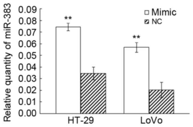
Confirmation of transfection with a
miR-383 mimic
The RT-qPCR results demonstrated that following the
transfection with an rno-miR-383 mimic, the relative expression
miR-383 level was 7.43×10⁻²±3.31×10⁻³ in the
HT-29 experimental group, and
3.46×10⁻²±5.42×10⁻³ in the negative control
group (P<0.01); in LoVo cells, the relative expression level was
5.68x10⁻²±4.25x10⁻³ in the
experimental group, and
2.03x10⁻²±6.57x10⁻³ in the
control group (Fig. 2; P<0.01).
These results suggest that transfection with an rno-miR-383 mimic
effectively upregulated miR-383 expression in colorectal cancer
cell lines.
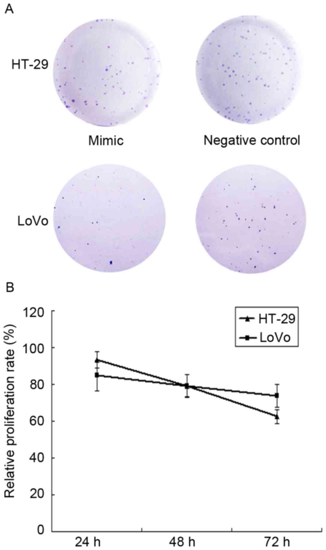
Effect of miR-383 on the proliferation
of HT-29 and LoVo cells
A colony formation assay was used to determine the
cell proliferation ability at 2 weeks. HT-29 and LoVo cell lines
with overexpressed miR-383 exhibited a relative colony formation
ability of 71.2±12.7, 26.4±7.8, respectively (P<0.05) compared
with the negative control groups; the colony formation ability of
the negative control groups were 125.3±18.9, 59.6±11.3; Compared
with the negative group, the colony formation ability of the
experimental group was significantly decreased (HT-29, P=0.0328;
LoVo, P=0.0162), as demonstrated in Fig.
3A.
Based on the MTT assay, subsequent to being cultured
for 24, 48 and 72 h, the relative proliferation rates of HT-29
cells in the experimental group were 93.3±4.27%, 78.9±6.23% and
62.5±3.62%, respectively (P<0.05) compared with the negative
control group; the relative proliferation rates in the LoVo cells
experimental group were 84.7±8.23%, 79.2±5.97% and 73.6±6.24%
(Fig. 3B; P<0.05).
These results indicated that the upregulation of
miR-383 expression inhibited the proliferation of HT-29 and LoVo
colon cancer cells.
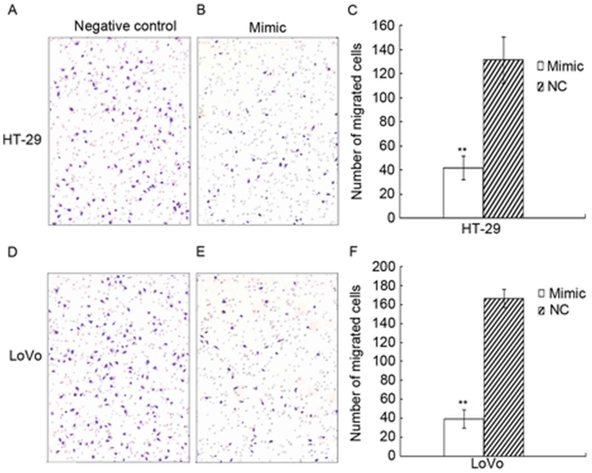
Effect of miR-383 on the migration and
invasion of HT-29 and LoVo cells
In the migration assay, the number of cells on the
lower membrane surface in the HT-29 experimental group was
41.5±9.6, whereas it was 131.3±19.2 for the control group;
therefore, the migration capacity decreased by 68.4% (P=0.031). The
number of lower surface cells in the LoVo experimental group was
39.6±9.7, whereas it was 166.2±23.9 in the control group;
therefore, the migtarion capacity decreased by 76.2% (P=0.016;
Fig. 4).
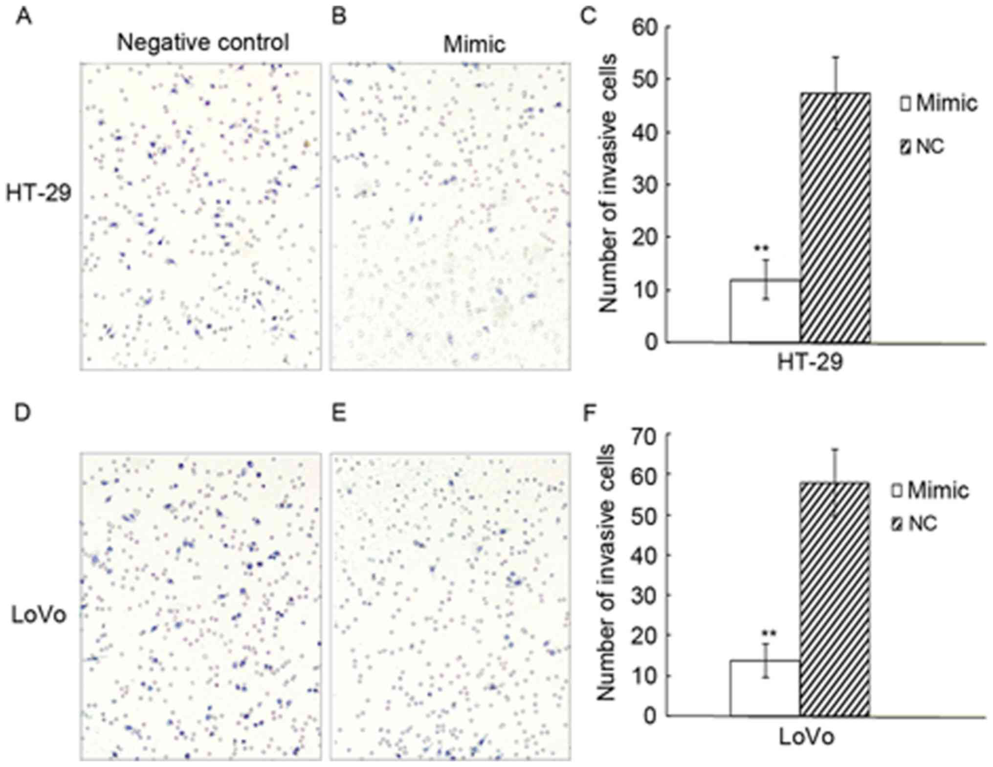
In the invasion assay, the number of cells invading
across the membrane in the HT-29 experimental group of was
11.9±3.7, whereas it was 47.3±6.9 in the negative control group;
the invasion capacity decreased by 74.8% (P=0.004). The number of
invading cells in the LoVo experimental group was 13.7±4.1, whereas
it was 57.9±8.2 in the negative control group; the invasion ability
decreased by 76.4% (P=0.006; Fig. 5).
In conclusion, the migratory and invasive capacity of HT-29 and
LoVo cells transfected with rno-miR-383 mimic was reduced compared
with the control group.
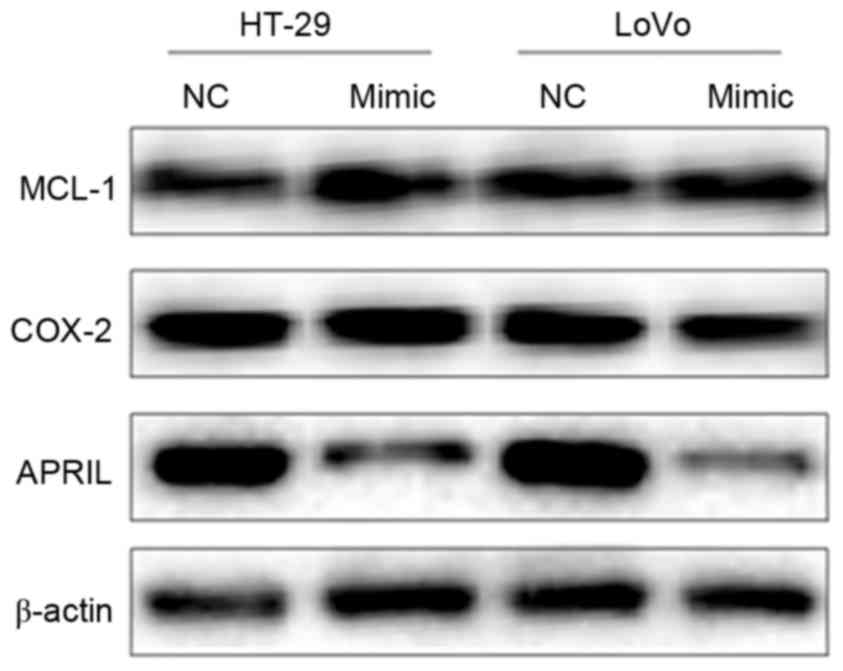
miR-383 inhibits the expression of
APRIL
Western blot analysis (Fig. 6) demonstrated that APRIL bands were
narrower and less intense in the HT-29 and LoVo experimental
groups, compared with the negative control groups and the β-actin
reference. By contrast, MCL-1 and COX-2 bands exhibited no clear
change. This suggested that the expression of APRIL decreased,
whereas the expression of MCL-1 and COX-2 remained unaltered,
indicating that miR-383 may regulate the expression of APRIL in
HT-29 and LoVo colon cancer cells.
Discussion
MiRNAs are small non-coding RNAs composed of 18–25
nucleotides. MiRNAs can bind the 3′UTR of their target mRNAs to
induce the degradation or inhibition of transcription, resulting in
the regulation of the target gene function (4). A number of miRNAs may regulate cell
migration, invasion and metastasis, including miR-1246 and miR-145
(26). MiRNA can also regulate the
differentiation, proliferation and apoptosis of cells; therefore,
alterations in expression can lead to disease, including gastric,
bladder and colon cancer (27).
MiRNAs serve a pivotal role in the occurrence and development of
colon cancer (5,6). For example, miR-34a can inhibit the
proliferation and metastasis of colon cancer cells by inhibiting
platelet growth factor receptor (28). Another study revealed that miR-381 was
significantly downregulated in human colon cancer tissues (29). A nuclear receptor, liver receptor
homolog 1, is a target gene of miR-381, and the downregulation of
miR-381 is associated with colon cancer progression (29).
MiR-383, with the sequence for has-miR-383 is
CACGAAAGATCAGAAGGTGATTG, has been the subject of previous studies,
although there is little evidence to demonstrate an association
between miR-383 expression and tumor development. miR-383 is
considered to be a tumor suppressor gene. For example, a previous
study demonstrated that miR-383 expression was suppressed in
hepatocellular carcinoma, and was significantly associated with
tumor size, lymph node metastasis and the overall survival of
patients with liver cancer (9).
Further experiments indicated that miR-383 may have inhibited the
growth of liver cancer cells by downregulating the expression of
APRIL (9). This result is similar to
the results of the present study; the present study confirmed that
miR-383 expression was relatively low in colon cancer tissue, and
that miR-383 expression was associated with the pathological type
and TNM stage of colon cancer.
In order to confirm that miR-383 can inhibit the
development of colon cancer, an rno-miR-383 mimic was transfected
into HT-29 and LoVo cells and the proliferation, migration and
invasion rates were determined. It was identified that the
upregulation of miR-383 suppressed proliferation and inhibited cell
migration and invasion in the cell lines. This is consistent with
the effect of miR-383 in medulloblastoma (7), glioma (8)
and hepatocellular carcinoma (9),
confirming the role of miR-383 as a tumor suppressor gene. The
further exploration of the biological significance of miR-383 in
colon cancer and the mechanism for interaction between miR-383 and
target genes may provide novel ideas for clinical treatment.
APRIL is a member of the tumor necrosis factor
family, and consists of 250 amino acids encoded by a gene located
at chromosome 17p13.1 (30). Previous
studies have confirmed that APRIL is highly expressed in malignant
tumors, and that its expression is associated with invasion and
metastasis (12,30). APRIL promotes the invasion and
metastasis of colon cancer by upregulating the expression of matrix
metalloproteinases (31). Chen et
al (9) revealed that miR-383 may
downregulate the expression of APRIL to inhibit the proliferation
of hepatocellular carcinoma cells. Previous studies have also
indicated that miR-383 may regulate COX-2 (22) and MCL-1 (23). In the present study, western blot
analysis revealed that the expression of APRIL was decreased in
cells with upregulated miR-383 compared with the control group,
whereas COX-2 and MCL-1 were not altered. At present, the
association between miR-383, and APRIL, MCL-1 and COX-2 expression
in colon cancer has not been confirmed. The results of the present
study suggest that miR-383 may be associated with the
downregulation of APRIL expression, which may inhibit the invasion,
migration and proliferation of colon cancer cells.
In conclusion, the expression of miR-383 in colon
cancer tissue was decreased, and its expression was associated with
the pathological type and TNM stage of colon cancer. The
upregulated expression of miR-383 in colon cancer cells inhibited
proliferation, migration and invasion. The mechanism for the effect
of miR-383 may be mediated by the downregulation of APRIL. In-depth
study of the interaction between miR-383 and APRIL may provide
insight into the mechanism of tumor suppression by miR-383.
References
|
1
|
Jemal A, Siegel R, Xu J and Ward E: Cancer
statistics, 2010. CA Cancer J Clin. 60:277–300. 2010. View Article : Google Scholar : PubMed/NCBI
|
|
2
|
Fritzmann J, Morkel M, Besser D, Budczies
J, Kosel F, Brembeck FH, Stein U, Fichtner I, Schlag PM and
Birchmeier W: A colorectal cancer expression profile that includes
transforming growth factor inhibitor BAMBI predicts metastatic
potential. Gastroenterology. 137:165–175. 2009. View Article : Google Scholar : PubMed/NCBI
|
|
3
|
Di Leva G, Garofalo M and Croce CM:
MicroRNAs in cancer. Annu Rev Pathol. 9:287–314. 2014. View Article : Google Scholar : PubMed/NCBI
|
|
4
|
Bartel DP: MicroRNAs: Genomics,
biogenesis, mechanism, and function. Cell. 116:281–297. 2004.
View Article : Google Scholar : PubMed/NCBI
|
|
5
|
Heinsbroek SE, Squadrito ML, Schilderink
R, Hilbers FW, Verseijden C, Hofmann M, Helmke A, Boon L,
Wildenberg ME, Roelofs JJ, et al: MiR-511-3p, embedded in the
macrophage mannose receptor gene, contributes to intestinal
inflammation. Mucosal Immunol. 9:960–973. 2016. View Article : Google Scholar : PubMed/NCBI
|
|
6
|
Akbari A, Ghahremani MH, Mobini GR,
Abastabar M, Akhtari J, Bolhassani M and Heidari M: Down-regulation
of miR-135b in colon adenocarcinoma induced by a TGF-β receptor I
kinase inhibitor (SD-208). Ran J Basic Med Sci. 18:856–861.
2015.
|
|
7
|
Wang XM, Zhang SF, Cheng ZQ, Peng QZ, Hu
JT, Gao LK, Xu J, Jin HT and Liu HY: MicroRNA-383 regulates
expression of PRDX3 in human medulloblastomas. Zhonghua Bing Li Xue
Za Zhi. 41:547–552. 2012.(In Chinese). PubMed/NCBI
|
|
8
|
Zhao L, Gu H, Chang J, Wu J, Wang D, Chen
S, Yang X and Qian B: MicroRNA-383 regulates the apoptosis of tumor
cells through targeting Gadd45g. PLoS One. 9:e1104722014.
View Article : Google Scholar : PubMed/NCBI
|
|
9
|
Chen L, Guan H, Gu C, Cao Y, Shao J and
Wang F: miR-383 inhibits hepatocellular carcinoma cell
proliferation via targeting APRIL. Tumour Boil. 37:2497–2507. 2016.
View Article : Google Scholar
|
|
10
|
Hahne M, Kataoka T, Schröter M, Hofmann K,
Irmler M, Bodmer JL, Schneider P, Bornand T, Holler N, French LE,
et al: APRIL, a new ligand of the tumor necrosis factor family,
stimulates tumor cell growth, stimulates tumor cell growth. J Exp
Med. 188:1185–1190. 1998. View Article : Google Scholar : PubMed/NCBI
|
|
11
|
Mhawech-Fauceglia P, Kaya G, Sauter G,
McKee T, Donze O, Schwaller J and Huard B: The source of APRIL
up-regulation in human solid tumor lesions. J Leukoc Biol.
80:697–704. 2006. View Article : Google Scholar : PubMed/NCBI
|
|
12
|
Mhawech-Fauceglia P, Allal A, Odunsi K,
Andrews C, Herrmann FR and Huard B: Role of the tumour necrosis
family ligand APRIL in solid tumour development: Retrospective
studies in bladder, ovarian and head and neck carcinomas. Eur J
Cancer. 44:2097–2100. 2008. View Article : Google Scholar : PubMed/NCBI
|
|
13
|
Lascano V, Zabalegui LF, Cameron K,
Guadagnoli M, Jansen M, Burggraaf M, Versloot M, Rodermond H, van
der Loos C, Carvalho-Pinto CE, et al: The TNF family member APRIL
promotes colorectal tumorigenesis. Cell Death Differ. 19:1826–1835.
2012. View Article : Google Scholar : PubMed/NCBI
|
|
14
|
Wang GH, Lu MH, Wang JC, Wang F, Ding WF,
Wang YG, Ju SQ and Wang HM: Abnormal expression of APRIL in
colorectal cancer cells promotes tumor growth and metastasis.
Zhonghua Zhong Liu Za Zhi. 35:249–255. 2013.(In Chinese).
PubMed/NCBI
|
|
15
|
Ding W, Wang J, Sun B, Ju S, Yuan H, Wang
X, Wang Y and Wang H: APRIL knockdown suppress migration and
invasion of human colon caner cells. Clin Biochem. 42:1694–1698.
2009. View Article : Google Scholar : PubMed/NCBI
|
|
16
|
Zhan J, Liu JP, Zhu ZH, Yao HR and Chen
CY: Relationship between COX-2 expression and clinicopathological
features of colorectal cancers. Chin Med J. 117:1151–1154.
2004.PubMed/NCBI
|
|
17
|
Basu GD, Liang WS, Stephan DA, Wegener LT,
Conley CR, Pockaj BA and Mukherjee P: A novel role for
cyclooxygenase-2 in regulating vascular channel formation by human
breast cancer cells. Breast Cancer Res. 8:R692006. View Article : Google Scholar : PubMed/NCBI
|
|
18
|
Tsujli M, Kawano S, Tsuji S, Swaoka H,
Hori M and DuBois RN: Cyclooxygenase regulates angiogenesis induced
by colon cancer cells. Cell. 93:705–716. 1998. View Article : Google Scholar : PubMed/NCBI
|
|
19
|
Akgul C: Mcl-1is a potential therapeutic
target in multiple types of cancer. Cell Mol Life Sci.
66:1326–1336. 2008. View Article : Google Scholar
|
|
20
|
Ma XT, Yu LW, Wang S, Du RY and Cui ZR:
Molecular mechanism of Stat5b signaling pathway in regulating the
expression of bcl-2 family members and promoting survival of human
colon cancer cells. Zhonghua Shi Yan Wai Ke Za Zhi. 22:1157–1169.
2005.(In Chinese).
|
|
21
|
Yoon JH, Werneburg NW, Higuchi H, Canbay
AE, Kaufmann SH, Akgul C, Edwards SW and Gores GJ: Bile acids
inhibit Mcl-1 protein turnover via an epidermal growth factor
receptor/Raf-1-dependent mechanism. Cancer Res. 62:6500–6505.
2002.PubMed/NCBI
|
|
22
|
Fan G, Jiang X, Wu X, Fordjour PA, Miao L,
Zhang H, Zhu Y and Gao X: Anti-Inflammatory Activity of Tanshinone
IIA in LPS-Stimulated RAW264.7 Macrophages via miRNAs and
TLR4-NF-κB pathway. Inflammation. 39:375–384. 2016. View Article : Google Scholar : PubMed/NCBI
|
|
23
|
Liao XH, Zheng L, He HP, Zheng DL, Wei ZQ,
Wang N, Dong J, Ma WJ and Zhang TC: STAT3 regulated ATR via
microRNA-383 to control DNA damage to affect apoptosis in A431
cells. Cell Signal. 27:2285–2295. 2015. View Article : Google Scholar : PubMed/NCBI
|
|
24
|
Sobin LH and Fleming ID: TNM
classification of malignant tumors, fifth edition (1997). Union
Internationale Contre le Cancer and the American Joint Committee on
Cancer. Cancer. 80:1803–1804. 1997. View Article : Google Scholar : PubMed/NCBI
|
|
25
|
Livak KJ and Schmittgen TD: Analysis of
relative gene expression data using real-time quantitative PCR and
the 2(-Delta Delta C(T)) method. Methods. 25:402–408. 2001.
View Article : Google Scholar : PubMed/NCBI
|
|
26
|
Lu J, Getz G, Miska EA, Alvarez-Saavedra
E, Lamb J, Peck D, Sweet-Cordero A, Ebert BL, Mak RH, Ferrando AA,
et al: MicroRNA expression profiles classify human cancer. Nature.
435:834–838. 2005. View Article : Google Scholar : PubMed/NCBI
|
|
27
|
Orellana EA and Kasinski AL: MicroRNAs in
cancer: A historical perspective on the path from discovery to
therapy. Cancers (Basel). 7:1388–1405. 2015. View Article : Google Scholar : PubMed/NCBI
|
|
28
|
Li C, Wang Y, Lu S, Zhang Z, Meng H, Liang
L, Zhang Y and Song B: MiR-34a inhibits colon cancer proliferation
and metastasis by inhibiting platelet-derived growth factor
receptor α. Mol Med Rep. 12:7072–7078. 2015. View Article : Google Scholar : PubMed/NCBI
|
|
29
|
Liang Y, Zhao Q, Fan L, Zhang Z, Tan B,
Liu Y and Li Y: Down-regulation of microRNA-381 promotes cell
proliferation and invasion in colon cancer through up-regulation of
LRH-1. Biomed Pharmacother. 75:137–141. 2015. View Article : Google Scholar : PubMed/NCBI
|
|
30
|
Roosnek E, Burjanadze M, Dietrich PY,
Matthes T, Passweg J and Huard B: Tumors that look for their
springtime in APRIL. Crit Rev Oncol Hematol. 72:91–97. 2009.
View Article : Google Scholar : PubMed/NCBI
|
|
31
|
Wang G, Wang F, Ding W, Wang X, Xu J, Li H
and Wang H: APRIL mediates migration and invasion of colorectal
cancer cell via regulation of matrix metalloproteinases. Chin J
Microbiol Immunol. 32:128–133. 2012.(In Chinese).
|