Introduction
Pancreatic cancer is a highly malignant cancer type
with a median survival of 3–6 months and a 5-year survival rate of
<5% worldwide (1–4). In 2015, compared with Western countries,
the incidence of pancreatic cancer in China increased
significantly. In 2010, 34,509 men and 23,226 women succumbed to
pancreatic cancer in China (5).
Furthermore, in 2015, 62.9% of pancreatic cancer cases are
diagnosed at stages I or II, and 37.1% of cases at stages III and
IV (6). Chemotherapy is one of the
main first-line treatments for advanced pancreatic cancer (7). However, controversy remains regarding
its use due to its invasive nature (8). Hence, it is necessary to develop
strategies, and identify novel potential pharmacological targets at
the molecular level, which are involved in the signaling pathways
associated with the occurrence, development and metastasis of
pancreatic cancer.
S100A11 is a member of S100 calcium-binding protein
family. Interaction of S100A11 with calcium leads to conformational
changes of S100A11 protein resulting in the exposure of its
hydrophobic region. This region in turn binds to the target protein
to transduce the regulated signals of calcium-dependent cells.
Thus, S100A11 protein is involved in multiple biological processes,
including cell proliferation, apoptosis, signal transduction, cell
adhesion, extracellular matrix remodeling and cell mobility
(9–11). At present, it has been demonstrated
that S100 protein is associated with the occurrence, development
and metastasis of tumors (12). The
S100 gene is located in the chromosome region Iq21. Tumor genes in
this region are frequently recombined, which may easily cause
loss-of-control of S100 gene expression (13). Furthermore, it has been demonstrated
that the expression of S100A11 is upregulated in the early stage of
pancreatic cancer and decreases with the progression of pancreatic
cancer (13). S100A11 may serve as a
significant tumor marker for pancreatic cancer and high expression
of S100A11 is an unfavorable predictor for prognosis of patients
with pancreatic cancer who have undergone surgical resection
(14–16).
The phosphoinositide 3-kinase (PI3K)/AKT
serine/threonine kinase (AKT) pathway is involved in the growth,
differentiation, and proliferation of somatic and cancer cells
(17). Activation of the PI3K/AKT
signaling pathway may result in increased tumor cell proliferation
and the inhibition of tumor cell apoptosis (17,18). As a
key protein in the pathway, AKT serves an important role in
regulating cell viability, growth and proliferation (17,18). PI3K
is an upstream effector of the PI3K/AKT signaling pathway, and may
induce the activation of AKT through phosphorylating threonine
and/or serine residues. PI3K inhibitor LY294002 is able to inhibit
AKT phosphorylation (18). It has
been reported that the PI3K/AKT signaling pathway is significantly
active in pancreatic cancer cells (19). Additionally, immunohistochemical
analysis demonstrated that the phosphorylated (p)-AKT positive rate
was 46% in pancreatic cancer tissues, which was significantly
higher compared with that in normal pancreatic tissues (17). Therefore, it has been considered that
p-AKT is associated with poor prognosis of patients with pancreatic
cancer (20). In vitro and
in vivo experiments have suggested that the blockade of the
PI3K/AKT signaling pathway significantly impairs the proliferation
of pancreatic cancer cells, promotes its apoptosis and induces cell
cycle arrest (21).
Furthermore, in normal human keratinocytes, AKT
phosphorylation is inhibited through downregulation of S100A11
expression in the cells, which leads to a decrease in AKT activity,
indicating that S100A11 is involved in AKT activation (22). Therefore, we hypothesized that S100A11
is associated with the PI3K/AKT signaling pathway in the occurrence
and development of pancreatic cancer. Thus, the present study aimed
to investigate the effects of S100A11 overexpression on cell
proliferation, cell apoptosis and cell cycle distribution in
pancreatic cancer cells, and to explore potential mechanisms
associated with the PI3K/AKT signaling pathway.
Materials and methods
Patients and tissue specimens
Pancreatic paraffin samples were provided by the
Department of Pathology, Affiliated Hospital of Nantong University
(Nantong, China). There were 30 resection specimens from patients
with pancreatic cancer hospitalized between January 2010 and June
2013 (male:female, 17:13; median age, 67 years; age range, 41–85
years. The incised margins were all >1 cm. Pathological
diagnosis of all the cases was clear, and all the
clinicopathological data were complete. None of the patients
recruited in the present study had received radiotherapy or
chemotherapy, or any other treatment prior to surgery. The Clinical
Research Ethics Committee of the Affiliated Hospital of Nantong
University approved the present study. All patients provided
written informed consent for the use of their medical records and
tissue specimens for research purposes.
Immunohistochemical analysis
The tissue sections were deparaffinized in xylene
twice for 5 min at room temperature and then rehydrated using a
graded ethanol series (100, 95, 80 and 70%; 5 min at room
temperature for each concentration). Subsequently, the endogenous
peroxidase activity was blocked by soaking in 0.3% hydrogen
peroxide for 10 min at room temperature. Thereafter, the sections
were processed in 10 mmol/l citrate buffer (pH 6.0) and were heated
to 121°C in an autoclave for 20 min to retrieve the antigen. After
being rinsed with PBS (pH 7.2), 10% goat serum (Seebio
Biotechnology Co. Ltd, Shanghai, China) was added and incubated at
room temperature for 1 h to block the non-specific reactions. The
sections were then incubated overnight at 4°C with mouse anti-human
S100A11 monoclonal antibody (cat. no. WH0006282M1; diluted 1:100;
Sigma-Aldrich; Merck KGaA, Darmstadt, Germany) and rabbit
anti-human p-Akt monoclonal antibody (cat. no. 4060; diluted 1:100;
Cell Signaling Technology Inc., Danvers, MA, USA). Negative control
slides were also processed in parallel using a non-specific IgG
(cat. no. 18015; Sigma-Aldrich; Merck KGaA) at the same
concentration as the primary antibody. All slides were processed
using the peroxidase antiperoxidase method (Dako; Agilent
Technologies, Inc., Santa Clara, CA, USA). After being rinsed with
PBS, the peroxidase reaction was visualized by incubating the
sections with 3,3-diaminobenzidine tetrahydrochloride for 5 min at
room temperature. The sections were then rinsed with water,
counterstained with hematoxylin for 1 min at room temperature, then
dehydrated and coverslipped.
Immunohistochemical evaluation
All of the immunostained sections were evaluated in
a blinded manner by three independent experienced observers without
any knowledge on the clinicopathological features of the patients.
For assessment of S100A11 and p-AKT, five fields (magnification,
×40) in each specimen were selected randomly, and nuclear staining
was examined using a light microscope. By counting the number of
cells per field of view, the rate of cells expressing S100A11 and
p-AKT was calculated; meanwhile, the staining intensity was
observed. The positively stained areas were noted as those with
light yellow to brown particles, and the specific expression in
each case was defined using the staining intensity score and
percentage of positively stained cells. The staining intensity
scoring (according to the degree of color) was as follows: 0, no
staining; 1, pale yellow diffused distribution from cytoplasm to
cytoplasmic membrane; 2, brown distribution with fine granular; 3,
brown or dark brown distribution with coarse granular from
cytoplasm to cytoplasmic membrane as well as nucleus. The
percentage of positively stained cells was categorized as follows:
0, <5%; 1, 5–25%; 2, 26–50%; 3, 51–75%; 4, >75%. The score of
each patient was calculated as the staining intensity score ×
positively stained cells score. In 50% of the samples, staining was
repeated twice to avoid technical errors, but similar results were
obtained in these samples.
Cell culture
The human pancreatic cancer PANC-1, Bxpc-1 and
AsPC-1 cell lines were purchased from the Type Culture Collection
of the Chinese Academy of Sciences (Shanghai, China). Cells were
maintained in RPMI-1,640 supplemented with 10% heat-inactivated
fetal calf serum, 2 mM L-glutamine, and 100 U/ml penicillin and
streptomycin mixture (all from Gibco, Thermo Fisher Scientific,
Inc., Waltham, MA, USA) at 37°C with 5% CO2.
S100A11 overexpression
RNA was extracted from PANC-1 using the RNeasy Mini
kit (Qiagen, Inc., Valencia, CA, USA), and cDNA was synthesized via
reverse transcription (RT) with the Sensiscript RT kit (Qiagen,
Inc.) according to the manufacturer's protocol. The cDNA was used
as the template to amplify the S100A11 gene using the S100A11
primers as follows: Forward,
5′-CTCGGATCCGCCACCATGGCAAAAATCTCCAGCCCTACAG-3′ (BamH I) and
reverse, 5′-GCCGCTCGAGGGGTCCTCAGGTCCGCTTCTG-3′ (Xhol I). The
control gene (β-actin) primers were as follows: Forward,
5′-AAGTACTCCGTGTGGATCGG-3′ and reverse, 5′-ATGCTATCACCTCCCCTGTG-3′.
The polymerase chain reaction (PCR) theremocycling parameters were
as follows: 95°C for 30 sec followed by 35 cycles of 94°C for 45
sec and 55°C for 45 sec. The PCR product was run using agarose gel
electrophoresis, and was then extracted and purified. The purified
DNA and pcDNA3.1/myc-HisA (eukaryotic expression vector,
Invitrogen; Thermo Fisher Scientific, Inc.) were digested using the
restriction enzymes BamH I-HF, Xhol I (both from New
England BioLabs, Inc., Ipswich, MA, USA). The digested insert (PCR
product) and vector (pcDNA3.1/myc-HisA) were ligated using T4 DNA
ligase (New England BioLabs, Inc.). Subsequently, the ligation
product was transformed into E.coli competent cells and the
competent cells were plated onto Luria-Bertani broth (LB) plates.
On the second day, the white colonies were picked from the LB
plates, followed by plasmid extraction and sequencing. Finally, the
plasmid was named as pcDNA3.1-S100A11. Lipofectamine 2000
(Invitrogen; Thermo Fisher Scientific, Inc.) was used to transfect
PANC-1 cells with 2.5 µg pcDNA3.1-S100A11 and pcDNA3.1 empty
vector.
Drug application
PANC-1 cells were seeded into 96-well plates
(5×10³ cells/well). After 24 h, the medium was removed
and replaced with LY294002 (Beyotime Institute of Biotechnology,
Haimen, China) dissolved in 50 µmol/l dimethylsulfoxide (DMSO)
according to a previous protocol (19). The concentration of LY294002 used in
experiments was 50 µmol/l, and was incubated with cells for 48 h at
37°C.
Cell proliferation assay
Cell proliferation was measured by a commercial Cell
Counting kit (CCK)-8 (Dojindo Molecular Technologies, Inc.,
Kumamoto, Japan) following the manufacturer's protocol. Briefly,
PANC-1 cells were seeded onto 96-well cell culture plates at a
concentration of 2×10⁴ cells/well in 100 µl culture
medium as aforementioned, and were grown overnight. CCK-8 reagent
(10 µl) was added to a subset of wells and incubated for 2 h at
37°C and the absorbance reading was taken using an automated plate
reader under the detection wavelength of 450 nm.
Cell cycle distribution determined by
flow cytometric analysis
For cell cycle analysis, cells were harvested,
washed once in PBS, fixed with 70% ethanol at −20°C overnight and
then incubated with 1 mg/ml RNase A for 30 min at 37°C.
Subsequently, the cells were stained with propidium iodide (PI; 50
µg/ml; BD Biosciences) in PBS-Triton X100 for an additional 20 min
at 4°C, and analyzed using the BD FACScan flow cytometer with
CellQuest acquisition and analysis programs (FlowJo 10; FlowJo LLC,
Ashland, OR, USA). Finally, the proportions of cells in the G0/G1,
S and G2/M phases were determined.
Cell apoptosis determined by flow
cytometric analysis
PANC-1 cell apoptosis was detected using an Annexin
V-FITC/PI staining assay. Flow cytometry analysis of apoptotic
cells was performed using an Annexin V-FITC/PI staining kit (BD
Biosciences). After washing with cold PBS, the cells were
resuspended in binding buffer (100 mM HEPES, pH 7.4, 100 mM NaCl,
and 25 mM CaCl2) followed by staining with Annexin
V-FITC/PI at room temperature in the dark for 15 min. Apoptotic
cells were then evaluated by gating PI and Annexin V-positive cells
on a FACS Calibur (BD Biosciences). Annexin V was set as the
horizontal axis and PI was set for the vertical axis. Early
apoptotic cells were located in the lower right quadrant of the
flow cytometric dot plot.
Semi-quantitative RT-PCR
Total RNA from PANC-1 cells was extracted using a
TRIzol extraction kit (Invitrogen; Thermo Fisher Scientific, Inc.)
according to the manufacturer's protocol. Total RNA was reverse
transcribed using the ThermoScript RT-PCR system (Invitrogen;
Thermo Fisher Scientific, Inc.). The primer sequences were as
follows: S100A11 forward, 5′-GAGTCCCTGATTGCTGTCTTC-3′ and reverse,
5′-AGGGTCCTTCTGGTTCTTTG-3′; AKT forward,
5′-GGTATTTTGATGAGGAGTTCACG-3′ and reverse,
5′-GAGTAGGAGAACTGGGGGAAGT-3′; β-actin forward,
5′-AAGTACTCCGTGTGGATCGG-3′ and reverse, 5′-ATGCTATCACCTCCCCTGTG-3′.
Thermocycling conditions were as follows: 94°C for 45 sec, 55°C for
45 sec, 72°C for 30 sec for a total of 30 cycles. After
amplification, the products were separated on a 1.5% agarose gel
(cast in the presence of ethidium bromide) and the intensity of the
bands was analyzed with Quantity One software (version 4.62;
Bio-Rad Laboratories, Inc., Hercules, CA, USA).
Western blot analysis
PANC-1 cells were lysed in the lysis buffer (1 M
Tris-HCl at pH 7.5, 1% Triton X-100, 1% Nonidet P-40, 10% SDS, 0.5%
sodium deoxycholate, 0.5 M EDTA, 10 µg/ml leupeptin, 10 µg/ml
aprotinin, and 1 mM phenylmethylsulfonyl fluoride) and then
centrifuged at 4°C, 10,000 × g for 30 min to collect the
supernatant. Protein concentration was determined using the Bio-Rad
protein assay kit (Bio-Rad Laboratories, Inc.). Equal amounts (0.6
mg/lane) of protein were separated with 10% gel using SDS-PAGE and
transferred to polyvinylidine difluoride filter membranes (EMD
Millipore, Billerica, MA, USA). Membranes were then blocked with 5%
non-fat milk in TBS with 0.05% Tween 20 for 2 h at room
temperature. Subsequently, the membrane was incubated for 12 h at
4°C with primary antibodies, including anti-S100A11 (1:500; cat.
no. WH0006282M1; Sigma-Aldrich; Merck KGaA), anti-p-AKT (1:500;
cat. no. 4060; Cell Signaling Technology, Inc.), anti-AKT (1:1,000;
cat. no. 4685; Cell Signaling Technology, Inc.) and anti-β-actin
(1:500; Santa Cruz Biotechnology Inc., Dallas, TX, USA). Next,
membranes were incubated with horseradish peroxidase-conjugated IgG
(1:4,000; anti-mouse IgG, cat. no. 7076; anti-rabbit IgG, cat. no.
7074; Santa Cruz Biotechnology, Inc.) for 1 h at room temperature.
Immunoblotting bands were visualized by chemiluminescence (NEN Life
Science Products, Inc., Boston, MA, USA). The band density was
measured using a computer-assisted image-analysis system (Image Lab
Software 5.2.x; Bio-Rad Laboratories, Inc.) and was normalized
against the β-actin level.
Statistical analysis
All analyses were performed using SPSS 19.0 software
(IBM, Corp., Armonk, NY, USA). Data are presented as the mean ±
standard error of mean of at least three independent experiments.
For statistical analysis, data analysis was performed using a
Student's t-test or one-way analysis of variance with the
Student-Newman-Keuls test. The expression levels of S100A11 and
p-AKT in the pancreatic tissues were analyzed using Pearson's
correlation analysis. P<0.05 was considered to indicate a
statistically significant difference.
Results
Correlation between S100A11 and p-AKT
expression in human pancreatic paraffin tissue samples
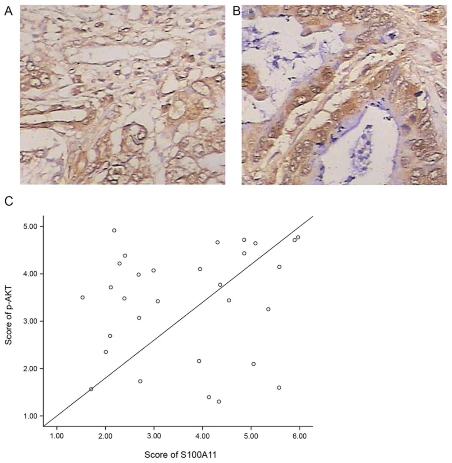
Immunohistochemical analysis revealed that S100A11
was highly expressed in the cytoplasm and to a less extent in the
nucleus, while p-AKT was expressed primarily in the cytoplasm with
brown yellow or brown staining (Fig. 1A
and B). Pearson correlation analysis revealed that the levels
of S100A11 and p-AKT were positively correlated (r, 0.802;
P<0.05; Fig. 1C) among the 30
cases of pancreatic tissues.
Effects of S100A11 overexpression on
cell proliferation, cell apoptosis and cell cycle distribution in
PANC-1 cells
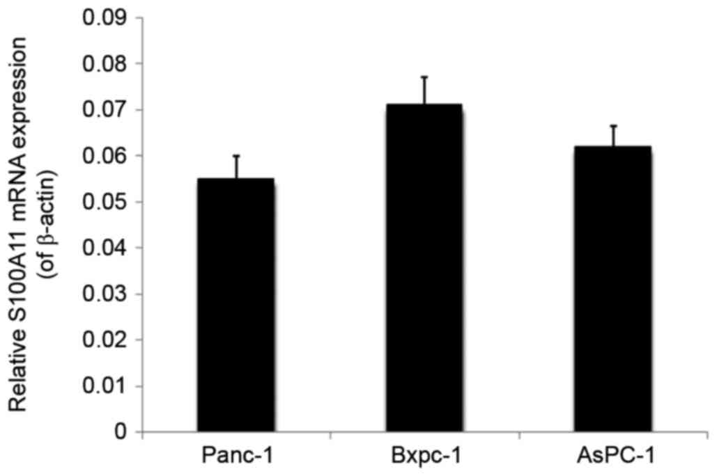
To examine the role of S100A11 on the proliferation,
apoptosis and cell cycle regulation of pancreatic cancer cells,
semi-quantitative RT-PCR was used to analyze the expression profile
of S100A11 in different pancreatic cancer cell lines including
PANC-1, Bxpc-1 and AsPC-1 cells. The results demonstrated that
PANC-1 had a relative low expression of endogenous S100A11 among
these cell lines (Fig. 2).
Accordingly, PANC-1 cell line was chosen for subsequent S100A11
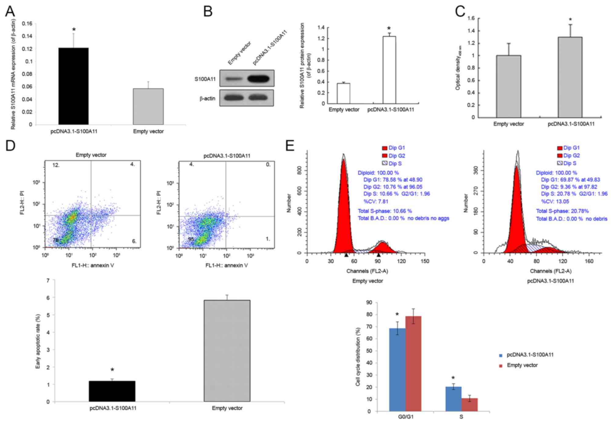
overexpression experiments. Compared with the control group, the
expression of S100A11 mRNA and protein in the cells transfected
with pcDNA3.1-S100A11 was significantly increased (Fig. 3A and B). Through the CCK-8 experiment,
the optical density was measured. Compared with cells transfected
with the pcDNA3.1 empty vector, transfection with 2.5 µg
pcDNA3.1-S100A11 significantly promoted the proliferation of PANC-1
cells (P<0.05; Fig. 3C).
Furthermore, flow cytometry analysis was used to determine the
level of apoptotic cell death of control and S100A11-overexpressing
PANC-1 cells. The early apoptotic rate (%) in the lower right
quadrant of the flow cytometric dot plot in the cells transfected
with pcDNA3.1-S100A11 was significantly reduced (1.18±0.12 vs.
5.83±0.30% in the control; P<0.05; Fig. 3D). To further explore the involvement
of S100A11 in the proliferation of pancreatic cancer cells, the
cell cycle distribution of control and S100A11-overexpressing
PANC-1 cells was detected (Fig. 3E).
The proportion of cells in G0/G1 phase in pcDNA3.1-S100A11 and
pcDNA3.1 groups was 68.81±5.42, and 78.68±6.21%, respectively
(P<0.05), while the proportion of cells in the S phase was
20.48±2.37 and 10.81±2.65%, respectively (P<0.05).
Effects of S100A11 overexpression on
AKT mRNA expression, p-AKT and total AKT protein expression in
PANC-1 cells
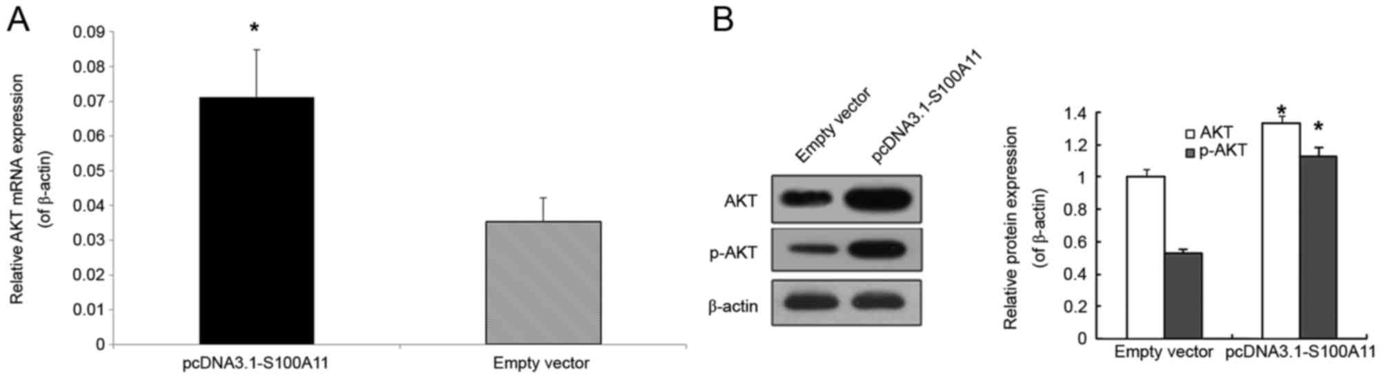
To further investigate the association between
S100A11 and AKT, the mRNA and protein expression levels of AKT were
analyzed. Compared with the empty vector group, AKT mRNA expression
was significantly higher in the pcDNA3.1-S100A11 group (Fig. 4A; P<0.05). In addition, the protein
levels of p-AKT and total AKT were significantly higher in the
S100A11 overexpression group (Fig.
4B; P<0.05).
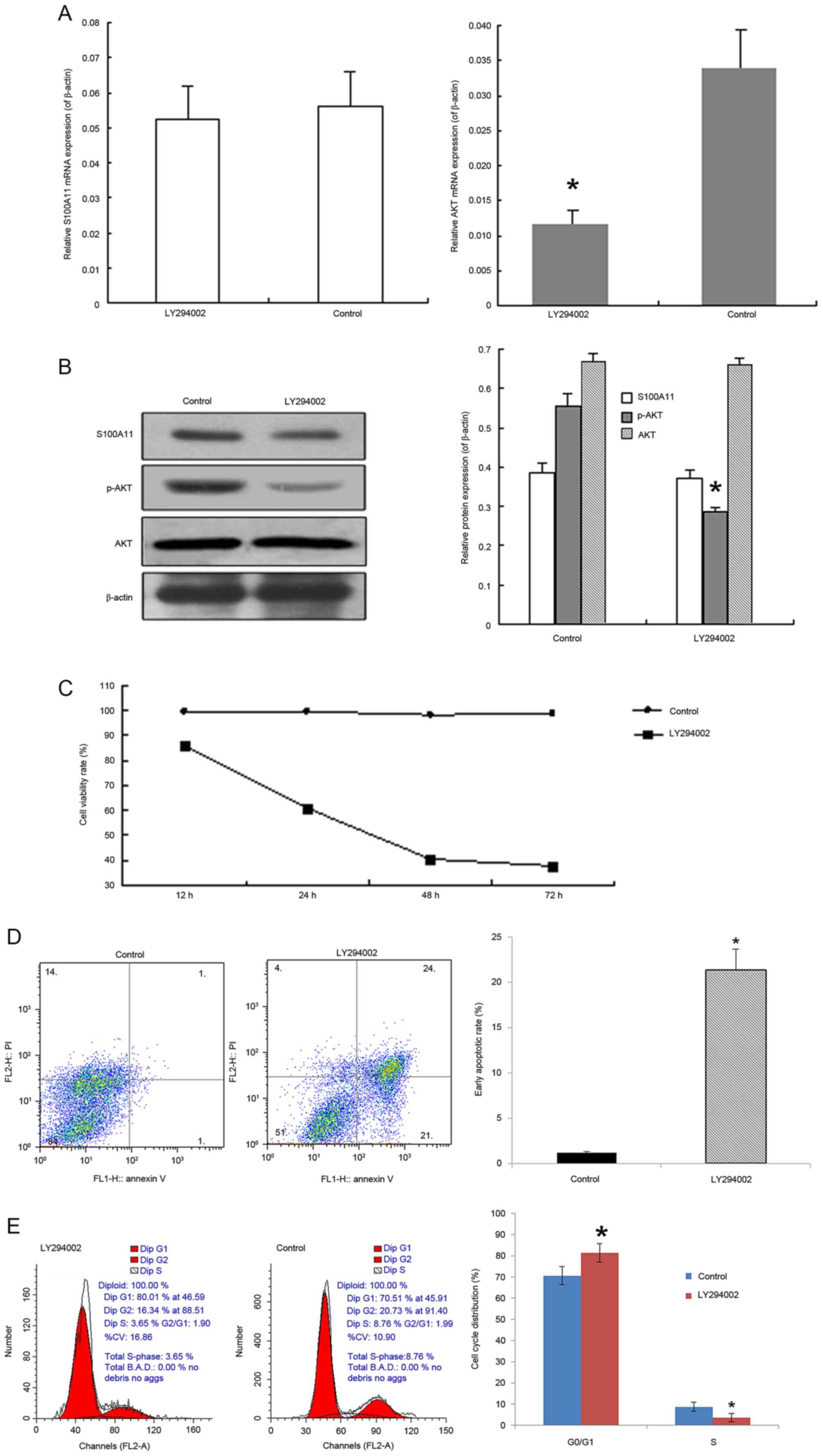
Effects of PI3K inhibitor
LY294002
LY294002 significantly inhibited the AKT mRNA
expression and p-AKT protein expression when compared with the
corresponding negative controls (Fig. 5A
and B; P<0.05). However, no significant differences were
noted in the mRNA of S100A11, and protein expression levels of
S100A11 and total AKT (Fig. 5A and
B). PANC-1 cells were treated with 50 µmol/l of LY294002 for
12, 24, 48 and 72 h, followed by CCK-8 analysis. It was
demonstrated that LY294002 treatment resulted in a dose-dependent
decrease in cell viability from 86.2% at 12 h to 37.4% at 72 h
(Fig. 5C). As LY294002 treatment
inhibited cell viability, the effect of LY294002 on PANC-1 cell
apoptosis and cell cycle distribution was further examined. The
proportion of early apoptotic cells was 21.36±2.34% in the lower
right quadrant of the scatter plot in the LY294002 group, which was
significantly higher compared with the proportion of early
apoptotic cells in the control group (1.18±0.12%; P<0.05;
Fig. 5D). Compared with the control
group, LY294002 treatment led to an increase of cells in the G0/G1
phase from 70.62±4.21 to 81.51±4.33%, while the proportion of cells
in the S phase significantly decreased from 8.75±2.13 to 3.61±2.05%
(both P<0.05; Fig. 5E).
Discussion
S100A11 is widely expressed in multiple tissues and
exists as a noncovalent homodimer with an anti-parallel
conformation. The binding of Ca2+ triggers
conformational changes of S100A11, which facilitates its
interaction with target proteins. S100A11 has been reported to be
overexpressed in numerous different types of human cancer,
including renal cell carcinoma, lung adenocarcinoma and papillary
thyroid carcinoma (23–26). Our previous study revealed that
S100A11 serves an important role in the development and prognosis
of pancreatic cancer (15). However,
the exact role of S100A11 in the pancreatic cancer remained
unknown. In order to investigate the molecular mechanism underlying
the tumor-promoting role of S100A11 in the occurrence and
development of pancreatic cancer, an in vitro model of
S100A11 overexpression in pancreatic cancer PANC-1 cells was
successfully constructed. High expression levels of S100A11 are
able to regulate cell cycle progression and the proliferation of
pancreatic cancer cells (12,14). The results of the present study
revealed that the overexpression of S100A11 significantly promoted
human pancreatic cancer PANC-1 cell proliferation, reduced the
percentage of early apoptotic cells, and enhanced the G1/S phase
transition of pancreatic cancer cells. These results were
consistent with previous studies of ovarian and lung tumors
(27,28).
The PI3K/AKT signaling pathway serves an important
function in the development of pancreatic cancer (19–21).
However, it remains unclear whether the PI3K/AKT signaling pathway
participated in the action of S100A11 in pancreatic cancer.
Firstly, immunohistochemistry was used to analyze the correlation
between S100A11 and p-AKT, which suggested a positive correlation
between of S100A11 and p-AKT expression in the pancreatic cancer
tissues of 30 cases. Furthermore, using an in vitro cell
model, it was demonstrated that the overexpression of S100A11
significantly increased AKT mRNA expression, and total AKT and
p-AKT protein expression. In order to further investigate the
association between S100A11 and the PI3K/AKT signaling pathway in
pancreatic cancer, PANC-1 cells were treated with LY294002, a
specific inhibitor of PI3K. Through the specific inhibition of PI3
catalytic activity, LY294002 treatment leads to the
dephosphorylation of downstream AKT at T308 and S473 residues
(29). It has been acknowledged that
the dephosphorylation of AKT inhibits cell growth and induces cell
apoptosis. In the current study, LY294002 was employed to inhibit
the phosphorylation of AKT, thereby specifically inhibiting the
PI3K/AKT pathway. The results from the present study revealed that
LY294002 treatment significantly inhibited cell proliferation, and
promoted cell apoptosis and cell cycle arrest in the G0/G1 phase in
PANC-1 cells. These results were supported by previous studies in
human keratinocyte cells, which reported that the PI3K/AKT
signaling pathway is involved in S100A11-triggered signal
transduction (22,30). Of note, it was demonstrated that
treatment with LY294002 significantly attenuated AKT mRNA
expression and the protein expression of p-AKT. However, the mRNA
of S100A11, and protein expression levels of S100A11 and total AKT
were not affected. Through AKT is rapidly activated through protein
phosphorylation, certain studies have also documented that
increased levels of total AKT is a hallmark of tumor cells
(31–33). The mRNA and protein levels of AKT are
regulated by complex mechanisms, and involve numerous signaling
molecules, including glycogen synthase kinase (GSK)-3β, Twist,
microRNA (miR)-185 and miR-203 (34–37).
Therefore, we hypothesized that LY294002 may regulate the
activities of these molecules, particularly GSK-3β, which is a
well-known AKT substrate, thus exerting an influence on AKT mRNA
expression, but not on the total AKT protein expression. However,
the potential mechanism underlying this effect remains elusive and
warrants further investigation.
To conclude, the results of the current study
demonstrated that S100A11 promoted human pancreatic cancer PANC-1
cell proliferation through the PI3K/AKT signaling pathway. LY294002
was able to significantly inhibit the proliferation of PANC-1
cells, and induce apoptosis through the PI3K/AKT pathway without
affecting the expression of S100A11 mRNA and protein levels,
suggesting that existence of S100A11 activity upstream of the
PI3K/Akt signaling pathway. However, the specific mechanism
regarding the role of S100A11 in the development of pancreatic
cancer requires further studies. S100A11 may serve as a potentially
effective target in pancreatic cancer therapy.
Acknowledgements
The present study was supported by grants from the
Natural Youth Science Foundation of China (grant no. 81502055), The
Health Project of Jiangsu Province (grant no. H201624), The Natural
Science Foundation of Jiangsu Province (grant no. BK20161286) and
The Social Development Foundation of Nantong City (grant nos.
MS22016056, MS22015062, HS2014072 and MS22015044).
References
|
1
|
Ryan DP, Hong TS and Bardeesy N:
Pancreatic adenocarcinoma. N Engl J Med. 371:1039–1049. 2014.
View Article : Google Scholar : PubMed/NCBI
|
|
2
|
Zell JA, Rhee JM, Ziogas A, Lipkin SM and
Anton-Culver H: Race, socioeconomic status, treatment and survival
time among pancreatic cancer cases in California. Cancer Epidemiol
Biomarkers Prev. 16:546–552. 2007. View Article : Google Scholar : PubMed/NCBI
|
|
3
|
Lau MK, Davila JA and Shaib YH: Incidence
and survival of pancreatic head and body and tail cancers: A
population-based study in the United States. Pancreas. 39:458–462.
2010. View Article : Google Scholar : PubMed/NCBI
|
|
4
|
Quaresma M, Coleman MP and Rachet B:
40-year trends in an index of survival for all cancers combined and
survival adjusted for age and sex for each cancer in England and
Wales, 1971–2011: A population-based study. Lancet. 385:1206–1218.
2015. View Article : Google Scholar : PubMed/NCBI
|
|
5
|
Lin QJ, Yang F, Jin C and Fu DL: Current
status and progress of pancreatic cancer in China. World J
Gastroenterol. 21:7988–8003. 2015. View Article : Google Scholar : PubMed/NCBI
|
|
6
|
Yu J, Blackford AL, Dal Molin M, Wolfgang
CL and Goggins M: Time to progression of pancreatic ductal
adenocarcinoma from low-to-high tumour stages. Gut. 64:1783–1789.
2015. View Article : Google Scholar : PubMed/NCBI
|
|
7
|
Polyak K, Lee MH, Erdjument-Bromage H,
Koff A, Roberts JM, Tempst P and Massagué J: Cloning of p27Kip1, a
cyclin-dependent kinase inhibitor and a potential mediator of
extracellular antimitogenic signals. Cell. 78:59–66. 1994.
View Article : Google Scholar : PubMed/NCBI
|
|
8
|
Yang F, Di Y, Li J, Wang XY, Yao L, Hao
SJ, Jiang YJ, Jin C and Fu DL: Accuracy of routine multidetector
computed tomography to identify arterial variants in patients
scheduled for pancreaticoduodenectomy. World J Gastroenterol.
21:969–976. 2015. View Article : Google Scholar : PubMed/NCBI
|
|
9
|
Arumugam T and Logsdon CD: S100P: A novel
therapeutic target for cancer. Amino Acids. 41:893–899. 2011.
View Article : Google Scholar : PubMed/NCBI
|
|
10
|
Hung KW, Chang YM and Yu C: Resonance
assignments of Ca2+-bound human S100A11. Biomol NMR
Assign. 7:211–214. 2013. View Article : Google Scholar : PubMed/NCBI
|
|
11
|
Sakaguchi M and Huh NH: S100A11, a dual
growth regulator of epidermal keratinocytes. Amino Acids.
41:797–807. 2011. View Article : Google Scholar : PubMed/NCBI
|
|
12
|
Ji YF, Huang H, Jiang F, Ni RZ and Xiao
MB: S100 family signaling network and related proteins in
pancreatic cancer (Review). Int J Mol Med. 33:769–776. 2014.
View Article : Google Scholar : PubMed/NCBI
|
|
13
|
Ohuchida K, Mizumoto K, Ohhashi S,
Yamaguchi H, Konomi H, Nagai E, Yamaguchi K, Tsuneyoshi M and
Tanaka M: S100A11, A putative tumor suppressor gene, is
overexpressed in pancreatic carcinogenesis. Clin Cancer Res.
12:5417–5422. 2006. View Article : Google Scholar : PubMed/NCBI
|
|
14
|
Chen H, Xu C, Jin Q and Liu Z: S100
protein family in human cancer. Am J Cancer Res. 4:89–115.
2014.PubMed/NCBI
|
|
15
|
Xiao MB, Jiang F, Ni WK, Chen BY, Lu CH,
Li XY and Ni RZ: High expression of S100A11 in pancreatic
adenocarcinoma is an unfavorable prognostic marker. Med Oncol.
29:1886–1891. 2012. View Article : Google Scholar : PubMed/NCBI
|
|
16
|
Donato R, Cannon BR, Sorci G, Riuzzi F,
Hsu K, Weber DJ and Geczy CL: Functions of S100 proteins. Curr Mol
Med. 13:24–57. 2013. View Article : Google Scholar : PubMed/NCBI
|
|
17
|
Yamamoto S, Tomita Y, Hoshida Y, Morooka
T, Nagano H, Dono K, Umeshita K, Sakon M, Ishikawa O, Ohigashi H,
et al: Prognostic significance of activated Akt expression in
pancreatic ductal adenocarcinoma. Clin Cancer Res. 10:2846–2850.
2004. View Article : Google Scholar : PubMed/NCBI
|
|
18
|
Jiang H, Fan D, Zhou G, Li X and Deng H:
Phosphatidylinositol 3-kinase inhibitor (LY294002) induces
apoptosis of human nasopharyngeal carcinoma in vitro and in vivo. J
Exp Clin Cancer Res. 29:342010. View Article : Google Scholar : PubMed/NCBI
|
|
19
|
Asano T, Yao Y, Zhu J, Li D, Abbruzzese JL
and Reddy SA: The PI 3-kinase/Akt signaling pathway is activated
due to aberrant Pten expression and targets transcription factors
NF-kappaB and c-Myc in pancreatic cancer cells. Oncogene.
23:8571–8580. 2004. View Article : Google Scholar : PubMed/NCBI
|
|
20
|
Chadha KS, Khoury T, Yu J, Black JD, Gibbs
JF, Kuvshinoff BW, Tan D, Brattain MG and Javle MM: Activated Akt
and Erk expression and survival after surgery in pancreatic
carcinoma. Ann Surg Oncol. 13:933–939. 2006. View Article : Google Scholar : PubMed/NCBI
|
|
21
|
Roy SK, Srivastava RK and Shankar S:
Inhibition of PI3K/AKT and MAPK/ERK pathways causes activation of
FOXO transcription factor, leading to cell cycle arrest and
apoptosis in pancreatic cancer. J Mol Signal. 5:102010. View Article : Google Scholar : PubMed/NCBI
|
|
22
|
Sakaguchi M, Sonegawa H, Murata H, Kitazoe
M, Futami J, Kataoka K, Yamada H and Huh NH: S100A11, an dual
mediator for growth regulation of human keratinocytes. Mol Biol
Cell. 19:78–85. 2008. View Article : Google Scholar : PubMed/NCBI
|
|
23
|
Gabril M, Girgis H, Scorilas A, Rotondo F,
Wala S, Bjarnason GA, Ding Q, Evans A, Tawedrous E, Pasic M, et al:
S100A11 is a potential prognostic marker for clear cell renal cell
carcinoma. Clin Exp Metastasis. 33:63–71. 2016. View Article : Google Scholar : PubMed/NCBI
|
|
24
|
Woo T, Okudela K, Mitsui H, Tajiri M, Rino
Y, Ohashi K and Masuda M: Up-regulation of S100A11 in lung
adenocarcinoma-its potential relationship with cancer progression.
PLoS One. 10:e01426422015. View Article : Google Scholar : PubMed/NCBI
|
|
25
|
Anania MC, Miranda C, Vizioli MG, Mazzoni
M, Cleris L, Pagliardini S, Manenti G, Borrello MG, Pierotti MA and
Greco A: S100A11 overexpression contributes to the malignant
phenotype of papillary thyroid carcinoma. J Clin Endocrinol Metab.
98:E1591–E1600. 2013. View Article : Google Scholar : PubMed/NCBI
|
|
26
|
Wang G, Wang X, Wang S, Song H, Sun H,
Yuan W, Cao B, Bai J and Fu S: Colorectal cancer progression
correlates with upregulation of S100A11 expression in tumor
tissues. Int J Colorectal Dis. 23:675–682. 2008. View Article : Google Scholar : PubMed/NCBI
|
|
27
|
Liu Y, Han X and Gao B: Knockdown of
S100A11 expression suppresses ovarian cancer cell growth and
invasion. Exp Ther Med. 9:1460–1464. 2015. View Article : Google Scholar : PubMed/NCBI
|
|
28
|
Hao J, Wang K, Yue Y, Tian T, Xu A, Hao J,
Xiao X and He D: Selective expression of S100A11 in lung cancer and
its role in regulating proliferation of adenocarcinomas cells. Mol
Cell Biochem. 359:323–332. 2012. View Article : Google Scholar : PubMed/NCBI
|
|
29
|
Gong C, Liao H, Wang J, Lin Y, Qi J, Qin
L, Tian LQ and Guo FJ: LY294002 induces G0/G1 cell cycle arrest and
apoptosis of cancer stem-like cells from human osteosarcoma via
down-regulation of PI3K activity. Asian Pac J Cancer Prev.
13:3103–3107. 2012. View Article : Google Scholar : PubMed/NCBI
|
|
30
|
Foertsch F, Teichmann N, Kob R, Hentschel
J, Laubscher U and Melle C: S100A11 is involved in the regulation
of the stability of cell cycle regulator p21 (CIP1/WAF1) in human
keratinocyte HaCaT cells. FEBS J. 280:3840–3853. 2013. View Article : Google Scholar : PubMed/NCBI
|
|
31
|
Vara JA Fresno, Casado E, de Castro J,
Cejas P, Belda-Iniesta C and Gonzalez-Baron M: PI3K/Akt signalling
pathway and cancer. Cancer Treat Rev. 30:193–204. 2004. View Article : Google Scholar : PubMed/NCBI
|
|
32
|
Vivanco I and Sawyers CL: The
phosphatidylinositol 3-kinase AKT pathway in human cancer. Nat Rev
Cancer. 2:489–501. 2002. View
Article : Google Scholar : PubMed/NCBI
|
|
33
|
Le Page C, Koumakpayi IH, Alam-Fahmy M,
Mes-Masson AM and Saad F: Expression and localisation of Akt-1,
Akt-2 and Akt-3 correlate with clinical outcome of prostate cancer
patients. Br J Cancer. 94:1906–1912. 2006. View Article : Google Scholar : PubMed/NCBI
|
|
34
|
Nemoto T, Kanai T, Yanagita T, Satoh S,
Maruta T, Yoshikawa N, Kobayashi H and Wada A: Regulation of Akt
mRNA and protein levels by glycogen synthase kinase-3beta in
adrenal chromaffin cells: Effects of LiCl and SB216763. Eur J
Pharmacol. 586:82–89. 2008. View Article : Google Scholar : PubMed/NCBI
|
|
35
|
Tsitoura E, Wells AU, Karagiannis K,
Lasithiotaki I, Vasarmidi E, Bibaki E, Koutoulaki C, Sato H,
Spandidos DA, Siafakas NM, et al: MiR-185/AKT and miR-29a/Collagen
1a pathways are activated in IPF BAL cells. Oncotarget.
7:74569–74581. 2016.PubMed/NCBI
|
|
36
|
Li J, Chen Y, Zhao J, Kong F and Zhang Y:
miR-203 reverses chemoresistance in p53-mutated colon cancer cells
through downregulation of Akt2 expression. Cancer Lett. 304:52–59.
2011. View Article : Google Scholar : PubMed/NCBI
|
|
37
|
Cheng GZ, Chan J, Wang Q, Zhang W, Sun CD
and Wang LH: Twist transcriptionally up-regulates AKT2 in breast
cancer cells leading to increased migration, invasion and
resistance to paclitaxel. Cancer Res. 67:1979–1987. 2007.
View Article : Google Scholar : PubMed/NCBI
|