Introduction
Gastric cancer (GC) ranks as the second most
frequently diagnosed cancer worldwide (1). Although the incidence and mortality rate
of GC is decreasing in many developed countries owing to the widely
used of the latest diagnostic and therapeutic technologies, yet GC
is still the second-leading cause of cancer-related mortalities
(2,3).
At the same time, the prevalence of GC in Eastern Asia including
China, Japan and Korea is still at a relative high level (4). Therefore, indentifying novel biomarkers
with high sensitivity and specificity for GC early detection and
prognosis prediction are still urgently needed.
MicroRNAs (miRNAs) are a class of small, endogenous,
single-strand non-coding RNAs with a length of 17–25 nucleotides
(5), which exerted their function
through binding to the 3′ untranslated region (3′ UTR) of their
target mRNAs (6). Accumulating
evidences have revealed that altered expression of miRNAs play an
important role in tumorigenesis of many human cancers (7–9). In the
mean time, studies have demonstrated that a single cancer can be
driven by different miRNAs (7,10,11), while a single miRNA can be found
aberrant expressed in different cancers (11,12). As
for GC, a number of miRNAs have reported to participate in a
variety of physiological processes such as cell proliferation,
differentiation, and apoptosis via regulating several target genes
(13–15). miR-100, one member of the miR-100
family, is located at chromosome 11 at 11q24.1 (16). Extensive studies have been performed
to evaluate the role of miR-100 in tumors but the results seemed to
be controversial. For example, miR-100 was found to acting as tumor
suppressor by deregulation its target genes in hepatocellular
carcinoma (17), epithelial ovarian
cancer (18), and breast cancer
(19). However, miR-100 was also
found to acting as oncogene in renal cell carcinoma (20) and acute myeloid leukemia (16). Therefore, the clinical significance of
miR-100 in GC and the underlying mechanism still remained to be
elucidated.
Chemokine (CXC motif) receptor 7 (CXCR7), also
called as RDC-1, plays a key role in cell survival and tumor
development (21). Growing evidences
have found CXCR7 was highly expressed in a variety of human
cancers, including GC (22–24). In particular, CXCR7 functions as a
tumor promoter in esophageal squamous cancer and its expression can
be regulated by miR-100 (25).
However, whether or not miR-100 can regulate the expression of
CXCR7 in GC requires more extensive investigations.
In the present study, the expression of miR-100 in
GC tissues and cell lines was evaluated and its association with
the clinicopathological features was explored. The effect of
miR-100 on the overall survival of patients with GC was
investigated. Furthermore, the role and mechanism of miR-100 on
cell proliferation in vitro was explored. Understanding the
clinical significance of miR-100 in GC may point to a new potential
therapeutic target for GC.
Materials and methods
Cell lines and clinical samples
The normal gastric epithelial cell line GES-1 and
the gastric cancer cell line BGC823 were obtained from the Chinese
Institute of Biochemistry and Cell Biology (Shanghai, China). Cells
were incubated in RPMI 1640 medium (Invitrogen, Carlsbad, CA, USA)
supplemented with 10% fetal bovine serum, 100 U/ml penicillin, and
100 U/ml streptomycin in a humidified atmosphere with 5%
CO2 at 37°C.
A total of 39 pairs of human gastric cancer tissues
and adjacent noncancerous tissues were obtained from the gastric
cancer patients, who underwent gastrectomy at Nantong Cancer
Hospital between September 2005 and October 2010. The tissues were
immediately frozen in liquid nitrogen and stored at −80°C until
further processing. No patients have ever received any adjuvant
treatments before surgery. The clinicopathological features were
collected from medical records and summarized in Table I. This study was approved by the
Ethics Committee of Nantong Cancer Hospital. Informed consent form
has been obtained from all the participated patients.
 | Table I.Relationship between miR-100
expression and clinicopathological features. |
Table I.
Relationship between miR-100
expression and clinicopathological features.
|
|
| miR-100 expression
level |
|
|---|
|
|
|
|
|
|---|
| Variable | Cases | High | Low | P-value |
|---|
| Age |
|
|
| 0.224 |
| ≥50 | 22 | 5 | 17 |
|
|
<50 | 17 | 4 | 13 |
|
| Gender |
|
|
| 0.655 |
| Male | 19 | 4 | 15 |
|
|
Female | 20 | 5 | 15 |
|
| Tumor diameter
(cm) |
|
|
| 0.034 |
| ≥5 | 21 | 3 | 18 |
|
|
<5 | 18 | 6 | 12 |
|
| Differentiation |
|
|
| 0.071 |
|
Well/moderate | 20 | 6 | 14 |
|
| Poor | 19 | 3 | 16 |
|
| Tumor stage |
|
|
| 0.008 |
| I–II | 13 | 2 | 11 |
|
| III | 26 | 7 | 19 |
|
| Lymph node
metastasis |
|
|
| 0.047 |
|
Negative | 19 | 6 | 13 |
|
|
Positive | 20 | 3 | 17 |
|
Cell transfection
The miR-100 mimic, inhibitor, and negative control
(NC) miRNA were purchased from RibiBio (Guangzhou, China). The
siRNA targeting CXCR7 and negative control siRNA were designed and
synthesized by RibiBio. Lipofectamine 2000 (Invitrogen, USA) was
used to transfect siRNA, miR-100 inhibitor, miR-100 mimic and
negative controls into the GES-1 and BGC823 cell line. All the
procedures were performed following the manufacturer's
instructions. The resulted cell lines were incubated in the
aforementioned conditions. After 48 h incubation, the cell lines
were harvested for RT-qPCR, western blot, and cell proliferation
analyses.
RNA isolation and quantitative
real-time PCR (RT-qPCR)
The expression level of miR-100 and CXCR7 was
analyzed using the RT-qPCR method. Total RNA was extracted from the
cultured cells using the Trizol reagent (Beyotime, Jiangsu. China)
based on the provided protocol. The concentration and purity of RNA
were spectrophotometrically determined by measuring the optical
density (A260/280>2.0, A260/230>1.8) using a NanoDrop ND-2000
spectrophotometer (Thermo Fisher Scientifc, Wilmington, DE, USA).
To measure the expression of miR-100, 1 µg total RNA was used for
reverse transcription to synthesize miRNA cDNA using miRNA cDNA
synthesis kit (Invitrogen, USA) according to the instructions.
RT-qPCR was performed using the Express SYBR-Green miRNA RT-qPCR
kit (Invitrogen, USA). The PCR procedure was as follows:
denaturation at 95°C for 2 min (1 cycle) and 95°C for 10 sec and
60°C for 1 min (40 cycles). U6 snRNA was used as an internal
control for normalization.
To measure the expression of CXCR7, 1 µg total RNA
was reverse transcribed to first-strand cDNA using the BeyoRT II
cDNA first-strand synthesis kit (Beyotime, China). RT-qPCR was
carried out using the BeyoFast SYBR Green qPCR Mix (Beyotime,
China) with the optimized procedure (denaturation at 95°C for 10
min at 1 cycle, 95°C for 30 sec and 58°C for 30 sec (40 cycles)).
β-actin was used as an endogenous control. The primers used in this
study were as follows: miR-100: forward:
5′-GCTCTGAACCGTAGATCCGAAC-3′, reverse: 5′-GTGCAGGGTCCGAGGT-3′; U6
snRNA: forward: 5′-CTCGCTTCGGCAGCACA-3′, reverse:
5′-AACGCTTCACGAATTTGCGT-3′, CXCR7: forward:
5′-AGCAGCAGGAGGAAGATGGT-3′, reverse: 5′-TCTCATTGTTGGACGCAGAC-3′,
β-actin: forward: 5′-AGAAAATCTGGCACCACACC-3′, reverse:
5′-TAGCACAGCCTGGATAGCAA-3′. Fold-change in expression was
calculated using the 2−ΔΔCt method (26).
Western blot analysis
Total protein was extracted from the cells using
RIPA lysis buffer (Beyotime, China) according to the instructions
provided by the manufacturer. The protein concentration was
quantified using the Bradford protein concentration determination
kit (Beyotime, China) according to the recommended protocol. Equal
amount of protein samples were separated using 10% SDS-PAGE gel and
then transferred to PVDF membrane. The membrane was then incubated
with primary antibody (anti-CXCR7: 1:1,000, bs-4897R, Bioss
antibody Inc., Woburn, MA, USA; anti-β-actin: 1:1,000, bs-50545R,
Bioss antibody Inc., USA) at 4°C overnight after blocked by 5%
fat-free milk in TBST. After that, the membrane was incubated with
TBST for three times. Following that, the membrane was incubated
with horseradish peroxidase (HRP) conjugated secondary antibody
(1:1,000, bs-0296G-HRP, Bioss antibody Inc., USA) for 1 h at room
temperature. The band was then developed using the enhanced
chemiluminescence kit (Beyotime, USA) according to the supplier's
recommendation. The results of protein expression were quantified
with Quantity One software (Bio-Rad, Hercules, CA, USA). The
expression of β-actin was used as an internal control. Each sample
was repeated in triplicate.
Luciferase assay
The CXCR7-3′ UTR (both wild type and mutant type)
was obtained from GenePharma Co. Ltd., (Shanghai, China) and
inserted into pmirGLO plasmid (Promega, Madison, WI, USA).
Co-transfection was performed in BGC-823 cells with Lipofectamine
2000 (Invitrogen, USA). At 48 h after transfection, the relative
luciferase activity was analyzed using the Dual-luciferase reporter
assay kit (Promega, USA).
Cell proliferation analysis
The cell proliferation rate was analyzed using the
widely used MTT assay. In brief, the cells were incubated into
96-well plate at a density of about 1×10⁴ cells/ml. The
cell proliferation rate was analyzed at the selected time points
(0, 24, 48, and 72 h after seeding). At the selected time points,
20 µl MTT (5 mg/ml, Beyotime, USA) was added into each well and the
cells were cultured at the aforementioned condition for additional
4 h. After that, the medium was discarded and 150 µl DMSO was added
to each well. The optical density of each well was measured using a
multi-wavelength spectrophotometer (Powerwave XS, Bio-Tek, Taipei,
Taiwan, R.O.C.) at 490 nm. Each sample was repeated in
triplicate.
Statistical analysis
Data analysis was performed using SPSS 16.0 software
(SPSS Inc., Chicago, IL, USA). Data were presented as mean ± SD of
at least three independent experiments. The difference between
groups was analyzed using Chi-square test. The overall survival was
analyzed using the Kaplan-Meier method and log-rank test.
Prognostic factors analyses were estimated by multivariate Cox
regression. P-value less than 0.05 was considered to be
statistically significant.
Results
The expression of miR-100 in GC
tissues and cell line
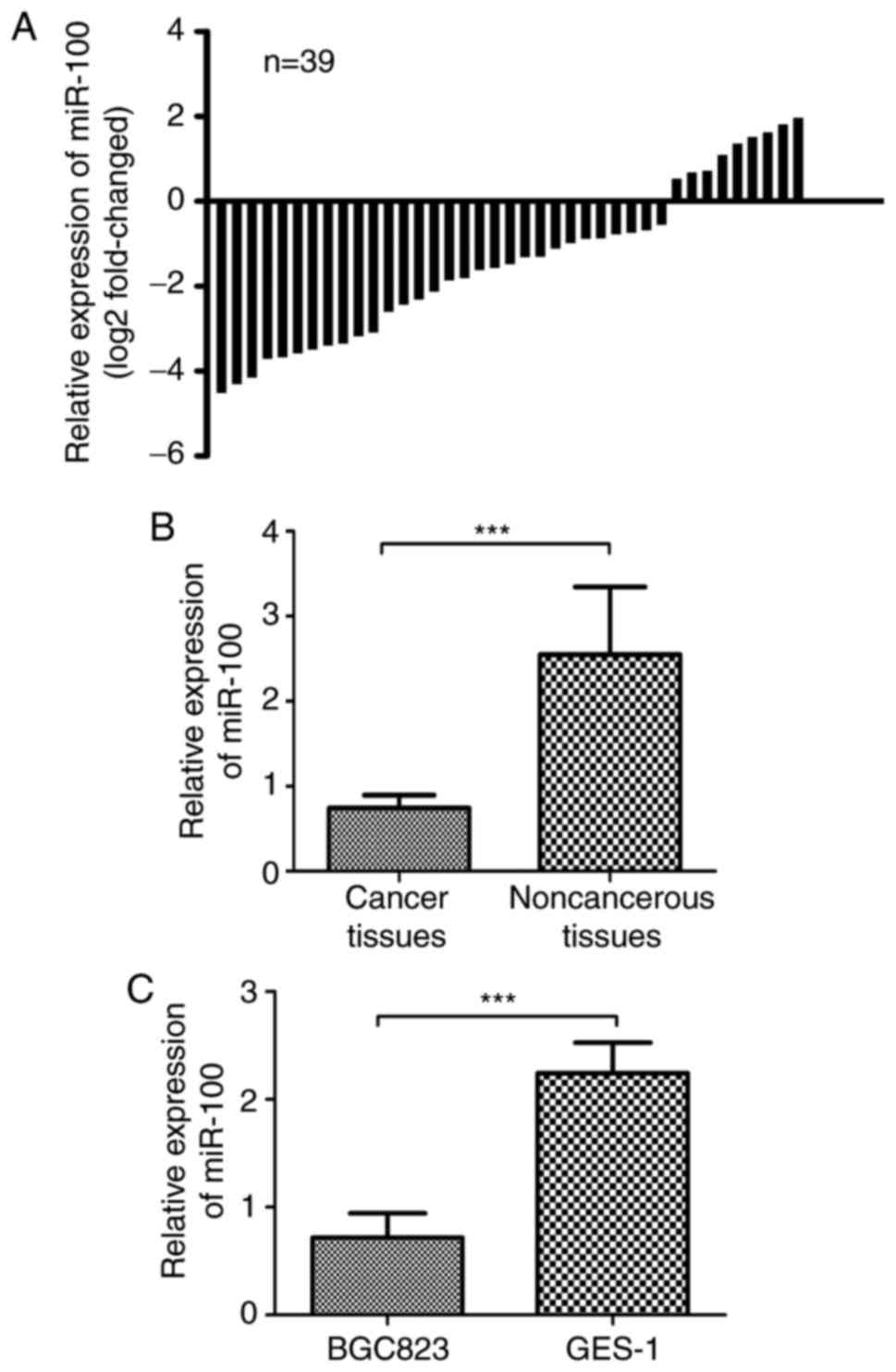
To examine the expression pattern of miR-100 in GC,
39 pairs of GC tissues and the adjacent noncancerous tissues were
quantified by RT-qPCR. As shown in Fig.
1A, we found the expression level of miR-100 was down-regulated
in 30 of 39 (76.92%) GC tissues. Therefore, these patients were
classified into the low miR-100 expression group and the rest of
them were classified into the high miR-100 expression group
accordingly. Also, the average expression level of miR-100 in tumor
tissues was significantly lower in GC tissues than in adjacent
noncancerous tissues (P<0.05, Fig.
1B). In addition, the expression level of miR-100 in GC cell
line (BGC823) and normal gastric epithelial cell line (GES-1) was
measured by the same method. As expected, the expression of miR-100
in BGC823 cell line was significantly lower than in GES-1 cell line
(P<0.05, Fig. 1C). Collectively,
our results revealed the miR-100 was frequently down-regulated in
GC tissues and cell line.
The correlation between miR-100
expression and clinicopathological features of GC
We then analyzed the correlation between miR-100
expression and the clinicopathological features in GC patients. As
summarized in Table I, we found the
miR-100 expression was closely associated with lymph node
metastasis (P=0.047), tumor diameter (P=0.034) and tumor stage
(P=0.008). However, no significant association was observed between
miR-100 expression and age, gender, and differentiation (all
P>0.05).
Clinical significance of miR-100 in
GC
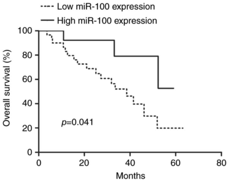
To explore the prognostic significance of miR-100 in
GC patients, we measured the correlation between miR-100 expression
and the overall survival of 39 GC patients using Kaplan-Meier
analysis and log-rank test. We found the miR-100 expression was
significantly correlated with the GC patients' overall survival
(P=0.041, Fig. 2). In other words,
the patients with low miR-100 expression had a poorer overall
survival compared with the patients with high miR-100 expression.
Furthermore, we found the miR-100 expression showed unfavorable
prognosis in GC patients (P=0.024), along with lymph node
metastasis (P=0.042), tumor diameter (P=0.031) and tumor stage
(P=0.014) (Table II). Besides that,
the miR-100 expression was proven to be a poor independent
predictor for GC patients through multivariate analysis (P=0.019,
Table II).
| Table II.Univariate and multivariate analyses
of overall survival rate. |
Table II.
Univariate and multivariate analyses
of overall survival rate.
|
| Univariate
analysis |
| Multivariate
analysis |
|
|---|
|
|
|
|
|
|
|---|
| Variables | HR | 95% CI | P-value | HR | 95% CI | P-value |
|---|
| miR-100 | 2.704 | 1.140–6.417 | 0.024 | 2.764 | 1.186–6.441 | 0.019 |
| Age | 2.309 | 0.867–6.150 | 0.094 | – | – | – |
| Gender | 2.044 | 0.701–5.960 | 0.191 | – | – | – |
| Tumor diameter | 2.643 | 1.093–6.395 | 0.031 | 2.637 | 1.090–6.383 | 0.032 |
|
Differentiation | 2.489 | 0.985–6.293 | 0.054 | – | – | – |
| Tumor stage | 2.832 | 1.237–6.483 | 0.014 | 3.232 | 1.335–7.823 | 0.009 |
| Lymph node
metastasis | 2.560 | 1.036–6.329 | 0.042 | 2.578 | 1.044–6.367 | 0.040 |
Upregulation of miR-100 inhibits cell
proliferation in vitro
To understand the role of miR-100 expression in
tumor progression of GC, we analyzed the effect of miR-100
expression on GC cell proliferation through miR-100 mimic, miR-100
inhibitor, negative control miRNA transfection. It is noteworthy
the BGC823 cell line with miR-100 mimic transfection had the
highest miR-100 expression compared with the cell line with miR-100
inhibitor or negative control miRNA transfection (Fig. 3A). Then, the cell proliferation rate
of the above-mentioned cell lines was analyzed. Conversely, we
found the BGC823 cell line with miR-100 mimic had the lowest cell
proliferation rate compared with the cell line with miR-100
inhibitor or negative control miRNA transfection (Fig. 3B). Taken together, our results
suggested that the miR-100 expression could inversely regulate cell
proliferation rate and thus we deduct the miR-100 might play a
tumor suppressor role in GC.
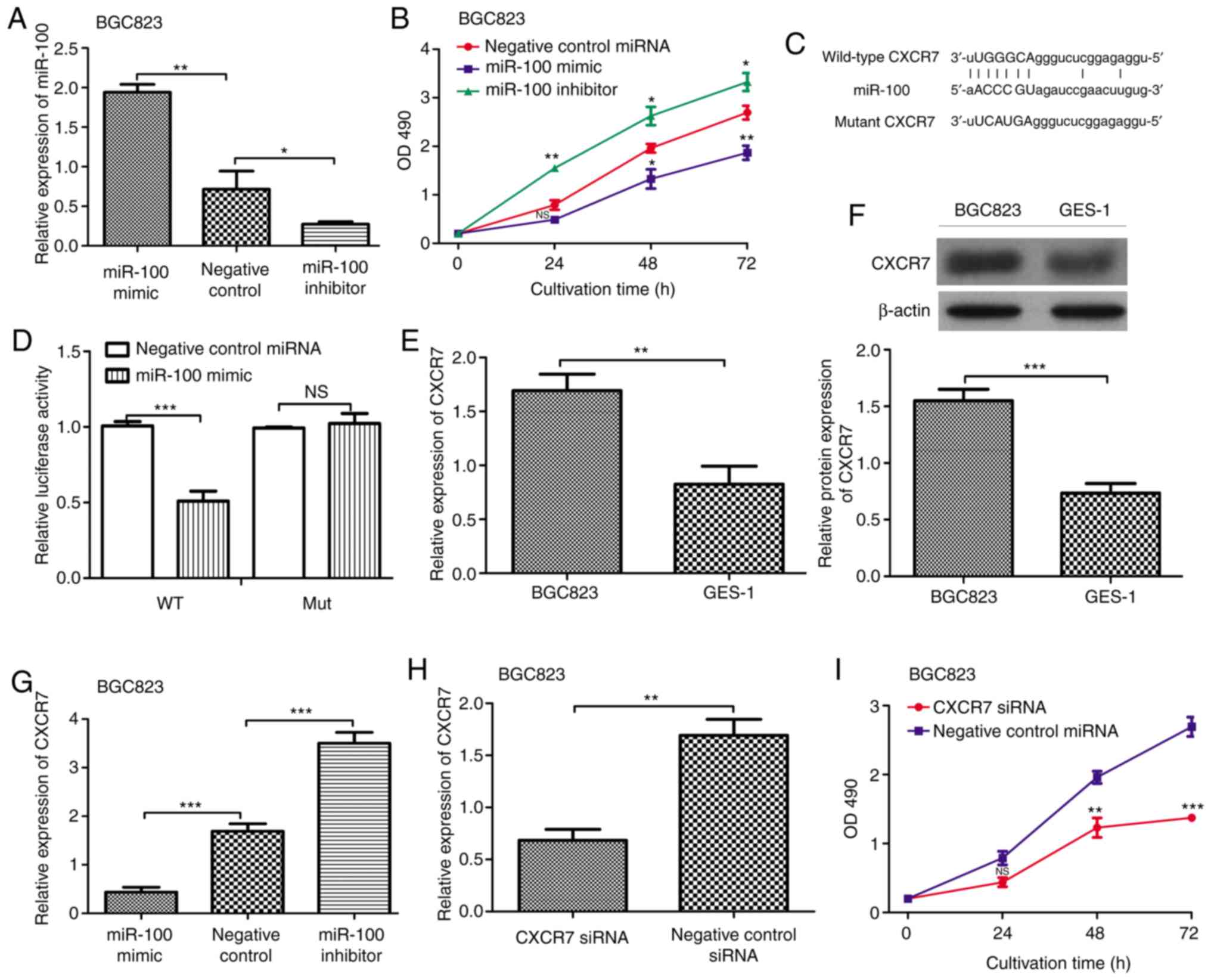 | Figure 3.miR-100 regulates the expression of
CXCR7 in vitro. (A) RT-qPCR analysis of the expression of
miR-100 in gastric cancer cell line (BGC823) after miR-100 mimic,
inhibitor, and negative control miRNA transfection. (B) MTT assay
to determine the level of cell proliferation in gastric cancer cell
line (BGC823) after miR-100 mimic, inhibitor, and negative control
miRNA transfection. (C) Putative miR-100-binding sequence within
the 3′-UTR of CXCR7 mRNA. (D) Luciferase gene reporter gene assays
were performed to verify the relationship between miR-100 and
CXCR7. (E) RT-qPCR and (F) Western blot analysis of the expression
of CXCR7 in gastric cancer cell line (BGC823) and normal gastric
epithelial cell line (GES-1). (G) RT-qPCR analysis of the
expression of CXCR7 in gastric cancer cell line (BGC823) after
miR-100 mimic, inhibitor, and negative control miRNA transfection.
(H) RT-qPCR analysis of the expression of CXCR7 in gastric cancer
cell line (BGC823) after CXCR7-sepcific siRNA and negative control
siRNA transfection. (I) MTT assay to determine the level of cell
proliferation in gastric cancer cell line (BGC823) after
CXCR7-sepcific siRNA and negative control siRNA transfection (NS:
Not significance, *P<0.05, **P<0.01, ***P<0.001). miR-100,
microRNA-100; CXCR7, Chemokine (CXC motif) receptor 7; siRNA,
small-interfering RNA; OD490, optical density at 490
nm. |
CXCR7 is a target gene of miR-100
To further explore the mechanisms underlying the
inhibitory role of miR-100 in the proliferation of GC cells, the
computational prediction website TargetScan was used to identify
functionally targets of miR-100. Interestingly, we found the CXCR7
contains a putative target sequence of miR-100 in 3′-UTR (Fig. 3C). Meanwhile, the luciferase reporter
assay demonstrated that the luciferase activity in BGC-823 cells
with wild-type 3′-UTR CXCR7 and miR-100 mimic transfection was
significantly lower than those with mutant 3′-UTR CXCR7 and miR-100
mimic transfection (Fig. 3D). Then,
the expression of CXCR7 in GC cell line BGC823 and normal gastric
epithelial cell line GES-1 was examined by RT-qPCR and western
blot. The data showed that the expression of CXCR7 in the GES-1
cell line was significantly decreased compared to that in the
BGC823 cell line (Fig. 3E and F).
Furthermore, the expression of CXCR7 in the miR-100 mimic, miR-100
inhibitor, and negative control miRNA transfection BGC823 cell line
was examined. The results presented in Fig. 3G illustrated that the transfection of
miR-100 mimic could decrease CXCR7 mRNA expression level, which
means the expression of CXCR7 could be inversely regulated by
miR-100. In addition, the effect of CXCR7 expression on BGC823 cell
proliferation was analyzed through CXCR7-specific siRNA and
negative control siRNA transfection. The results demonstrated that
the CXCR7-specific siRNA could impair both the expression of CXCR7
and the cell proliferation of BGC823 cell line (Fig. 3H and I). These results demonstrated
that up-regulation of miR-100 expression could suppress the cell
proliferation of GC cell line BGC823, at least partly through
down-regulating the expression of CXCR7.
Discussion
Dysregulation of miRNAs in diseases especially in
tumor has attracted extensive attentions in recent years (7–12).
Mounting evidences have demonstrated that aberrant expression of
several miRNAs is associated with survival outcome of cancer
patients (27,28) and therefore could be regarded as
potential therapeutic targets for these cancers. Take GC as an
example, numerous miRNAs have been identified involved in GC
development, but their underlying molecular mechanism in GC
development is still poorly understood (7,14).
Therefore, in this present study, we aimed to identify new miRNA
biomarker that could be used as predictor for the overall survival
of GC patients and also with the hope to reveal the underlying
mechanism of how miRNA participated in the development of GC.
miR-100 has been found aberrant expressed in several
human cancers but its role in tumorigenesis is still controversial
(16–20). The reason for that might be it can
regulate different downstream target genes and therefore it can
have different and even opposite functions in tumors. Therefore, in
this study, we first examined the expression pattern of miR-100 in
GC tissues. Our results showed that the expression of miR-100 was
clearly downregulated in GC tissues compared with the surrounding
noncancerous tissues (P<0.05), which implies the miR-100 might
play an important role in the progression of GC. Following, we
analyzed the expression of miR-100 in GC cell line. Not
surprisingly, the same trend was found when compared with the
expression of miR-100 in GC cell line and the normal gastric
epithelial cell line.
Since miR-100 is frequently down-regulated in GC
tissues, we then analyzed the association between miR-100
expression and clinicopathological features. We found the miR-100
expression was closely associated with lymph node metastasis
(P=0.047), tumor diameter (P=0.034) and tumor stage (P=0.008). The
survival analysis demonstrated that the low miR-100 expression
predicts poor prognosis in GC patients, which highlighted the
importance of miR-100 in GC. Finally, we identified the miR-100
could be used as an independent predictor for the overall survival
of GC patients through the univariate and multivariate analyses.
Taken together, we explored the expression pattern of miR-100 in GC
and revealed the clinical importance of miR-100 in GC. However, the
biological function of miR-100 in GC is still unknown.
Previous studies demonstrated that miR-100 could
inhibit cell proliferation, migration, and differentiation in human
cancers (19,29–30).
Therefore, the effect of miR-100 on cell proliferation was
evaluated in vitro. We found the level of cell proliferation
in GC cell line was obviously higher than in normal gastric
epithelial cell line (P<0.05). By transferring the miR-100 mimic
and inhibitor into GC cell line, we found forced the expression of
miR-100 could inhibit GC cell proliferation. However, the
transfection of miR-100 inhibitor could promote GC cell
proliferation. Collectively, we deducted that miR-100 expression
could suppress the cell proliferation in GC.
Numerous miR-100 target genes have been identified
in previous literatures (17,19,25,29,30).
Using the bioinformatical tools, we found the 3′-UTR of CXCR7 has a
putative binding site for miR-100 and the luciferase reporter assay
confirmed CXCR7 is a direct target of miR-100. Importantly, the
CXCR7 was found overexpressed in GC in previous study (24). Therefore, we first examined the
expression of CXCR7 in GC cell line and normal gastric epithelial
cell line. In accordance with the previous finding, we found the
CXCR7 was overexpressed in GC cell line. Besides that, the
expression of CXCR7 was found inversely correlated with the
expression of miR-100. Following, the forced downregulate
expression of CXCR7 in GC cell line could impair the cell
proliferation. Taken together, we deducted the CXCR7 might be a
downstream target for miR-100 in GC and the miR-100 could exerts
its oncogenic role be targeting CXCR7.
In summary, we found the miR-100 was downregulated
in GC and was correlated with the poor prognosis of GC. Another
important finding in this study was we found CXCR7 was the
downstream target gene of miR-100. These results indicate that
miR-100 deregulation may play important roles in tumor growth and
that miR-100 may be a potential therapeutic target for the
treatment of GC.
References
|
1
|
Tinico A, Gottardi LF and Boechat ED:
Gastric cancer in the excluded stomach 10 years after gastric
bypass. Case Rep Surg. 2015:4682932015.PubMed/NCBI
|
|
2
|
Siegel RL, Miller KD and Jemal A: Cancer
Statistics, 2016. CA Cancer J Clin. 66:7–30. 2016. View Article : Google Scholar : PubMed/NCBI
|
|
3
|
Malvezzi M, Bonifazi M, Bertuccio P, Levi
F, La Vecchia C, Decarli A and Negri E: An age-period-cohort
analysis of gastric cancer mortality from 1950 to 2007 in Europe.
Ann Epidemiol. 20:898–905. 2010. View Article : Google Scholar : PubMed/NCBI
|
|
4
|
Torre LA, Bray F, Siegel RL, Ferlay J,
Lortet-Tieulent J and Jemal A: Global cancer statistics, 2012. CA
Cancer J Clin. 65:87–108. 2015. View Article : Google Scholar : PubMed/NCBI
|
|
5
|
Sionov RV: MicroRNAs and
glucocorticoid-induced apoptosis in lymphoid maligancies. ISRN
hematol. 2013:3482122013. View Article : Google Scholar : PubMed/NCBI
|
|
6
|
Santpere G, Lopez-Valenzuela M,
Petit-Marty N, Navarro A and Espinosa-Parrila Y: Differences in
molecular evolutionary rates among microRNAs in the human and
chimpanzee genomes. BMC Genomics. 17:5282016. View Article : Google Scholar : PubMed/NCBI
|
|
7
|
Shen Y, Gong JM, Zhou LL and Sheng JH:
Mir-451 as a new tumor marker for gastric cancer. Oncotarget.
8:56542, 565452017.
|
|
8
|
Adlakha YK and Saini N: Brain microRNAs
and insights into biological functions and therapeutic potential of
brain enriched miRNA-128. Mol Cancer. 13:332014. View Article : Google Scholar : PubMed/NCBI
|
|
9
|
Donohoe OH, Henshilwood K, Way K,
Hakimjavadi R, Stone DM and Walls D: Identification and
characterization of cyprinid herpesvirus-3 (CyHV-3) encoded
MicroRNAs. PLoS One. 10:e01254342015. View Article : Google Scholar : PubMed/NCBI
|
|
10
|
Wang Q and Yu JH: MiR-129-5p suppresses
gastric cancer cell invasion and proliferation by inhibiting
COL1A1. Biochem Cell Biol. May 8–2017.(Epub ahead of print).
|
|
11
|
Xie M, Dart DA, Guo T, Xing XF, Cheng XJ,
Du H, Jiang WG, Wen XZ and Ji JF: MicroRNA-1 acts as a tumor
suppressor microRNA by inhibiting angiogenesis-related growth
factors in human gastric cancer. Gastric Cancer. May 10–2017.(Epub
ahead of print). View Article : Google Scholar
|
|
12
|
Datta J, Kutay H, Nasser MW, Nuovo GJ,
Wang B, Majumder S, Liu CG, Volinia S, Croce CM, Schmittgen TD, et
al: Methylation mediated silencing of microRNA-1 gene and its role
in hepatocellular carcinogenesis. Cancer Res. 68:5049–5058. 2008.
View Article : Google Scholar : PubMed/NCBI
|
|
13
|
Zang Y, Wang T, Pan J and Gao F: miR-215
promotes cell migration and invasion of gastric cancer cell lines
by targeting FOXO1. Neoplasma. 64:579–587. 2017. View Article : Google Scholar : PubMed/NCBI
|
|
14
|
Jafarzadeh-Samani Z, Sohrabi S,
Shirmohammadi K, Effatpanah H, Yadegarazari R and Saidijam M:
Evaluation of miR-22 and miR-20a as diagnostic biomarkers for
gastric cancer. Chin Clin Oncol. 6:162017. View Article : Google Scholar : PubMed/NCBI
|
|
15
|
Hu CE, Du PZ, Zhang HD and Huang GJ: Long
noncoding RNA CRNDE promotes proliferation of gastric cancer cells
by targeting miR-145. Cell Physiol Biochem. 42:13–21. 2017.
View Article : Google Scholar : PubMed/NCBI
|
|
16
|
Qin C, Huang RY and Wang ZX: Potential
role of miR-100 in cancer diagnosis, prognosis, and therapy. Tumor
Biol. 36:1403–1409. 2015. View Article : Google Scholar
|
|
17
|
Zhou HC, Fang JH, Luo X, Zhang L, Yang J,
Zhang C and Zhuang SM: Downregulation of microRNA-100 enhances the
ICMT-Rac1 signaling and promotes metastasis of hepatocellular
carcinoma cells. Oncotarget. 5:12177–12188. 2014. View Article : Google Scholar : PubMed/NCBI
|
|
18
|
Azizmohammadi S, Azizmohammadi S, Safari
A, Kosari N, Kaghazian M, Yahaghi E and Seifoleslami M: The role
and expression of miR-100 and miR-203 profile as prognostic markers
in epithelial ovarian cancer. Am J Tansl Res. 8:2403–2410.
2016.
|
|
19
|
Jiang Q, He M, Guan S, Ma M, Wu H, Yu Z,
Jiang L, Wang Y, Zong X, Jin F and Wei M: MicroRNA-100 suppresses
the migration and invasion of breast cancer cells by targeting
FZD-8 and inhibiting Wnt/β-catenin signaling pathway. Tumor Biol.
37:5001–5011. 2016. View Article : Google Scholar
|
|
20
|
Gu L, Li H, Chen L, Ma X, Gao Y, Li X,
Zhang Y, Fan Y and Zhang X: MicroRNAs as prognostic molecular
signatures in renal cell carcinoma: A systematic review and
meta-analysis. Oncotarget. 6:32545–32560. 2015. View Article : Google Scholar : PubMed/NCBI
|
|
21
|
Burns JM, Summers BC, Wang Y, Melikian A,
Berahovich R, Miao Z, Penfold ME, Sunshine MJ, Littman DR, Kuo CJ,
et al: A novel chemokine receptor for SDF-1 and I-TAC involved in
cell survival, cell adhesion, and tumor development. J Exp Med.
203:2201–2213. 2006. View Article : Google Scholar : PubMed/NCBI
|
|
22
|
Liu Z, Teng XY, Meng XP and Wang BS:
Expression of stromal cell-dervied factor 1 and CXCR7 ligand
receptor system in pancreatic adenocarcinoma. World J Surg Oncol.
12:3482014. View Article : Google Scholar : PubMed/NCBI
|
|
23
|
Hattermann K, Held-Feindt J, Lucius R,
Müerköster SS, Penfold ME, Schall TJ and Mentlein R: The chemokine
receptor CXCR7 is highly expressed in human glioma cells and
mediates antiapoptoic effects. Cancer Res. 70:3299–3308. 2010.
View Article : Google Scholar : PubMed/NCBI
|
|
24
|
Nambara S, Iguchi T, Oki E, Tan P, Maehara
Y and Mimori K: Overexpression of CXCR7 is a novel prognostic
indicator in gastric cancer. Dig Surg. 34:312–318. 2017. View Article : Google Scholar : PubMed/NCBI
|
|
25
|
Zhou SM, Zhang F, Chen XB, Jun CM, Jing X,
Wei DX, Xia Y, Zhou YB, Xiao XQ, Jia RQ, et al: miR-100 suppresses
the proliferation and tumor growth of esophageal squamous cancer
cells via targeting CXCR7. Oncol Rep. 35:3453–3459. 2016.
View Article : Google Scholar : PubMed/NCBI
|
|
26
|
Livak KJ and Schmittgen TD: Analysis of
relative gene expression data using real-time quantitative PCR and
the 2(-Delta Delta C(T)) method. Methods. 25:402–408. 2001.
View Article : Google Scholar : PubMed/NCBI
|
|
27
|
White NM and Yousef GM: MicroRNAs:
Exploring a new dimension in the pathogenesis of kidney cancer. BMC
Med. 8:652010. View Article : Google Scholar : PubMed/NCBI
|
|
28
|
Jamali Z, Asl Aminabadi N, Attaran R,
Pournagiazar F, Ghertasi Oskouei S and Ahmadpour F: MicroRNAs as
prognostic molecular signatures in human head and neck squamous
cell carcinoma: A systematic review and meta-analysis. Oral Oncol.
51:321–331. 2015. View Article : Google Scholar : PubMed/NCBI
|
|
29
|
Fu HL, Pan HX, Zhao B, Dong BC, Shao L, Fu
GS, Wang Q and Li M: MicroRNA-100 inhibits bone morphogenetic
protein-induced osteoblast differentiation by targeting Smad1. Eur
Rev Med Pharmacol Sci. 20:3911–3919. 2016.PubMed/NCBI
|
|
30
|
Luan YX, Zhang S, Zuo L and Zhou L:
Overexpression of miR-100 inhibits cell proliferation, migration,
and chemosensitivity in human glioblastoma through FGFR3. Onco
Targets Ther. 8:3391–3400. 2015.PubMed/NCBI
|