Introduction
Lung cancer is caused by the interaction among
smoking, environmental and genetic factors and is currently the
most clinically common respiratory system tumor, and it is the
shortened form of primary bronchogenic carcinoma (1). Lung cancer has a high mortality rate,
and the 5-year survival rate of lung cancer patients is <20%.
Surgery is currently the major treatment means of lung cancer, and
the clinical treatment of this disease is unsatisfactory at
present, mainly because lung cancer is prone to spread, and the
patients have often missed the best opportunity of surgical
resection when diagnosed (2,3). Patients lacking the surgical conditions
are often treated with the combined therapy of chemotherapy and
radiotherapy. The commonly-used chemotherapy methods are the
platinum drugs combined with cytotoxic drugs, such as lobaplatin
combined with gemcitabine used for the treatment of lung cancer
(4,5).
Platinum drugs have a strong effect of killing lung cancer cells,
but lung cancer cells will produce resistance to platinum drugs,
resulting in decreased sensitivity of lung cancer cells and finally
loss of the therapeutic effect (6).
Paclitaxel is a kind of antitumor drug that acts on tubulin, and
its effective rate in the treatment of lung cancer is <15%. Ueno
and Mamounas (7) found that
paclitaxel can increase the sensitivity of carboplatin to breast
cancer, enhance the effect of carboplatin and effectively prolong
the survival time of breast cancer patients. Zou et al
(8) found that paclitaxel can
effectively reduce the migration and invasion capacities of colon
cancer cells, and inhibit the proliferation of colon cancer cells
through reducing the expression level of phosphorylated-Akt
(p-Akt). A large number of studies have shown that
phosphatidylinositol 3-kinase (PI3K)/Akt is involved in the
proliferation process of multiple tumors, and a variety of
antitumor drugs can play an antitumor role through acting on the
PI3K/Akt signaling pathway (9,10).
In the present study, the sensitivity of cancer
cells to lobaplatin in the treatment of lung cancer cells with
paclitaxel combined with lobaplatin, and whether PI3K/Akt signaling
pathway was involved in the effect of paclitaxel on lung cancer
cells was investigated, so as to provide a theoretical basis for
the clinical treatment of lung cancer with paclitaxel combined with
lobaplatin.
Materials and methods
Reagents and instruments
Lung cancer cell line NCI-H446 (Kunming Cell Bank,
Chinese Academy of Sciences); methyl thiazolyltetrazolium (MTT),
dimethylsulfoxide (DMSO), paclitaxel, lobaplatin and LY294002
(Sigma, St. Louis, MO, USA); RPMI-1640 medium (Gibco; Thermo Fisher
Scientific, Inc., Waltham, MA, USA); rabbit anti-human PI3K, p-Akt,
AKT, phosphorylated-glycogen synthase kinase 3β (p-GSK3β), GSK3β,
β-actin monoclonal antibody and goat anti-rabbit secondary
polyclonal antibody (cat. nos. 4249, 4060, 4685, 9323, 9315, 4970
and 14708) (Cell Signaling Technology, Danvers, MA, USA); Annexin
V-FITC apoptosis assay kit (BD Biosciences, Heidelberg, Germany);
inverted fluorescence microscope (Thermo Fisher Scientific, Inc.,
Waltham, MA, USA); cell culture bottle (Corning Inc., Corning, NY,
USA); Transwell chamber (EMD Millipore, Billerica, MA, USA) and
pipettor (Eppendorf AG, Hamburg, Germany).
Detection of cell survival rate
The lung cancer cell line NCI-H446 purchased from
Kunming Cell Bank, Chinese Academy of Sciences were cultured at
37°C and 5% CO2 after the replacement of medium,
followed by passage at 80% cell confluency. The cells continued to
be cultured until the logarithmic phase for the experiments. After
cells were placed in the 96-well plate overnight, lobaplatin in
different concentrations (5, 10, 20, 30, 50 and 80 µg/ml) was added
for incubation at 37°C and 5% CO2 for 24 h. Then MTT was
added for incubation for 4 h. The culture solution was adsorbed and
DMSO was added, followed by detection of absorbance value using the
microplate reader and calculation of cell survival rate. Lobaplatin
in appropriate concentration was selected and added into the cells
based on the time gradient for incubation for 6, 12, 24 and 48 h,
respectively. The effects of different treatment time on cell
survival rates were detected via MTT assay. According to the above
experimental results, lobaplatin group (group L), 2 µg/ml
paclitaxel combined with lobaplatin group (group LP) and lobaplatin
combined with 10 µmol/ml LY294002 group (group LL) were set up. The
cell survival rate in each group was detected via MTT assay after
incubation for 24 h.
Detection of cell apoptosis
The density of cells in the logarithmic growth phase
was adjusted to 1×10⁶ cells/ml and spread evenly on the
6-well plate. Group L, group LP and group LL were set up. At 2 h
before lobaplatin was added, 2 µg/ml paclitaxel and 10 µmol/ml
LY294002 were added into group LP and group LL for incubation at
37°C and 5% CO2 for 24 h. The culture solution was
absorbed and the cells were washed with pre-cooled phosphate
buffered saline (PBS) three times. Then the cells were digested and
collected into the centrifuge tube for centrifugation for 5 min at
800 × g. After that, the cells were washed with PBS again,
resuspended and centrifuged under the same conditions three times.
According to instructions of Annexin V-FITC apoptosis detection
kit, 200 µl staining fluid was added into each group for staining
in the dark for 15 min, then 800 µl buffer was added for machine
inspection, preferably within 60 min.
Detection of cell migration
capacity
The density of cells in the logarithmic growth phase
was adjusted to 1×10⁶ cells/ml and spread evenly on the
medium plate. Group L, group LP and group LL were set up. After the
cells adhered to the wall, cell culture dish in each group was
marked using the marking-off pin. After the observation area was
marked, the same area was observed again after incubation for 12 h
and photographed under the microscope. The cell migration and
migration distance in each group were recorded (unit/µm).
Detection of cell invasion
capacity
The density of cells in the logarithmic growth phase
was adjusted to 5×10⁵ cells/ml and added into the
Transwell chamber. Group L, group LP and group LL were set up.
After the cells in each group were incubated at 37°C and 5%
CO2 for 24 h, stained and fixed, the number of cells
passing through the chamber was observed under the microscope and
recorded.
Protein level detection
The cells in the logarithmic growth phase were
spread on the 6-well plate. Group L, group LP and group LL were set
up and treated for 24 h. The protein in each group was extracted
and quantified using the bicinchoninic acid protein quantification
kit. The total loading quantity in each group was determined as 2
µg, and the loading buffer was added to prepare the loading sample.
The sample was boiled under high temperature to inactivate protein,
followed by 12% sodium dodecyl sulfate-polyacrylamide gel
electrophoresis for 120 min under 80 V. Then the gel was
transferred to the membrane transfer tank for preparation of
membrane-transfer ‘sandwich’ and membrane transfer for 90 min under
100 V. After that, the membrane was sealed using 5% skim milk
powder at room temperature for 1 h, and PI3K, p-Akt, Akt, p-GSK3β
and GSK3β specific monoclonal antibodies (dilution, 1:1,000) were
incubated at 4°C overnight, with β-actin as the internal reference.
Then the membrane was washed with TBSP for 10 min for a total of
three times. After the secondary antibody was incubated (dilution,
1:2,000) at room temperature for 2 h, the membrane was washed again
three times (10 min/time). After the tabletting time was selected
according to the fluorescence intensity, the developing liquid was
added onto the protein band in the dark for exposure to obtain the
corresponding protein bands. After the bands were scanned, the
expression level of corresponding protein in each group was
detected with β-actin as the internal reference.
Statistical analysis
The data in the present study were analyzed using
SPSS 19.0 software (SPSS Inc., Chicago, IL, USA). The t-test was
used for intergroup comparison, while analysis of variance was used
for comparison among groups. A value of p<0.05 was considered to
indicate a statistically significant difference.
Results
Effect of lobaplatin on the growth of
lung cancer cells
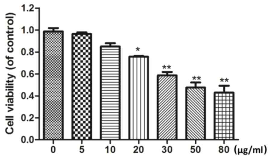
The growth of lung cancer cell line NCI-H446 after
being treated with lobaplatin in different concentrations was
detected via MTT assay. The results showed that the stability of
NCI-H446 could be reduced by lobaplatin in a
concentration-dependent manner. Lobaplatin (30 µg/ml) could
significantly inhibit the growth of NCI-H446 (p<0.01), so 30
µg/ml lobaplatin was used for subsequent experimental study
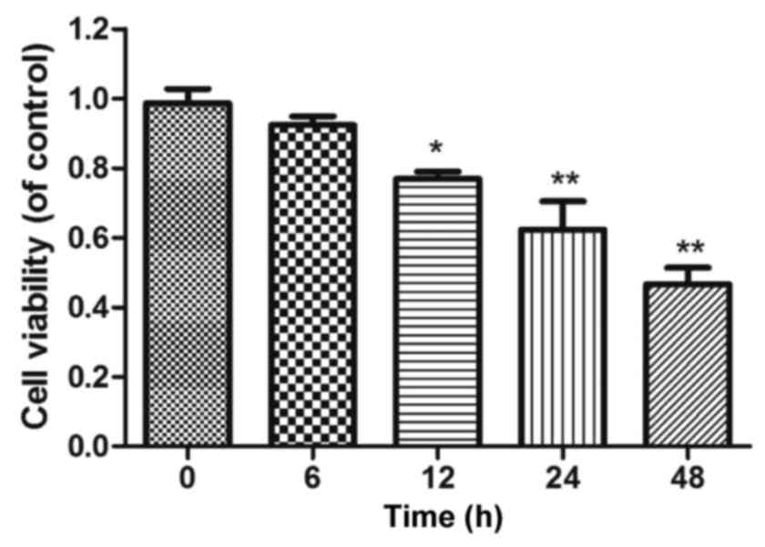
(Fig. 1). The growth of cells was
detected via MTT assay after they were treated with 30 µg/ml
lobaplatin for 6, 12, 24 and 48 h, respectively. The results
revealed that the effect of 30 µg/ml lobaplatin on cell growth was
in a time-dependent manner; the survival rate of cells could be
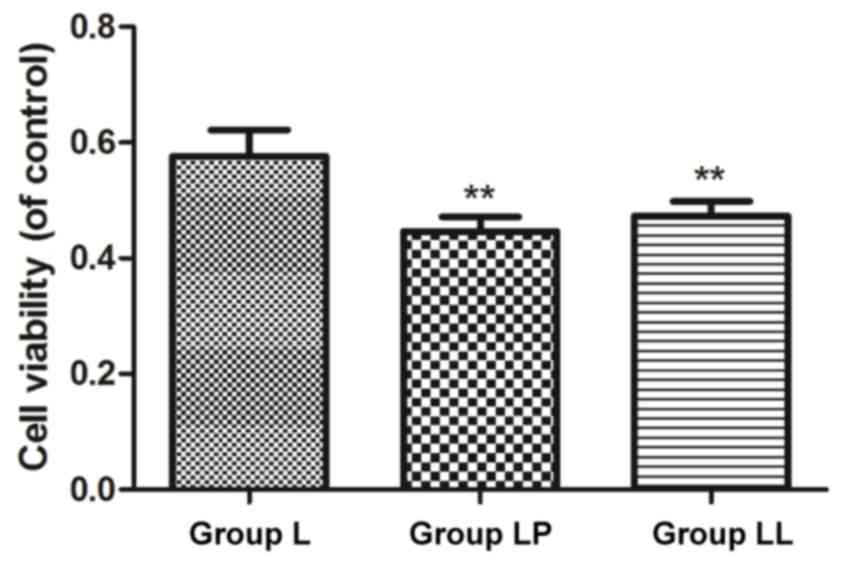
significantly reduced after treatment for 12 h (Fig. 2). Group L, group LP and group LL were
set up and the survival rate in each group was detected. The
survival rates in group LP and group LL were significantly lower
than that in group L, and the differences were statistically
significant (p<0.01), and there was no statistically significant
difference in the survival rate between group LP and group LL
(p>0.05) (Fig. 3).
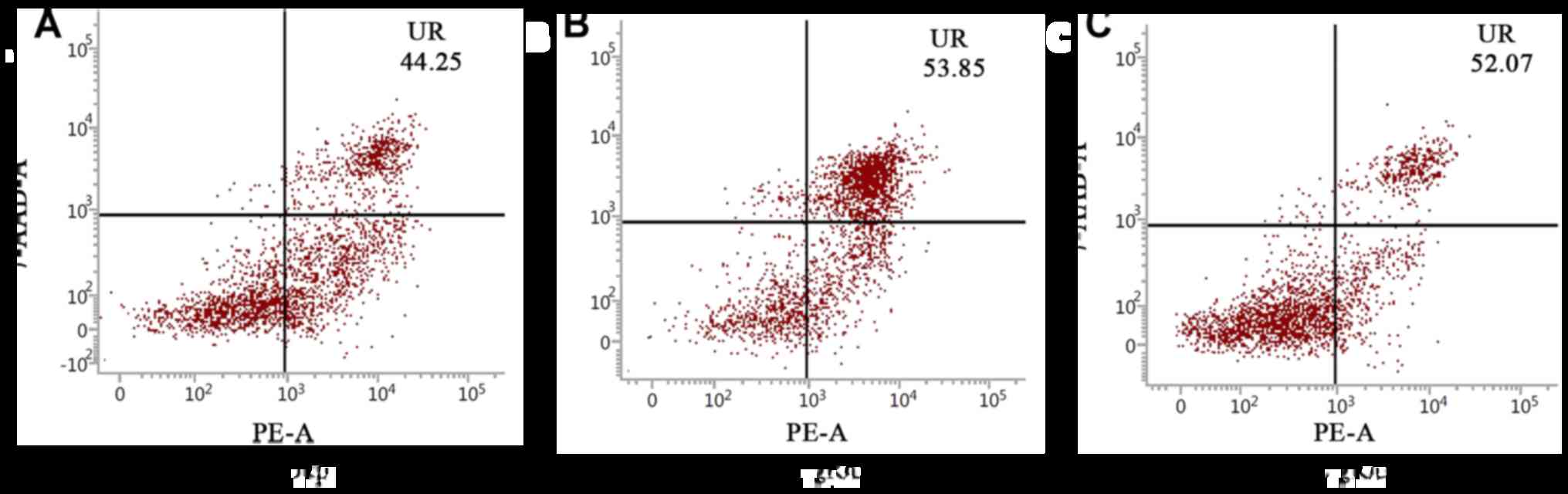
Detection of cell apoptosis level
Cell apoptosis in each group was detected using flow
cytometry. The results showed that the number of cells in the 2nd
and 3rd quadrants in group LP and group LL were significantly more
than that in group L; in other words, the apoptosis levels in group
LP and the group LL were higher than that in group L, and there was
no significant difference in the apoptosis level between group LP
and group LL (Fig. 4).
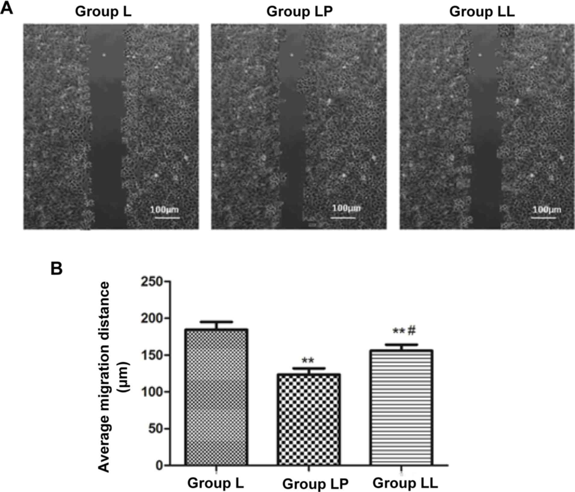
Detection of cell migration
capability
The cell migration capability in each group was
detected via cell wound scratch assay. The results showed that the
cell migration capability in group L was obviously higher than
those in group LP and group LL, and the differences were
statistically significant (p<0.01). The cell migration
capability in group LL was higher than that in group LP (p<0.05)
(Fig. 5).
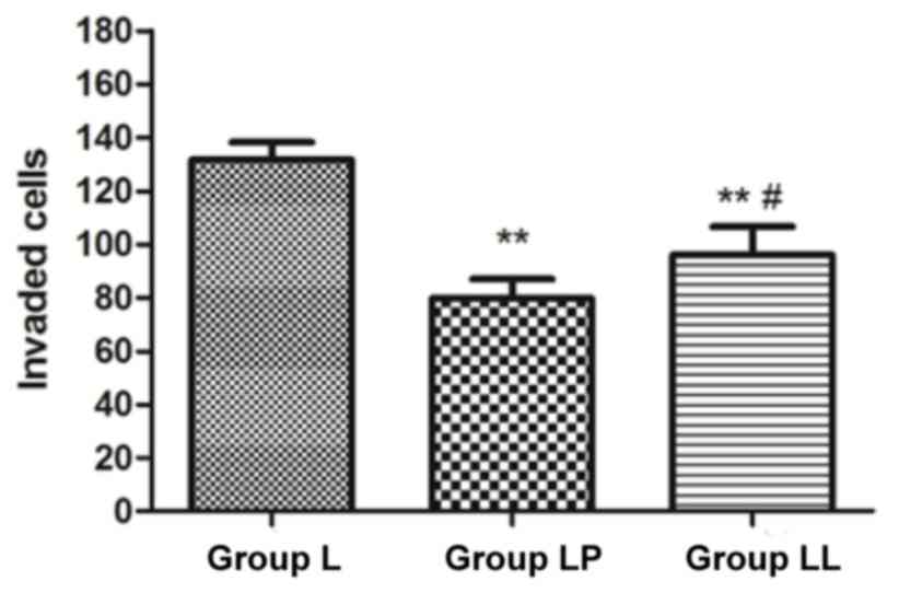
Detection of cell invasion
capability
The cell invasion capability in each group was
detected via Transwell assay. The results revealed that the number
of invasive cells in group LP and group LL were obviously lower
than that in group L (p<0.01), and the number of invasive cells
in group LP was significantly lower than that in group LL
(p<0.05) (Fig. 6).
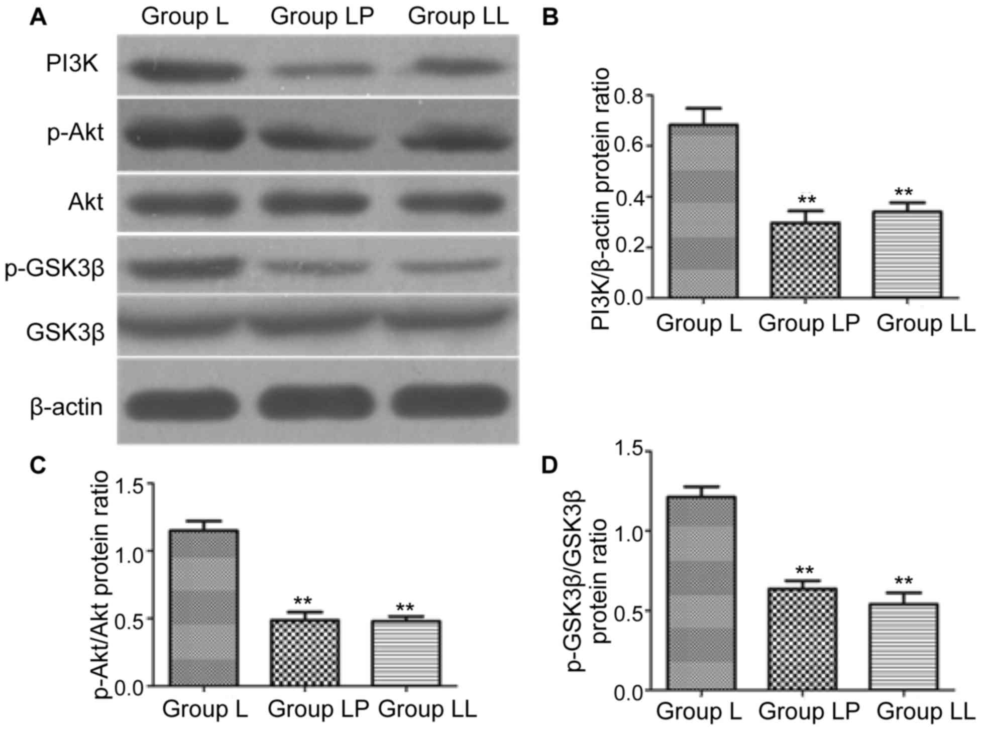
Detection of protein expression
level
In order to investigate whether PI3K/Akt signaling
pathway was involved in the effect of paclitaxel on NCI-H446, the
expression levels of PI3K/Akt signaling pathway-related proteins
were detected via western blotting. The results showed that
compared with that in group L, the protein expression levels of
PI3K in group LP and group LL were significantly decreased
(p<0.01), and there was no statistically significant difference
between group LP and group LL (p>0.05). The protein levels of
p-Akt and p-GSK3β in group LP and group LL were significantly lower
than those in group L (p<0.01), and there was no statistically
significant difference between group LP and group LL (Fig. 7).
Discussion
Lung cancer seriously threatens human life and
health, and the epidemiological survey showed that the incidence
rate of lung cancer shows an increasing trend year by year, and the
morbidity and mortality rates in male patients are significantly
higher than those in female patients (11). Platinum drugs, as the first-line
treatment drug of lung cancer, have high clinical value,
characterized by the high specificity and slight side effects, but
the resistance of tumor cells to platinum compounds is an important
factor affecting the treatment with such compounds, so the combined
administration is often clinically used to reduce the resistance of
tumor cells to platinum compounds (12,13).
Paclitaxel is an antitumor drug extracted from the traditional
Chinese medicine, bark of Taxus chinensis, which can promote
the microtubule aggregation and inhibit the microtubule
disaggregation, thus affecting the cell cycle and killing cells
(14). The study of Zhang et
al (15) showed that carboplatin
combined with paclitaxel can significantly reduce the resistance of
colon cancer cells to carboplatin in the treatment of colon cancer.
PI3K/Akt signaling pathway exists in a variety of cells, and is
involved in cell apoptosis. A number of studies have shown that
this signaling pathway plays a vital role in the regulation of
tumor cell apoptosis (16). The study
of Cheng et al (17) found
that compared with those in normal cells, the expression levels of
PI3K and p-Akt in lung cancer cells are significantly increased,
and the application of PI3K inhibitor can promote lung cancer cell
apoptosis.
In the present study, it was found that lobaplatin
could inhibit the growth of lung cancer cells. Within the range of
effective concentration, the inhibitory effect on the cell growth
was gradually enhanced with the increase of drug dose, as well as
with the prolongation of action time; in other words, the effect of
lobaplatin on lung cancer cells is concentration- and
time-dependent. Lobaplatin, is a third-generation platinum
antitumor drug, and one of the most widely-used antitumor drugs in
clinical practice, which, with tumor cells, can form the
intra-chain cross-linking between platinum and DNA bases, thus
affecting the transcription and translation processes of DNA in
tumor cells and killing tumor cells (18). Flow cytometry showed that the combined
application of paclitaxel and lobaplatin could significantly
increase apoptosis of lung cancer cells and inhibit the migration
and invasion of lung cancer cells. These results indicated that
paclitaxel can increase the sensitivity of lobaplatin to lung
cancer cells, which can further promote apoptosis of lung cancer
cells and inhibit the migration and invasion of lung cancer cells.
Paclitaxel, as a kind of tubulin polymerase inhibitor, it can
effectively inhibit cell mitosis and inhibit the cell
proliferation, and has a certain killing effect on tumor cells,
which has a synergistic effect with the antitumor effect of
platinum drugs. The study of Owonikoko et al (19) reported that paclitaxel combined with
carboplatin can significantly reduce the onset dose of carboplatin
in the treatment of NSCLC. The effect of paclitaxel combined with
lobaplatin on lung cancer cells was similar to that of lobaplatin
combined with PI3K inhibitor LY294002. Besides, the detection of
PI3K expression level via western blotting revealed that the
expression level of PI3K in group LP was significantly lower than
that in group L, but was similar to that in group LL; the
expression levels of p-Akt and p-GSK3β in group LP and group LL
were also obviously lower than those in group L, and there were no
statistically significant differences in the expression levels
between group LP and group LL. Paclitaxel can inhibit the
phosphorylation of Akt and GSK3β through inhibiting PI3K, and
regulate the PI3K/Akt signaling pathway, thus regulating the
proliferation of lung cancer cells and inducing lung cancer cell
apoptosis. P-GSK3β is a necessary protein for cell survival. When
PI3K is inhibited, the phosphorylation of Akt can be further
inhibited, thus affecting the phosphorylation of GSK3β, inducing
cell apoptosis and inhibiting the cell migration and invasion
(20).
In conclusion, paclitaxel can significantly increase
the sensitivity of lobaplatin to lung cancer cell line NCI-H446,
increase lung cancer cell apoptosis and decrease the onset
concentration of lobaplatin through inhibiting PI3K/Akt pathway.
The above results provide a theoretical basis for the clinical
combination of lobaplatin and paclitaxel in the treatment of lung
cancer.
Competing interests
The authors declare that they have no competing
interests.
References
|
1
|
Chambers SK, Dunn J, Occhipinti S, Hughes
S, Baade P, Sinclair S, Aitken J, Youl P and O'Connell DL: A
systematic review of the impact of stigma and nihilism on lung
cancer outcomes. BMC Cancer. 12:1842012. View Article : Google Scholar : PubMed/NCBI
|
|
2
|
Lee PN, Forey BA and Coombs KJ: Systematic
review with meta-analysis of the epidemiological evidence in the
1900s relating smoking to lung cancer. BMC Cancer. 12:3852012.
View Article : Google Scholar : PubMed/NCBI
|
|
3
|
Gilham C, Rake C, Burdett G, Nicholson AG,
Davison L, Franchini A, Carpenter J, Hodgson J, Darnton A and Peto
J: Pleural mesothelioma and lung cancer risks in relation to
occupational history and asbestos lung burden. Occup Environ Med.
73:290–299. 2016. View Article : Google Scholar : PubMed/NCBI
|
|
4
|
Larsen JE and Minna JD: Molecular biology
of lung cancer: Clinical implications. Clin Chest Med. 32:703–740.
2011. View Article : Google Scholar : PubMed/NCBI
|
|
5
|
Chen W, Li Z, Bai L and Lin Y: NF-kappaB
in lung cancer, a carcinogenesis mediator and a prevention and
therapy target. Front Biosci. 1:1172–1185. 2011. View Article : Google Scholar
|
|
6
|
Zhang B-Y, Wang Y-M, Gong H, Zhao H, Lv
X-Y, Yuan GH and Han SR: Isorhamnetin flavonoid synergistically
enhances the anticancer activity and apoptosis induction by
cis-platin and carboplatin in non-small cell lung carcinoma
(NSCLC). Int J Clin Exp Pathol. 8:25–37. 2015.PubMed/NCBI
|
|
7
|
Ueno NT and Mamounas EP: Neoadjuvant
nab-paclitaxel in the treatment of breast cancer. Breast Cancer Res
Treat. 156:427–440. 2016. View Article : Google Scholar : PubMed/NCBI
|
|
8
|
Zou H, Li L, Garcia Carcedo I, Xu ZP,
Monteiro M and Gu W: Synergistic inhibition of colon cancer cell
growth with nanoemulsion-loaded paclitaxel and PI3K/mTOR dual
inhibitor BEZ235 through apoptosis. Int J Nanomed. 11:1947–1958.
2016.
|
|
9
|
Chen QY, Jiao DM, Wu YQ, Chen J, Wang J,
Tang XL, Mou H, Hu HZ, Song J, Yan J, et al: MiR-206 inhibits
HGF-induced epithelial-mesenchymal transition and angiogenesis in
non-small cell lung cancer via c-Met/PI3k/Akt/mTOR pathway.
Oncotarget. 7:18247–18261. 2016.PubMed/NCBI
|
|
10
|
Mateen S, Raina K and Agarwal R:
Chemopreventive and anti-cancer efficacy of silibinin against
growth and progression of lung cancer. Nutr Cancer. 65 Suppl
1:3–11. 2013. View Article : Google Scholar : PubMed/NCBI
|
|
11
|
Jin S, Deng Y, Hao J-W, Li Y, Liu B, Yu Y,
Shi FD and Zhou QH: NK cell phenotypic modulation in lung cancer
environment. PLoS One. 9:e1099762014. View Article : Google Scholar : PubMed/NCBI
|
|
12
|
Samanta D, Kaufman J, Carbone DP and Datta
PK: Long-term smoking mediated down-regulation of Smad3 induces
resistance to carboplatin in non-small cell lung cancer. Neoplasia.
14:644–655. 2012. View Article : Google Scholar : PubMed/NCBI
|
|
13
|
Ma T, Fuld AD, Rigas JR, Hagey AE, Gordon
GB, Dmitrovsky E and Dragnev KH: A phase I trial and in vitro
studies combining ABT-751 with carboplatin in previously treated
non-small cell lung cancer patients. Chemotherapy. 58:321–329.
2012. View Article : Google Scholar : PubMed/NCBI
|
|
14
|
Khongkow P, Gomes AR, Gong C, Man EPS,
Tsang JWH, Zhao F, Monteiro LJ, Coombes RC, Medema RH, Khoo US and
Lam EW: Paclitaxel targets FOXM1 to regulate KIF20A in mitotic
catastrophe and breast cancer paclitaxel resistance. Oncogene.
35:990–1002. 2016. View Article : Google Scholar : PubMed/NCBI
|
|
15
|
Zhang Q, Si S, Schoen S, Chen J, Jin XB
and Wu G: Suppression of autophagy enhances preferential toxicity
of paclitaxel to folliculin-deficient renal cancer cells. J Exp
Clin Cancer Res. 32:992013. View Article : Google Scholar : PubMed/NCBI
|
|
16
|
Zhang C, Lan T, Hou J, Li J, Fang R, Yang
Z, Zhang M, Liu J and Liu B: NOX4 promotes non-small cell lung
cancer cell proliferation and metastasis through positive feedback
regulation of PI3K/Akt signaling. Oncotarget. 5:4392–405. 2014.
View Article : Google Scholar : PubMed/NCBI
|
|
17
|
Cheng H, Zou Y, Ross JS, Wang K, Liu X,
Halmos B, Ali SM, Liu H, Verma A and Montagna C: RICTOR
amplification defines a novel subset of patients with lung cancer
who may benefit from treatment with mTORC1/2 inhibitors. Cancer
Discov. 5:1262–1270. 2015. View Article : Google Scholar : PubMed/NCBI
|
|
18
|
Wu Q, Qin SK, Teng FM, Chen CJ and Wang R:
Lobaplatin arrests cell cycle progression in human hepatocellular
carcinoma cells. J Hematol Oncol. 3:432010. View Article : Google Scholar : PubMed/NCBI
|
|
19
|
Owonikoko TK, Ramalingam SS, Kanterewicz
B, Balius TE, Belani CP and Hershberger PA: Vorinostat increases
carboplatin and paclitaxel activity in non-small-cell lung cancer
cells. Int J Cancer. 126:743–755. 2010. View Article : Google Scholar : PubMed/NCBI
|
|
20
|
Tang X, Zheng D, Hu P, Zeng Z, Li M,
Tucker L, Monahan R, Resnick MB, Liu M and Ramratnam B: Glycogen
synthase kinase 3 beta inhibits microRNA-183-96-182 cluster via the
β-Catenin/TCF/LEF 1 pathway in gastric cancer cells. Nucleic Acids
Res. 42:2988–98. 2014. View Article : Google Scholar : PubMed/NCBI
|