Introduction
Hepatocellular carcinoma (HCC) has one of the
highest cancer-associated mortality rates worldwide, with a higher
incidence observed more frequently in males than females (1). Although the prognosis for HCC has
improved during the last two decades, HCC remains the sixth most
common type of cancer globally and the second leading cause of
cancer-associated mortality among males globally, with China alone
accounting for ~50% of the total number of cases and mortalities
(1). The traditional treatment method
for HCC is surgery, including partial hepatectomy, transarterial
chemoembolization and liver transplantation, followed by systemic
postoperative chemotherapy; however, its clinical benefits remain
uncertain (2–4). As the majority of patients with HCC are
diagnosed at an advanced stage, with lymphatic or hematogenous
metastasis to distal organs, curative surgical treatment at this
time is no longer beneficial (5).
Consequently, it is an ongoing effort to identify the metastatic
behavior of HCC in clinical studies and the specific molecular
biomarkers that may serve as potential diagnostic and prognostic
indicators.
MicroRNAs (miRNAs/miRs) are a small class of
endogenous non-coding RNAs (length, 19–22 nucleotides) that
regulate the expression of protein-coding genes. In general, miRNAs
suppress mRNA translation or degradation by binding to the
3′-untranslated region (3′-UTR) of target mRNAs (6). Numerous studies have demonstrated that
miRNAs serve pivotal regulatory functions in cell cycle control
(7), proliferation (8), differentiation (9), metastasis (10) and carcinogenesis (11). In human cancer, it has been observed
that miRNAs function as tumor oncogenes or suppressor genes in the
occurrence and progression of tumors (12,13).
Dysregulation of miR-638 has been reported in several different
types of cancer. For example, miR-638 has been demonstrated to be
involved in colorectal carcinoma (14), gastric cancer (15), breast cancer (16) and osteosarcoma (17), through dysregulation of its target
genes as a tumor suppressor. Furthermore, miR-638 has also been
demonstrated to promote melanoma progression and metastasis by
suppressing tumor protein p53-mediated apoptosis pathways and
autophagy as an oncogene (18). These
inconsistent observations suggested that the function of miR-638 in
tumorigenesis is cancer-specific.
The sex-determining region Y (SRY)-box 2 (SOX2) gene
is a key transcriptional regulator associated with the maintenance
of cell pluripotency and self-renewal in embryonic stem cells,
encoding a member of the SRY-associated high mobility group
(HMG)-box (SOX) family of transcription factors (19). Over time, accumulating evidence has
established that the pro-oncogenic roles of SOX2 vary among
different types of human malignant tumors, including breast
(20), colorectal (21), prostate (22) and lung cancer (23). However, there has been contradictory
evidence regarding SOX2 in certain types of tumor, with a number of
studies suggesting that SOX2 may suppress tumors and that increased
SOX2 expression inhibits cell proliferation and metastasis
(24–26). In line with this, several studies have
reported that miR-638 suppressed cell invasion, proliferation and
epithelial-mesenchymal transition by downregulating SOX2 gene
expression in non-small cell lung cancer (NSCLC) and colorectal
carcinoma cells (27,28). As for HCC, there are limited studies
that have investigated miR-638 and SOX2 expression and the clinical
significance of this (29,30). Notably, whether or not an association
exists between these two molecules remains to be elucidated.
The aim of the present study was to investigate
miR-638 and SOX2 expression in human HCC tissues and their matched
non-cancerous tissues, and to assess their association with
clinicopathological features. The results of the present study
demonstrated that miR-638 was downregulated and that SOX2 was
upregulated in HCC, with a significant inverse correlation.
Furthermore, reduced miR-638 expression and elevated SOX2
expression was associated with tumor stage, portal vascular
invasion and poor postoperative survival. Consequently, they may be
regarded as potential biomarkers for predicting HCC progression and
prognosis.
Materials and methods
Clinical specimens and follow-up
data
A total of 78 fresh cancerous tissues and their
matched adjacent non-cancerous controls (≥3 cm from the tumor
margin) were obtained from patients with HCC who had undergone
routine curative surgical removal of the tumor at The First
Affiliated Hospital of Wenzhou Medical University (Zhejiang, China)
between January 2011 and July 2012. The histological diagnoses of
the HCC specimens were independently confirmed by two senior
pathologists at the First Affiliated Hospital of Wenzhou Medical
University (Wenzhou, China), and the relevant clinicopathological
information and follow-up data were retrieved from patient hospital
records. Some of the surgically resected specimens were stored in
liquid nitrogen at −80°C for further reverse
transcription-quantitative polymerase chain reaction (RT-qPCR) or
western blot analysis. The remaining specimens were fixed in a
fixation solution containing 4% paraformaldehyde for 24 h at 4°C
and were embedded in paraffin blocks for immunohistochemical
analysis. Ethical approval was obtained from the Ethics Committee
of The First Affiliated Hospital of Wenzhou Medical University, and
written informed consent was obtained from all patients. Clinical
specimens were collected from 67 males and 11 females aged between
30 and 83 years (mean, 57 years). Tumor size was classified
according to the maximum size of the tumor detected by magnetic
resonance imaging. Tumors were staged between I and IV, according
to the latest edition of Tumor-Node-Metastasis (TNM) classification
system of the American Joint Committee Cancer and Union for
International Cancer Control (31).
Tumor differentiation was assigned using the World Health
Organization classification and grading system (32,33). The
detailed clinicopathological data regarding the specimens are
summarized in Table I.
 | Table I.Association between miR-638 or SOX2
expression and clinicopathological characteristics in 78 patients
with hepatocellular carcinoma. |
Table I.
Association between miR-638 or SOX2
expression and clinicopathological characteristics in 78 patients
with hepatocellular carcinoma.
|
| Expression of
miR-638 | Expression of
SOX2 |
|---|
|
|
|
|
|---|
| Variable | n=78 | Low (n=39) | High (n=39) | P-value | Negative
(n=33) | Positive
(n=45) | P-value |
|---|
| Age, years |
|
|
| 0.820 |
|
| 0.710 |
|
<57 | 35 | 17 | 18 |
| 14 | 21 |
|
|
≥57 | 43 | 22 | 21 |
| 19 | 24 |
|
| Sex |
|
|
| 0.104 |
|
| 0.188 |
|
Male | 67 | 36 | 31 |
| 26 | 41 |
|
|
Female | 11 | 3 | 8 |
| 7 | 4 |
|
| Tumor size, cm |
|
|
| 0.712 |
|
| 0.009b |
|
<5 | 51 | 23 | 28 |
| 27 | 24 |
|
| ≥5 | 27 | 11 | 16 |
| 6 | 21 |
|
| Tumor number |
|
|
| 0.711 |
|
| 0.456 |
| 1 | 70 | 34 | 36 |
| 31 | 39 |
|
| ≥2 | 8 | 5 | 3 |
| 2 | 6 |
|
| Hepatitis B
virus |
|
|
| 0.411 |
|
| 0.224 |
|
Positive | 61 | 29 | 32 |
| 28 | 33 |
|
|
Negative | 17 | 10 | 7 |
| 5 | 12 |
|
| Cirrhosis |
|
|
| 0.745 |
|
| 0.751 |
|
Yes | 67 | 34 | 33 |
| 29 | 38 |
|
| No | 11 | 5 | 6 |
| 4 | 7 |
|
| AFP |
|
|
| 0.784 |
|
| 0.915 |
| <400
ng/ml | 61 | 31 | 30 |
| 26 | 35 |
|
| ≥400
ng/ml | 17 | 8 | 9 |
| 7 | 10 |
|
| Tumor
differentiation |
|
|
| 0.751 |
|
| 0.675 |
|
Well | 31 | 16 | 15 |
| 15 | 16 |
|
|
Moderately | 31 | 14 | 17 |
| 12 | 19 |
|
|
Poorly | 16 | 9 | 7 |
| 6 | 10 |
|
| TNM stage |
|
|
| 0.001b |
|
| 0.002b |
|
I–II | 48 | 17 | 31 |
| 27 | 21 |
|
|
III–IV | 30 | 22 | 8 |
| 6 | 24 |
|
| Portal vascular
invasion |
|
|
| 0.005b |
|
| 0.012a |
|
Yes | 21 | 16 | 5 |
| 4 | 17 |
|
| No | 57 | 23 | 34 |
| 29 | 28 |
|
| Distant
metastasis |
|
|
| 0.156 |
|
| 0.071 |
|
Yes | 9 | 7 | 2 |
| 1 | 8 |
|
| No | 69 | 32 | 37 |
| 32 | 37 |
|
RT-qPCR
Total RNA was extracted from tumor tissues and
adjacent non-cancerous tissues using the E.Z.N.A miRNA kit (Omega
Bio-Tek, Inc., Norcross, GA, USA), according to the manufacturer's
protocol. miRNA expression analysis, synthesis of first strand cDNA
and RT-qPCR were performed using the All-in-One™ miR RT-qPCR
detection kit (GeneCopoeia, Inc., Rockville, MD, USA), according to
the manufacturer's protocol. The reverse transcriptase reaction
mixture (25 µl), containing 2 µg total RNA, was applied by
incubating mixtures at 37°C for 60 min, 85°C for 5 min, and 4°C for
30 min. The 20 µl PCR reaction mixture contained 1.2 µl RT product,
10 µl 2× All-in-One qPCR Master mix, 2 µl of each primer and 4.8 µl
ddH2O. The primer sequences for miR-638 (cat. no.
HmiR0295) and the reference gene small nuceloular RNA, C/D box 44
(RNU44; cat. no. HmiRQP9011) were designed and purchased from
GeneCopoeia Inc. Following an initial denaturation step at 95°C for
10 min, the PCR samples were run for 40 cycles of 95°C for 10 sec,
60°C for 20 sec and 72°C for 10 sec. The relative expression of
miR-638 was calculated using the 2−∆∆Cq method (34), based upon the quantification cycle
(Cq) method with RNU44 small nuclear RNA molecule as an endogenous
reference.
For the measurement and quantification of SOX2 mRNA,
cDNA was synthesized using the Hiscript® Q RT SuperMix
for qPCR (Vazyme, Piscataway, NJ, USA). The PCR reaction (20 µl)
contained 2 µl reverse transcriptase product, 10 µl
AceQ® qPCR SYBR®-Green Master mix (Vazyme),
and 0.4 µl of each primer. The PCR samples were subsequently
incubated at 95°C for 3 min, followed by 49 cycles at 95°C for 15
sec, 60°C for 1 min and 50°C for 30 sec. Relative expression levels
of SOX2 were calculated based on the 2−∆∆Cq method and
GAPDH was used as an internal control. All PCR analyses were
performed using a CFX96™ Real-Time PCR detection system
(Bio-Rad Laboratories, Inc., Hercules, CA, USA).
The primers for SOX2 and GAPDH were as follows: SOX2
forward, 5′-CGAGATAAACATGGCAATCAAAAT-3′; and reverse,
5′-AATTCAGCAAGAAGCCTCTCCTT-3′; GAPDH forward,
5′-TGCACCACCAACTGCTTAGC-3′ and reverse,
5′-GGCATGGACTGTGGTCATGAG-3′. The primer sequences for miR-638 (cat.
no. HmiR0295) and RNU44 (cat. no. HmiRQP9011) were purchased from
GeneCopoeia Inc.
Immunohistochemistry (IHC) and
assessment of IHC
Briefly, paraffin-embedded tissue blocks were
sectioned (5 µm thick) using a microtome, transferred onto tissue
anti-off slides and heated at 60°C for 4 h. Tissue sections were
dewaxed with dimethylbenzene and rehydrated in a descending ethanol
series (100, 95, 85 and 75%). Endogenous peroxidase activity was
blocked with 0.3% hydrogen peroxide for 10 min at room temperature,
and the sections were subsequently boiled by microwave in 0.01
mol/l citrate antigen retrieval solution (pH 6.0) for 15 min at
100°C. Following 3 washes with phosphate-buffered saline, the
sections were blocked with 10% goat serum (cat. no C0265; Beyotime
Institute of Biotechnology, Haimen, China) in phosphate-buffered
saline (PBS) for 15 min at 37°C, and incubated with a rabbit
anti-human SOX2 polyconal antibody (dilution, 1:200; cat. no.
ab97959; Abcam, Cambridge, UK) in a humidified chamber overnight at
4°C. The sections were subsequently incubated with a horseradish
peroxidase (HRP)-labeled goat anti-rabbit secondary antibody
(dilution, 1:100; cat. no. pv-6001; OriGene Technologies, Inc.,
Rockville, MD, USA) for 30 min at 37°C and washed with PBS to
remove excess antibody. The sections were stained with
3,3′-Diaminobenzidine tetrahydrochloride for 1 min at room
temperature and observed using an optical microscope at a
magnification, ×40. Finally, the sections were counterstained with
hematoxylin (OriGene Technologies, Inc.) for 3 min at room
temperature, dehydrated, cleared, mounted and examined. In the
present study, a PBS-only stained liver sample was used as a
negative control, and human gastric cancer tissue was used as a
positive control.
Semi-quantitative analysis
SOX2 expression in tumor tissues was
semi-quantitatively evaluated and scored on the basis of the
staining extent of positively stained cells, as described
previously by Chen et al (24). Only nuclei stained brown were defined
as SOX2-positive. For all samples, the staining intensity and
percentage of positive tumor cells were evaluated and classified
under double-blind conditions. Briefly, the staining intensities
were scored as follows: 0, negative staining; 1, weak staining; 2,
medium staining; or 3, strong staining. The staining percentage
score (positively-stained cells/total tumor cells ×100%) was
defined as follows: 0, 0; 1, 1–9; 2, 10–29; 3, 30–49; or 4,
50–100%. Consequently, a final semi-quantitative immunoreactivity
score (IRS) of SOX2 staining was obtained by multiplying the
staining percentage score by the staining intensity score,
resulting in scores ranging between 0 and 12. For statistical
analyses, tumors with a final IRS of <5 were categorized as a
low protein expression group and those with a final IRS of ≥6 were
categorized as a high protein expression group.
Western blot analysis
The cancerous liver tissue samples and their
adjacent non-cancerous tissue samples were homogenized using a
radioimmunoprecipitation acid lysis buffer kit (Beyotime Institute
of Biotechnology), and the lysates were subsequently centrifuged at
13,400 × g for 15 min at 4°C. The total protein concentration was
measured by using the BCA method (Beyotime Institute of
Biotechnology). Protein samples (40 µg of each sample) were
separated by 12% SDS-PAGE, prior to being transferred onto
polyvinylidene difluoride membranes. Following blocking with 5%
skimmed milk buffer for 120 min at room temperature, the membrane
was incubated with anti-SOX2 (dilution, 1:2,000; cat. no. ab97957;
Abcam, Cambridge, UK) and GAPDH (dilution, 1:2,000; cat. no. 5174;
Cell Signaling Technology, Inc., Danvers, MA, USA) primary
antibodies overnight at 4°C. Following three washes with 0.1% TBST
for 5 min each time, the membranes were incubated with a
HRP-conjugated goat anti-rabbit IgG secondary antibody (dilution,
1:5,000; cat. no 774P2; Cell Signaling Technology, Inc.) for 2 h at
room temperature, prior to being washed again with 0.1% TBST 3
times. Finally, the protein complexes on the band were detected
using a SuperSignal Western Femto kit (Pierce; Thermo Fisher
Scientific, Inc., Waltham, MA, USA), and were subsequently
quantified using the ChemiDoc XRS+ system with Image
Lab™ 3.0 Software (cat. no. 1708265; Bio-Rad Laboratories,
Inc.).
Statistical analysis
Data are presented as the mean ± standard deviation,
and were analyzed using SPSS 20.0 software (IBM Corp., Armonk, NY,
USA) and GraphPad Prism 5.01 statistical software (GraphPad
Software, Inc., La Jolla, CA, USA). The Pearson χ2 and
Fisher's exact tests were performed in order to test the
significance of observed clinicopathological variables among
different groups. Student's t-test was performed to compare the
miR-638 and SOX2 expression levels between different groups.
Comparisons among multiple groups were performed using one-way
analysis of variance and a Tukey's test. The association between
miR-638 and SOX2 in the matched HCC tumor specimens was determined
using Pearson's correlation analysis, and r-values represent the
Pearson's correlation coefficient. The Kaplan-Meier method,
followed by the log-rank test, and Cox proportional hazards
regression model analysis were performed in order to plot survival
curves and to calculate the hazard ratios (HRs) and the 95%
confidence intervals (CIs), respectively. P<0.05 was considered
to indicate a statistically significant difference.
Results
Patient characteristics
With regards to tumor size, 51 cases (65.4%) were
classified as small (<5 cm in maximum diameter) and 27 cases
(34.6%) were classified as large (≥5 cm in maximum diameter). With
respect to tumor grades, 31 cases (39.7%) were categorized as
well-differentiated, 31 cases (39.7%) as moderately-differentiated,
and 16 cases (20.6%) as poorly-differentiated (Table I). In terms of clinical TNM stage, 48
cases (61.5%) exhibited TNM stage I–II disease and 30 cases (38.5%)
exhibited TNM stage III–IV disease. With regards to tumor vascular
invasion, 21 cases (26.9%) exhibited portal vascular invasion and
57 cases (73.1%) exhibited no vascular invasion (Table I). There were 61 cases (78.2%) of
hepatitis B (HBV) positivity and 9 cases (11.5%) of distant
metastasis. The overall survival period was defined as the time
period between the date of surgical resection of HCC to the last
follow-up or patient mortality. Mortalities caused by other events
were considered to be censored (Table
I).
Expression of miR-638 and SOX2 mRNA in
human HCC
To assess miR-638 and SOX2 expression in HCC tumor
tissues and paired adjacent normal tissues, RT-qPCR was performed.
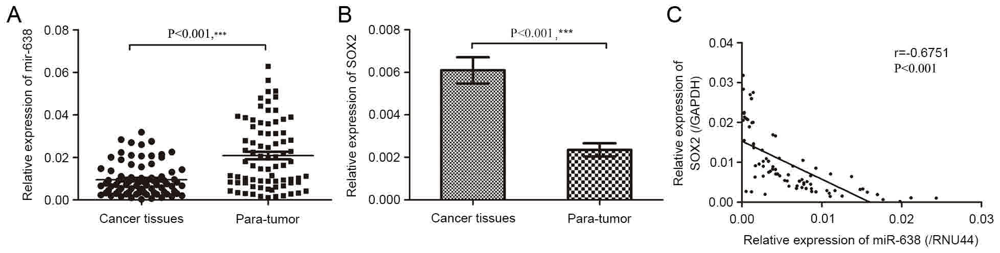
In the present study, it was demonstrated that miR-638 expression
in tumor tissues was significantly lower than that in paired
pericancerous healthy tissues (Fig.
1A; P<0.001). However, SOX2 mRNA expression was
significantly upregulated (Fig. 1B;
P<0.001). Furthermore, miR-638 expression in HCC tissues were
negatively correlated with that of SOX2 mRNA (r=−0.675; Fig. 1C; P<0.001). The median expression
of miR-638 and SOX2 were used as cut-off points to divide 78
samples into two groups. For statistical analysis, samples with
miR-638 expression levels equal to or above the cut-off point were
categorized as the high expression group (n=39) and samples with
miR-638 expression levels below the cut-off point were categorized
as the low expression group (n=39).
SOX2 protein expression is inversely
associated with miR-638 expression
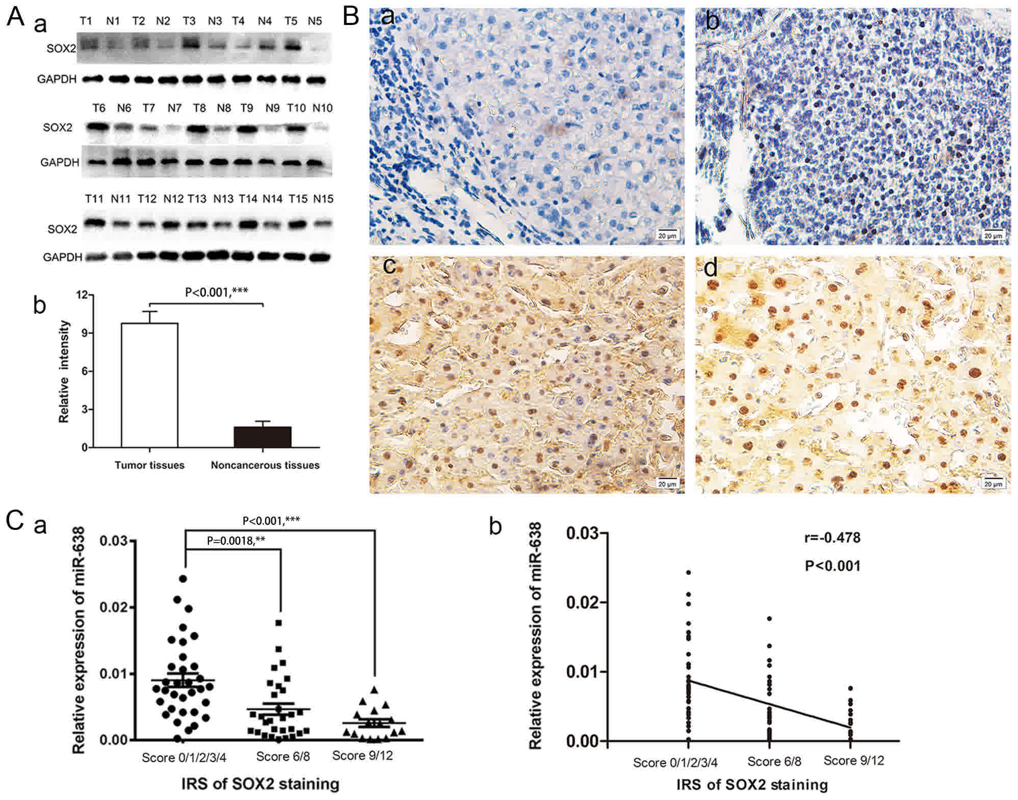
In order to study the association between miR-638
and SOX2 protein in HCC, SOX2 protein levels were measured in 78
pairs of tumor tissues and their adjacent non-cancerous tissues
using western blot and immunohistochemical analyses. Western blot
analysis demonstrated that SOX2 protein levels were significantly
increased in 15 paired HCC samples, compared with adjacent
non-cancerous samples (Fig. 2A;
P<0.001). Immunohistochemical staining results revealed that
SOX2-positive staining was only evident in the nuclei of tumor
cells but not in peritumoral tissues. The representative images of
SOX2 immunostaining in HCC tissues are presented in Fig. 2B. Of the 78 samples, 45 (57.7%)
displayed high expression of SOX2 in tumor tissues compared with 21
(26.9%) paracancerous tissues samples exhibiting high expression
(χ2=15.127, Table II;
P<0.001). Furthermore, in order to statistically analyze the
correlation between miR-638 and SOX2, a cross analysis was
performed and is presented in Table
III (χ2=15.551; P<0.0001), and the results
revealed that the semi-quantitative immunoreactivity scores (IRS)
of SOX2 staining in HCC tissues were negatively associated with
miR-638 expression level (one-way analysis of variance, P<0.05;
r=−0.478, P<0.001; Fig. 2C). The
aforementioned results indicated that increased SOX2 expression in
HCC may be due to miR-638 underexpression, suggesting a potential
functional link between these two molecules.
| Table II.Expression of SOX2 in HCC tissues and
paracancerous tissues. |
Table II.
Expression of SOX2 in HCC tissues and
paracancerous tissues.
|
|
| Expression of
SOX2 |
|
|
|---|
|
|
|
|
|
|
|---|
| Group | Total | Negative | Positive | Positive rate
(%) | χ2 | P-value |
|---|
| HCC tissues | 78 | 33 | 45 | 57.7 | 15.127 | 0.000 |
| Paracancerous
tissues | 78 | 57 | 21 | 26.9 |
|
|
| Table III.The association between miR-638 and
the IRS of SOX2 staining expression in 78 pairs of hepatocellular
carcinoma samples. |
Table III.
The association between miR-638 and
the IRS of SOX2 staining expression in 78 pairs of hepatocellular
carcinoma samples.
|
| IRS |
|
|
|
|---|
|
|
|
|
|
|
|---|
| Group | 0/1/2/3/4 | 6/8 | 9/12 | Total | χ2 | P-value |
|---|
| miR-638
expression |
|
|
|
|
|
|
|
High | 8 | 19 | 12 | 39 | 15.551 | 0.000 |
|
Low | 25 | 10 | 4 | 39 |
|
|
| Total | 33 | 29 | 16 | 78 |
|
|
Correlation between miR-638 or SOX2
expression and clinicopathological features
To observe whether miR-638 and SOX2 expression
levels were correlated with clinicopathological features of HCC,
clinicopathological analysis was performed (Table I). The data indicated that miR-638 and
SOX2 were not significantly correlated with age, sex, tumor size,
tumor number, serum a-fetoprotein (AFP) level, tumor
differentiation (all P>0.05). By contrast, a significant
association was observed between miR-638 or SOX2 expression and TNM
staging (P=0.001 and 0.002, respectively). It was also demonstrated
that patients with HCC with lower miR-638 expression and higher
SOX2 expression were more likely to be at a higher portal vascular
rate (P=0.005 and 0.012, respectively). The results from the
present study suggested that miR-638 and SOX2 expression may serve
a vital role in HCC progression and may have potential as novel
prognosis biomarkers for HCC.
Association between miR-638 or SOX2
expression and the prognosis of patients with HCC
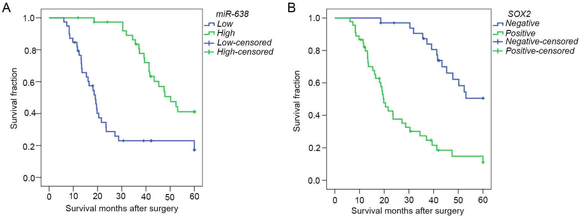
In order to further evaluate whether there was an
association between miR-638 or SOX2 expression and the overall
prognosis of patients with HCC, the Kaplan-Meier method and Cox
regression analysis were performed. Kaplan-Meier survival analysis
suggested that miR-638 expression was significantly associated with
a higher overall survival rate [median survival time (determined
from the date of operation to the last visit or death), 50.20 vs.
19.10 months; Fig. 3A; P<0.001].
Consistently, the overall survival rate of patients with exhibiting
SOX2-positive expression was significantly lower than in those with
SOX2-negative expression (median survival time, 19.80 vs. 50.75
months; Fig. 3B; P<0.001).
Furthermore, Univariate Cox proportional hazards regression model
analysis demonstrated that tumor size, tumor number, HBV status,
TNM stage, portal vascular invasion, distant metastasis, and
miR-638 and SOX2 expression were statistically significant risk
factors for the overall survival of patients with HCC (Table IV). However, age, sex, cirrhosis,
serum AFP level and tumor differentiation had no prognostic value.
Multivariate analysis, stratified for the known prognostic
variables, revealed that portal vascular invasion (P=0.029),
expression of SOX2 (P=0.013) and expression of miR-638 (P=0.008)
were independent prognostic factors of HCC. These observations
suggested that miR-638 and SOX2 may serve as novel prognostic
biomarkers for patients diagnosed with HCC.
| Table IV.Cox regression analysis of overall
survival for patients with hepatocellular carcinoma (n=78). |
Table IV.
Cox regression analysis of overall
survival for patients with hepatocellular carcinoma (n=78).
|
| Univariate
analysis | Multivariate
analysis |
|---|
|
|
|
|
|---|
| Variable | HR | (95% CI) | P-value | HR | (95% CI) | P-value |
|---|
| Age, years (≥57 vs.
<57) | 1.018 | 0.990–1.046 | 0.216 |
|
|
|
| Sex (female vs.
male) | 0.770 | 0.327–1.812 | 0.549 |
|
|
|
| Tumor size, cm (≥5
vs. <5 cm) | 2.615 | 1.475–4.634 | 0.001b | 1.560 | 0.907–1.824 | 0.347 |
| Tumor number (≥2
vs. 1) | 2.257 | 1.047–4.865 | 0.038a |
|
|
|
| Hepatitis B virus
(positive vs. negative) | 0.471 | 0.240–0.926 | 0.029a |
|
|
|
| Cirrhosis (yes vs.
no) | 0.813 | 0.365–1.811 | 0.612 |
|
|
|
| AFP, U/l (≥400 vs.
<400) | 1.581 | 0.822–3.040 | 0.170 |
|
|
|
| Tumor
differentiation (poorly vs. moderately vs. well) | 1.407 | 0.975–2.030 | 0.068 |
|
|
|
| TNM stage (III+IV
vs. I+II) | 4.565 | 2.536–8.220 |
<0.001c | 2.016 | 0.806–5.041 | 0.134 |
| Portal vascular
invasion (yes vs. no) | 5.672 | 3.049–10.549 |
<0.001c | 3.172 | 1.126–8.931 | 0.029a |
| Distant metastasis
(yes vs. no) | 4.030 | 1.893–8.579 |
<0.001c | 1.505 | 0.589–3.846 | 0.394 |
| SOX2 (positive vs.
negative) | 4.732 | 2.499–8.959 |
<0.001c | 2.812 | 1.896–3.780 | 0.013a |
| miR-638 expression
(high vs. low) | 0.269 | 0.269–0.149 |
<0.001c | 0.338 | 0.152–0.751 | 0.008b |
Discussion
HCC remains one of the leading causes of
cancer-associated mortality worldwide (1). Due to the fact that there are no
effective methods for prevention or early diagnosis, the lack of
effective treatment strategies means that patients diagnosed with
HCC exhibit a high mortality rate and a poor prognosis (2–5). In view
of this, identifying sensitive and specific biomarkers for early
diagnosis, therapy guidance and predicting prognosis is imperative.
In the present study, the data indicated that miR-638 may
negatively regulate SOX2 expression, and may be associated with
advanced TNM stages and portal vascular invasion. The results
provided evidence for the regulatory roles of miR-638 and SOX2 in
HCC progression, and suggested that miR-638 and SOX2 may be
potential biomarkers for predicting the prognosis of patients with
HCC.
Dysregulation of miR-638 has been described to be
involved in tumorigenesis and tumor progression in various types of
human cancer, by targeting specific genes. Notably, the roles of
miR-638 in different tumor types remains controversial as it is
able to behave either as a tumor suppressive gene or an oncogene.
For instance, Tan et al (16)
demonstrated that expression of miR-638 reduced cell proliferation
and decreased triple-negative breast cancer cell invasion, which in
turn contributed to esophageal squamous carcinoma proliferation
in vivo (35). Zhao et
al (36) also identified that
miRNA-638 acts as an anti-oncogene in human gastric cancer cells.
By contrast, it has been observed that miR-638 promotes
tumorigenesis and tumor development in human colon carcinoma cells
and human osteosarcoma cells (37),
and contributes to DNA damage in the benzo (a) pyrene-induced human
cell transformation (38). Although
inconsistent findings regarding miR-638 have been observed in
different types of tumors, the role of miR-638 in
hepatocarcinogenesis has not been clearly elucidated. Only one
previous report demonstrated that underexpression of miRNA-638
promoted the angiogenesis and proliferation of HCC cells by
targeting vascular endothelial growth factor (29), indicating that downregulation of
miR-638 may serve a vital role in HCC progression. In agreement
with this observation, the present study also confirmed that
miR-638 expression levels were downregulated in a large number of
HCC clinical samples. In addition, it was revealed that the reduced
miR-638 expression was negatively correlated with advanced TNM
stages and portal vascular invasion. Notably, it was confirmed that
patients with tumors with a low expression of miR-638 were
significantly more likely to exhibit a poorer overall survival. In
line with this, a Cox proportional hazards model, adjusted for the
possible prognostic variables, revealed that miR-638 may serve as
an independent and favorable prognostic factor for HCC.
SOX2 is a highly conserved transcriptional
regulator, which contributes an important role to the maintenance
of embryonic stem cell pluripotency and self-renewal (19). It is well documented that SOX2 is
expressed in various tissues and serves an important function in
the differentiation and morphogenesis of the esophagus and stomach
epithelial (39,40). The roles of SOX2 in these biological
processes implicates that it has the potential to modulate the
progression of cellular malignant transformation (41). Therefore, numerous studies have
demonstrated that SOX2 was aberrantly expressed in a wide variety
of solid malignant tumors, including breast, colorectal, prostate
and lung cancer (20–23), indicating that SOX2 may act as a key
factor in tumorigenesis and tumor development. In addition,
previous studies have demonstrated that the overexpression of SOX2
in tumor tissues was associated with a strong invasiveness and a
poor prognosis (42,43). Furthermore, SOX2 exhibits a close
interaction with numerous miRNAs and, in previous studies, SOX2
activity was attributable to regulation by several miRNAs,
including miR-126, miR-429 and miR-625 (44–46). In
2014, Ma et al (27)
demonstrated a functional association between miR-638 and SOX2.
Their study identified that downregulation of miR-638 promotes
colorectal carcinoma cell (CRC) invasion and proliferation by
influencing SOX2 expression. It was further demonstrated that
miR-638 expression levels were negatively correlated with SOX2
expression, and the invasive and differentiative potential of CRC
cells. In the same year, Xia et al (28) reported that the expression levels of
miR-638 and SOX2 were inversely associated in non-small-cell lung
cancer (NSCLC) tissues. The expression levels of miR-638 in the
highly aggressive NSCLC cells were much lower than those in normal
lung tissue cells. It was also observed that low miR-638 and high
SOX2 expression in NSCLC tissues was significantly associated with
tumor size, TNM stage and distant metastasis. Gain and loss of
function experiments revealed that miR-638 downregulated SOX2
protein expression in NSCLC cells and inhibited cell invasive
potential. Additionally, the regulation of SOX2 by miR-638 may
influence the epithelial-mesenchymal transition in NSCLC cells. An
investigation into SOX2 expression and its clinical significance in
HCC is therefore important for the management of the disease. The
present study confirmed that SOX2 was overexpressed in liver cancer
tissues and that SOX2 expression was positively correlated with
tumor size, tumor stage and portal vascular invasion, as compared
with negative controls. In addition, it was demonstrated that high
SOX2 expression levels were associated with a poorer prognosis in
patients with HCC, and served as an independent and unfavorable
prognostic factor for HCC. The results of the present study
supported the hypothesis that SOX2 is a key regulator in
tumorigenesis and tumor development, which is in accordance with
the results of previous studies on multiple types of cancer
(42,43).
In the present retrospective study of patients with
HCC, it was observed that miR-638 expression was markedly
downregulated and SOX2 presented significantly higher expression in
HCC tissue, compared with expression in adjacent non-cancerous
controls. It was further validated that reduced miR-638 expression
was negatively associated with overexpression of SOX2 protein in
HCC. These observations were, in part, consistent with the
conclusion of Zhang et al (30), that miR-638 may influence HCC
progression by negatively regulating SOX2 expression. In addition,
the present study demonstrated that miR-638 and SOX2 expression
were significantly associated with tumor stage, portal vascular
invasion and postoperative survival in patients with HCC. However,
the failure to validate the molecular rationale for the involvement
of miR-638 and SOX2 in the progression of HCC in vitro is
one limitation of the present study. Another limitation is that the
present study was retrospective and therefore, the results require
further validation with more extensive tests in future prospective
studies.
In summary, to the best of our knowledge, the
present study was the first to provide evidence regarding the
detailed expression pattern and clinical significance of miR-638
and SOX2 in a large number of patients with HCC. Furthermore, the
results of the present study suggested that miR-638 may serve an
important role in the occurrence and progression of HCC by
downregulating SOX2 expression. Consequently, these proteins may
serve as potential novel biomarkers, and may also be beneficial to
the currently available HCC indicators for predicting HCC
progression and poor prognosis. By identifying the patients who are
more likely to have a higher risk of mortality, there is a
possibility of implementing a more aggressive therapeutic regimen.
Present and future studies regarding miR-638 and SOX2 expression in
HCC progression may provide novel insights into the diagnosis and
prognosis of this devastating disease.
Acknowledgments
Not applicable.
Funding
The present study was funded by the National Natural
Science Foundation of Zhejiang Province of China (grant no.
LY13H030007).
Availability of data and materials
The datasets used and/or analyzed during the current
study are available from the corresponding author on reasonable
request.
Authors' contributions
WYe, XC and JL carried out the experiments, and the
data collection and interpretation. GF and YZ participated in the
design and coordination of experimental work, and data acquisition.
CZ and LC significantly contributed to analysis of data and
preparation of the manuscript. WYa designed the study, analyzed and
interpreted the data, and drafted the manuscript.
Ethics approval and consent to
participate
Ethical approval was obtained from the Ethics
Committee of The First Affiliated Hospital of Wenzhou Medical
University (Zhejiang, China), and written informed consent was
obtained from all patients.
Consent for publication
Written informed consent was obtained from all
patients.
Competing interests
The authors declare that they have no competing
interests.
References
|
1
|
Torre LA, Bray F, Siegel RL, Ferlay J,
Lortet-Tieulent J and Jemal A: Global cancer statistics, 2012. CA
Cancer J Clin. 65:87–108. 2015. View Article : Google Scholar : PubMed/NCBI
|
|
2
|
Bruix J and Sherman M: American
Association for the Study of Liver Diseases: Management of
hepatocellular carcinoma: An update. Hepatology. 53:1020–1022.
2011. View Article : Google Scholar : PubMed/NCBI
|
|
3
|
Hsu CY, Liu PH, Hsia CY, Lee YH, Nagaria
TS, Lee RC, Lin HC and Huo TI: Surgical resection is better than
transarterial chemoembolization for patients with hepatocellular
carcinoma beyond the milan criteria: A prognostic nomogram study.
Ann Surg Oncol. 23:994–1002. 2016. View Article : Google Scholar : PubMed/NCBI
|
|
4
|
Tremosini S, Reig M, de Lope CR, Forner A
and Bruix J: Treatment of early hepatocellular carcinoma: Towards
personalized therapy. Dig Liver Dis. 42 Suppl 3:S242–S248. 2010.
View Article : Google Scholar : PubMed/NCBI
|
|
5
|
Imamura H, Matsuyama Y, Tanaka E, Ohkubo
T, Hasegawa K, Miyagawa S, Sugawara Y, Minagawa M, Takayama T,
Kawasaki S and Makuuchi M: Risk factors contributing to early and
late phase intrahepatic recurrence of hepatocellular carcinoma
after hepatectomy. J Hepatol. 38:200–207. 2003. View Article : Google Scholar : PubMed/NCBI
|
|
6
|
Bartel DP: MicroRNAs: Genomics,
biogenesis, mechanism, and function. Cell. 116:281–297. 2004.
View Article : Google Scholar : PubMed/NCBI
|
|
7
|
Lin SL, Chang DC, Ying SY, Leu D and Wu
DT: MicroRNA miR-302 inhibits the tumorigenecity of human
pluripotent stem cells by coordinate suppression of the CDK2 and
CDK4/6 cell cycle pathways. Cancer Res. 70:9473–9482. 2010.
View Article : Google Scholar : PubMed/NCBI
|
|
8
|
Wang Z, Yin B, Wang B, Ma Z, Liu W and Lv
G: MicroRNA-210 promotes proliferation and invasion of peripheral
nerve sheath tumor cells targeting EFNA3. Oncol Res. 21:145–154.
2013. View Article : Google Scholar : PubMed/NCBI
|
|
9
|
Tome M, López-Romero P, Albo C, Sepúlveda
JC, Fernández-Gutiérrez B, Dopazo A, Bernad A and González MA:
miR-335 orchestrates cell proliferation, migration and
differentiation in human mesenchymal stem cells. Cell Death Differ.
18:985–995. 2011. View Article : Google Scholar : PubMed/NCBI
|
|
10
|
Zhang Y, He X, Liu Y, Ye Y, Zhang H, He P,
Zhang Q, Dong L, Liu Y and Dong J: microRNA-320a inhibits tumor
invasion by targeting neuropilin 1 and is associated with liver
metastasis in colorectal cancer. Oncol Rep. 27:685–694.
2012.PubMed/NCBI
|
|
11
|
Ma D, Tao X, Gao F, Fan C and Wu D:
miR-224 functions as an onco-miRNA in hepatocellular carcinoma
cells by activating AKT signaling. Oncol Lett. 4:483–488. 2012.
View Article : Google Scholar : PubMed/NCBI
|
|
12
|
Calin GA and Croce CM: MicroRNA signatures
in human cancers. Nat Rev Cancer. 6:857–866. 2006. View Article : Google Scholar : PubMed/NCBI
|
|
13
|
Xu J, Li J, Zheng TH, Bai L and Liu ZJ:
MicroRNAs in the occurrence and development of primary
hepatocellular carcinoma. Adv Clin Exp Med. 25:971–975. 2016.
View Article : Google Scholar : PubMed/NCBI
|
|
14
|
Zhang J, Fei B, Wang Q, Song M, Yin Y,
Zhang B, Ni S, Guo W, Bian Z, Quan C, et al: MicroRNA-638 inhibits
cell proliferation, invasion and regulates cell cycle by targeting
tetraspanin 1 in human colorectal carcinoma. Oncotarget.
5:12083–12096. 2014.PubMed/NCBI
|
|
15
|
Zhang J, Bian Z, Zhou J, Song M, Liu Z,
Feng Y, Zhe L, Zhang B, Yin Y and Huang Z: MicroRNA-638 inhibits
cell proliferation by targeting phospholipase D1 in human gastric
carcinoma. Protein Cell. 6:680–688. 2015. View Article : Google Scholar : PubMed/NCBI
|
|
16
|
Tan X, Peng J, Fu Y, An S, Rezaei K,
Tabbara S, Teal CB, Man YG, Brem RF and Fu SW: miR-638 mediated
regulation of BRCA1 affects DNA repair and sensitivity to UV and
cisplatin in triple-negative breast cancer. Breast Cancer Res.
16:4352014. View Article : Google Scholar : PubMed/NCBI
|
|
17
|
Wang XX, Liu J, Tang YM, Hong L, Zeng Z
and Tan GH: MicroRNA-638 inhibits cell proliferation by targeting
suppress PIM1 expression in human osteosarcoma. Tumour Biol. Jan
3–2017.(Epub ahead of print).
|
|
18
|
Bhattacharya A, Schmitz U, Raatz Y,
Schönherr M, Kottek T, Schauer M, Franz S, Saalbach A, Anderegg U,
Wolkenhauer O, et al: miR-638 promotes melanoma metastasis and
protects melanoma cells from apoptosis and autophagy. Oncotarget.
6:2966–2980. 2015. View Article : Google Scholar : PubMed/NCBI
|
|
19
|
Fong H, Hohenstein KA and Donovan PJ:
Regulation of self-renewal and pluripotency by Sox2 in human
embryonic stem cells. Stem cells. 26:1931–1938. 2008. View Article : Google Scholar : PubMed/NCBI
|
|
20
|
Leis O, Eguiara A, Lopez-Arribillaga E,
Alberdi MJ, Hernandez-Garcia S, Elorriaga K, Pandiella A, Rezola R
and Martin AG: Sox2 expression in breast tumours and activation in
breast cancer stem cells. Oncogene. 31:1354–1365. 2012. View Article : Google Scholar : PubMed/NCBI
|
|
21
|
Neumann J, Bahr F, Horst D, Kriegl L,
Engel J, Luque RM, Gerhard M, Kirchner T and Jung A: SOX2
expression correlates with lymph-node metastases and distant spread
in right-sided colon cancer. BMC Cancer. 11:5182011. View Article : Google Scholar : PubMed/NCBI
|
|
22
|
Jia X, Li X, Xu Y, Zhang S, Mou W, Liu Y,
Liu Y, Lv D, Liu CH, Tan X, et al: SOX2 promotes tumorigenesis and
increases the anti-apoptotic property of human prostate cancer
cell. J Mol Cell Biol. 3:230–238. 2011. View Article : Google Scholar : PubMed/NCBI
|
|
23
|
Nakatsugawa M, Takahashi A, Hirohashi Y,
Torigoe T, Inoda S, Murase M, Asanuma H, Tamura Y, Morita R,
Michifuri Y, et al: SOX2 is overexpressed in stem-like cells of
human lung adenocarcinoma and augments the tumorigenicity. Lab
Invest. 91:1796–1804. 2011. View Article : Google Scholar : PubMed/NCBI
|
|
24
|
Chen Y, Huang Y, Zhu L, Chen M, Huang Y,
Zhang J, He S, Li A, Chen R and Zhou J: SOX2 inhibits metastasis in
gastric cancer. J Cancer Res Clin Oncol. 142:1221–1230. 2016.
View Article : Google Scholar : PubMed/NCBI
|
|
25
|
Cho YY, Kim DJ, Lee HS, Jeong CH, Cho EJ,
Kim MO, Byun S, Lee KY, Yao K, Carper A, et al: Autophagy and
cellular senescence mediated by Sox2 suppress malignancy of cancer
cells. PLoS One. 8:e571722013. View Article : Google Scholar : PubMed/NCBI
|
|
26
|
Wang S, Tie J, Wang R, Hu F, Gao L, Wang
W, Wang L, Li Z, Hu S, Tang S, et al: SOX2, a predictor of survival
in gastric cancer, inhibits cell proliferation and metastasis by
regulating PTEN. Cancer Lett. 358:210–219. 2015. View Article : Google Scholar : PubMed/NCBI
|
|
27
|
Ma K, Pan X, Fan P, He Y, Gu J, Wang W,
Zhang T, Li Z and Luo X: Loss of miR-638 in vitro promotes cell
invasion and a mesenchymal-like transition by influencing SOX2
expression in colorectal carcinoma cells. Mol Cancer. 13:1182014.
View Article : Google Scholar : PubMed/NCBI
|
|
28
|
Xia Y, Wu Y, Liu B, Wang P and Chen Y:
Downregulation of miR-638 promotes invasion and proliferation by
regulating SOX2 and induces EMT in NSCLC. FEBS Lett. 588:2238–2245.
2014. View Article : Google Scholar : PubMed/NCBI
|
|
29
|
Cheng J, Chen Y, Zhao P, Liu X, Dong J, Li
J, Huang C, Wu R and Lv Y: Downregulation of miRNA-638 promotes
angiogenesis and growth of hepatocellular carcinoma by targeting
VEGF. Oncotarget. 7:30702–30711. 2016.PubMed/NCBI
|
|
30
|
Zhang Y, Zhang D, Jiang J and Dong L: Loss
of miR-638 promotes invasion and epithelial-mesenchymal transition
by targeting SOX2 in hepatocellular carcinoma. Oncol Rep.
37:323–332. 2017. View Article : Google Scholar : PubMed/NCBI
|
|
31
|
Poon RT and Fan ST: Evaluation of the new
AJCC/UICC staging system for hepatocellular carcinoma after hepatic
resection in Chinese patients. Surg Oncol Clin N Am. 12(35–50):
viii2003.
|
|
32
|
Li ZS and Li Q: The latest 2010 WHO
classification of tumors of digestive system. Zhonghua Bing Li Xue
Za Zhi. 40:351–354. 2011.(In Chinese). PubMed/NCBI
|
|
33
|
Flejou JF: WHO Classification of digestive
tumors: The fourth edition. Ann Pathol. 31 5 Suppl:S27–S31.
2011.PubMed/NCBI
|
|
34
|
Livak KJ and Schmittgen TD: Analysis of
relative gene expression data using real-time quantitative PCR and
the 2(-Delta Delta C(T)) method. Methods. 25:402–408. 2001.
View Article : Google Scholar : PubMed/NCBI
|
|
35
|
Zhang X, Wei J, Zhou L, Zhou C, Shi J,
Yuan Q, Yang M and Lin D: A functional BRCA1 coding sequence
genetic variant contributes to risk of esophageal squamous cell
carcinoma. Carcinogenesis. 34:2309–2313. 2013. View Article : Google Scholar : PubMed/NCBI
|
|
36
|
Zhao LY, Yao Y, Han J, Yang J, Wang XF,
Tong DD, Song TS, Huang C and Shao Y: miR-638 suppresses cell
proliferation in gastric cancer by targeting Sp2. Dig Dis Sci.
59:1743–1753. 2014. View Article : Google Scholar : PubMed/NCBI
|
|
37
|
Tay Y, Tan SM, Karreth FA, Lieberman J and
Pandolfi PP: Characterization of dual PTEN and p53-targeting
microRNAs identifies microRNA-638/Dnm2 as a two-hit oncogenic
locus. Cell Rep. 8:714–722. 2014. View Article : Google Scholar : PubMed/NCBI
|
|
38
|
Li D, Wang Q, Liu C, Duan H, Zeng X, Zhang
B, Li X, Zhao J, Tang S, Li Z, et al: Aberrant expression of
miR-638 contributes to benzo(a)pyrene-induced human cell
transformation. Toxicol Sci. 125:382–391. 2012. View Article : Google Scholar : PubMed/NCBI
|
|
39
|
Wegner M: From head to toes: The multiple
facets of Sox proteins. Nucleic Acids Res. 27:1409–1420. 1999.
View Article : Google Scholar : PubMed/NCBI
|
|
40
|
Ishii Y, Rex M, Scotting PJ and Yasugi S:
Region-specific expression of chicken Sox2 in the developing gut
and lung epithelium: Regulation by epithelial-mesenchymal
interactions. Dev Dyn. 213:464–475. 1998. View Article : Google Scholar : PubMed/NCBI
|
|
41
|
Liu K, Lin B, Zhao M, Yang X, Chen M, Gao
A, Liu F, Que J and Lan X: The multiple roles for Sox2 in stem cell
maintenance and tumorigenesis. Cell Signal. 25:1264–1271. 2013.
View Article : Google Scholar : PubMed/NCBI
|
|
42
|
Honing J, Pavlov KV, Meijer C, Smit JK,
Boersma-van Ek W, Karrenbeld A, Burgerhof JG, Kruyt FA and Plukker
JT: Loss of CD44 and SOX2 expression is correlated with a poor
prognosis in esophageal adenocarcinoma patients. Ann Surg Oncol. 21
Suppl 4:S657–S664. 2014. View Article : Google Scholar : PubMed/NCBI
|
|
43
|
Lundberg IV, Löfgren Burström A, Edin S,
Eklöf V, Öberg Å, Stenling R, Palmqvist R and Wikberg ML: SOX2
expression is regulated by BRAF and contributes to poor patient
prognosis in colorectal cancer. PLoS One. 9:e1019572014. View Article : Google Scholar : PubMed/NCBI
|
|
44
|
Li J, Du L, Yang Y, Wang C, Liu H, Wang L,
Zhang X, Li W, Zheng G and Dong Z: MiR-429 is an independent
prognostic factor in colorectal cancer and exerts its
anti-apoptotic function by targeting SOX2. Cancer Lett. 329:84–90.
2013. View Article : Google Scholar : PubMed/NCBI
|
|
45
|
Otsubo T, Akiyama Y, Hashimoto Y, Shimada
S, Goto K and Yuasa Y: MicroRNA-126 inhibits SOX2 expression and
contributes to gastric carcinogenesis. PLoS One. 6:e166172011.
View Article : Google Scholar : PubMed/NCBI
|
|
46
|
Wang Z, Qiao Q, Chen M, Li X, Wang Z, Liu
C and Xie Z: miR-625 down-regulation promotes proliferation and
invasion in esophageal cancer by targeting Sox2. FEBS Lett.
588:915–921. 2014. View Article : Google Scholar : PubMed/NCBI
|