Introduction
Urothelial cell carcinoma of the bladder (UCB) is a
common urogenital malignancy worldwide, and in the United States of
America ~17,000 patients succumb to UCB annually (1). There are 2 distinct complex pathways
that occur in the initiation/progression of UCB (2), and the present study aimed to identify
specific molecular marks or targets for UCB, particularly low
aggressive non-muscle-invasive bladder cancer (NMIBC) and the
highly aggressive muscle-invasive bladder cancer (MIBC) (1,3,4). Assessment of the risks of recurrence and
progression to MIBC remains a major problem for urologists
(5).
Meyer-Siegler et al (6), demonstrated that the inhibition of
macrophage migration inhibitory factor (MIF) may decrease UCB cells
proliferation and cytokine expression through the MIF-cluster of
differentiation (CD)74 pathway. Taylor et al (7), suggested that MIF may serve a role in
the progression to invasive bladder cancer. However, growing
evidence has suggested that MIF-targeted therapy may be potentially
hazardous to health, considering that MIF participates in various
host defense and immunological reactions to inflammation (8–11).
Conversely, CD74 has indicated limited expression in normal human
tissues, and was suggested to be the integrant chaperone for MIF
receptors including CD44, C-X-C chemokine receptor (CXCR)2, CXCR4
or Toll-like receptor 4 (TLR4), to compose receptor complexes,
modulate cell proliferation/apoptosis, initiate signal transduction
of the Nuclear Factor κB (NF-κB), Extracellular regulated protein
kinase 1/2 (Erk1/2) and the Phosphoinositide 3′-kinase/RAC-alpha
serine/threonine protein kinase (PI3K/Akt) pathway, and in turn
participate in a number of processes including inflammation and
carcinogenesis (12–16). At present, CD74 expression was
demonstrated to be increased only in high-grade UCB (16). In light of these data, experiments
investigating the effect of CD74-knockdown on UCB cells may be a
promising strategy for treatment.
In the present study, the potential association
between the expression levels of MIF and CD74 with clinical and
pathological characteristics were analyzed, and whether the
knockdown of CD74 would affect protein expression, proliferation,
apoptosis, invasion, angiogenesis and signal transduction
associated with UCB was also explored.
Materials and methods
Samples, cell lines and agents
Human tissue specimens were obtained from 108
patients with UCB (mean age, 63.4±11.3 years, age range 45–74
years) who underwent either transurethral resection or radical
cystectomy, and 20 patients who had received either cystoscopic
biopsy, ureteral re-implantation or cystoprostatectomy (mean age,
62.8±13.0 years, age range 41–82 years) in Beijing Chao-Yang
Hospital (Beijing, China) from August 2004 to March 2013. Informed
consent was obtained from all patients enrolled. Tumors staged as
carcinoma in situ were not included. The present study was
approved by the Beijing Chao-Yang Hospital Institutional Research
Ethical Board. All samples were confirmed and staged according to
the American Joint Committee on Cancer TNM standard, and graded by
2 independent experienced genitourinary pathologists of the Beijing
Chao-Yang Hospital (Beijing, China) (17). Table I
summarizes the clinical and pathological characteristics of all
patients enrolled.
 | Table I.Association between MIF and CD74
expression with clinical and pathological characteristics of
patient samples. |
Table I.
Association between MIF and CD74
expression with clinical and pathological characteristics of
patient samples.
|
|
| MIF |
| CD74 |
|
|---|
|
|
|
|
|
|
|
|---|
|
Characteristics | Total | − | + | ++ | P-value | − | + | ++ | P-value |
|---|
| Sex |
|
|
|
|
|
|
|
|
|
|
Male | 92 | – | – | – | – | – | – | – | – |
|
Female | 36 | – | – | – | – | – | – | – | – |
| Age, mean ± SD | 62.9±11.6 |
|
|
|
|
|
|
|
|
| Normal
patients (20) | 62.8±13.0 | 5 | 7 | 8 |
| 9 | 10 | 1 | – |
| UCB
patients (108) | 63.4±11.3 | 12 | 28 | 30 |
| 25 | 26 | 19 | – |
| UCB Pathology |
|
|
|
|
|
|
|
|
|
| Stage |
|
|
|
| 0.106 |
|
|
| <0.001 |
| NMIBC
(pTa, pT1) | 70 | 12 | 28 | 30 |
| 25 | 26 | 19 |
|
| MIBC
(pT2-T4) | 38 | 3 | 12 | 23 |
| 4 | 8 | 26 |
|
| Grade |
|
|
|
| 0.631 |
|
|
| <0.001 |
|
Low | 36 | 8 | 10 | 18 |
| 20 | 12 | 4 |
|
|
High | 72 | 7 | 30 | 35 |
| 10 | 22 | 40 |
|
| Lymphogenous
metastasis |
|
|
|
| 0.148 |
|
|
| 0.031 |
| − | 94 | 13 | 38 | 43 |
| 29 | 30 | 35 |
|
| + | 14 | 2 | 2 | 10 |
| 1 | 4 | 9 |
|
The human immortalized urothelial cell line
SV-HUC-1, and UCB SW780 (G1 stage), 5637 (G2), T24 (G3), J82 (G3)
and HT-1376 (G3) cell lines were purchased from the Type Culture
Collection of Chinese Academy of Sciences (Shanghai, China).
SV-HUC-1 was cultured in F-12K medium (Hyclone; GE Healthcare Life
Sciences, Logan, UT, USA). SW780 cells were cultured in Leibovitz's
L-15 Medium (Hyclone; GE Healthcare Life Sciences), 5637, T24, J82
and HT-1376 were all cultured in RPMI-1640 (Hyclone; GE Healthcare
Life Sciences) medium supplemented with 10% fetal bovine serum
(Gibco; Thermo Fisher Scientific, Inc., Waltham, MA, USA). All
cells were routinely cultured in a 37°C humidified incubator with
5% CO2 and 95% O2.
PBS, 3′-diaminobenzidine (DAB), EDTA and DAPI were
obtained from Sigma-Aldrich; Merck KGaA (Darmstadt, Germany).
Commercial CD74 short hairpin (sh)RNA lentiviral particles (cat.
no., sc-35023-v), scramble shRNA lentiviral particles (cat. no.,
sc-108080) and CD34 (cat. no., sc-65261; dilution, 1:400) and NF-κB
p65 antibodies (cat. no., sc-71677; dilution, 1:200) were purchased
from Santa Cruz Biotechnology, Inc., (Dallas, TX, USA). The ELISA
kits for vascular endothelial growth factor (VEGF; cat. no.,
03-0068-00), matrix metalloproteinase (MMP)-2 (cat. no., MAB13431)
and MMP-9 (cat. no., QIA56) were purchased from Calbiochem; EMD
Millipore (Billerica, MA, USA). Cyclin D1 (cat. no., 2922S;
dilution, 1:800), Cyclin B1 (cat. no., 4138S; dilution, 1:500),
Proliferating Cell Nuclear Antigen (cat. no., 13110S; dilution,
1:800), B-cell lymphoma 2 (Bcl-2; cat. no., 3498S; dilution,
1:500), Bcl-extra large (Bcl-xL; cat. no., 2762S; dilution, 1:500),
Bcl-2-associated X protein (Bax; cat. no., 2774S; dilution, 1:800),
Bcl-2-associated death protein (Bad; cat. no., 9292S; dilution,
1:500) and GAPDH (cat. no., 8884S; dilution, 1:1,000) antibodies
were sourced from Cell Signaling Technology (CST, Boston, MA).
Erk1/2 (cat. no., V114A; dilution, 1:2,000), phosphorylated Erk1/2
(pErk1/2; cat. no., 9101S; dilution, 1:500), cleaved poly
(adenosine 5′-diphosphate-ribose) polymerase (PARP) p85 (cat. no.,
G734A; dilution, 1:300) antibodies were purchased from Promega
Corporation (Madison, WI, USA).
ELISA of VEGF, MMP-2 and MMP-9
Following 48 h of incubation, the HT-1376 cells
culture media of the basal chamber of control and CD74-shRNA were
collected separately. Following centrifugation (12,000 × g at 4°C
for 15 min), the protein levels were measured prior to ELISA assay.
The concentrations of the VEGF, MMP-2 and MMP-9 cytokines were
determined by the corresponding ELISA kits according to the
manufacturer's protocol; the absorbance was measured at 450 and 595
nm using a microplate reader (Bio-Rad Laboratories, Inc., Hercules,
CA, USA).
Immunostaining image assay of MIF and
CD74
Bladder tissue samples were examined by
immunohistochemistry and immunofluorescence for the expression of
MIF and CD74. Paraffin-embedded tissue sections (4-µm thick) were
then dewaxed at room temperature for 5 min in 99.0% xylene and
rehydrated in graded ethanol solutions (two washes of 100% ethanol
for 10 min each and two washes of 95% ethanol for 10 min each).
Antigen retrieval was performed following standard procedures:
Sections were cooled and immersed in 3% hydrogen peroxide solution
for 15 min to block endogenous peroxidase activity at room
temperature, and rinsed in PBS for 5 min. Non-specific labeling was
blocked by incubation with 5% bovine serum albumin (Thermo Fisher
Scientific, Inc., Waltham, MA, USA) at room temperature for 30 min.
Sections were then incubated with primary antibodies against MIF
(cat. no., sc-271631; dilution, 1:200) or CD74 (cat. no., sc-70781;
1:200; Santa Cruz Biotechnology, Inc.) at 4°C overnight. They were
then rinsed with PBST three times, and incubated at 37°C for 15 min
with horseradish peroxidase (HRP)-conjugated goat IgG secondary
antibody (cat. no., sc-2354; dilution, 1:50; Santa Cruz
Biotechnology, Inc.), developed with HRP-conjugated streptavidin
and DAB (2.5 ml 10X DAB solution and 22.5 ml of stable peroxide
substrate buffer; Sigma-Aldrich; Merck KGaA) for 1 h at room
temperature and counterstained with hematoxylin for 2 min. The
immunohistochemical results were then scored by two independent
pathologists of Beijing Chao-Yang Hospital (Beijing, China).
Staining intensity (percentage of positively tumor cells among all
tumor cells) was scored as ‘−’ (negative), ‘+’ (moderate) or ‘++’
(strong). The extent of staining was scored as ‘−’ (<20% of
urothelial cells stained), ‘+’ (20–60% stained) or ‘++’ (>60%
stained).
UCB cell-covered slides were collected from the
media and washed twice using PBS, and then sections were incubated
for 60 min at 37°C with fluorescein isothiocyanate-anti-mouse
secondary antibody (Santa Cruz Biotechnology, Inc.). Finally,
4′,6-diamidino-2-phenylindole (Sigma-Aldrich; Merck KGaA) was used
to treat the slides. Images were captured using a
confocal-microscope in 3 fields of view (magnification, ×100;
Olympus, Tokyo, Japan).
Western blotting assay
Lysis buffer (Bio-Rad Laboratories, Inc.) was used
to prepare the tissue and cells, and protein concentrations were
measured by Bradford's method (18).
A total of 50 µg protein was placed in each lane of 12% SDS-PAGE
(Bio-Rad Laboratories, Inc.), and then transferred onto
nitrocellulose membranes (Bio-Rad Laboratories, Inc.). Fat-free
milk powder (5%) was used to block membranes, which was incubated
in primary antibodies overnight at 4°C, and then with
HRP-conjugated secondary antibodies (Santa Cruz Biotechnology,
Inc.). A electrochemiluminescent reagent (Pharmacia Corp., Basking
Ridge, NJ, USA) was used to detect signals and the Kodak Image
Station 4000MM Pro (Kodak, Rochester, NY, USA) was used for
analyzing and recording.
Reverse transcription polymerase chain
reaction (RT-PCR) assay
Total RNA of untreated, scramble shRNA and
CD74-targeted shRNA transfected HT-1376 cells were extracted for
the RT-PCR assay using an RNeasy kit (cat. no., 74104) (Qiagen
Sciences, Inc., Gaithersburg, MD, USA) according to the
manufacturer's protocol. The primers were as follows: CD74 Forward,
5′-CGGAAGATCAGAAGCCAGTC-3′; and CD74 reverse,
5′-GCGAGGAGCAGAGTCACCAG-3′; GAPDH forward,
5′-GTCAAGGCTGAGAACGGGAA-3′; and GAPDH reverse
5′-GCGAGGAGCAGAGTCACCAG-3′. A total of 35 cycles of PCR (95°C for
40 sec, 45°C for 40 sec, and 72°C for 60 sec) were completed for
CD74 and GAPDH, and the expression of CD74 was normalized to that
of GAPDH.
Transfection of lentivirus CD74 shRNA
in HT-1376 cells
The CD74 shRNA lentiviral particles (cat. no.,
sc-35023-V; 1.0×10⁶/200 µl; Santa Cruz Biotechnology,
Inc.) targeting the human CD74 transcript and control shRNA
lentiviral particles (cat. no., sc-108080; 1.0×10⁶/200
µl; Santa Cruz Biotechnology, Inc.) were used to knock down CD74 in
HT-1376 cells. Cells were seeded at 2×10⁵ cells/well in
6-well plates (Corning Incorporated, Corning, NY, USA) and grown to
60% confluence on the day of transfection. The media was removed
from the plate wells and replaced with 2 ml complete medium
containing Polybrene (cat. no., sc-134220; Santa Cruz
Biotechnology, Inc.) at a final concentration of 5 µg/ml. Cells
were transfected with control (scramble, 20 µl) or CD74 shRNA
lentiviral particles (20 µl) diluted in media according to the
manufacturer's protocol in 48 h. Infected cells were selected with
puromycin (5 µg/ml; cat. no., 11811-023; Gibco; Thermo Fisher
Scientific, Inc., Waltham, MA, USA). A total of 72 h after
transfection, transfected cells were verified by RT-PCR and western
blotting analysis, comparing untreated and scramble cells.
Cell vitality assay
A cell vitality test was performed using the
CellTiterTM 96 Aqueous assay kit (cat. no., G358C; Promega
Corporation) and performed according to the instructions of the
manufacturer. The HT-1376 cells (10,000/well) were seeded in
96-well plates (Corning Incorporated) and incubated at 37°C, and
transfected with control or CD74 shRNA lentiviral particles after
24 h. Cell proliferation was assessed after transfected 2, 4, 6, 8
and 10 days, with control or CD74 shRNA lentiviral particles using
a multiwall spectrophotometer (Bio-Rad Laboratories, Inc.).
Flow cytometry
Control or CD74-knockout HT-1376 cells were seeded
in 6-well plates (Corning Incorporated) and incubated in medium
without FBS at 37°C overnight. Then, the cells were rinsed 3 times
and incubated in medium containing 10% FBS. The cells were
collected and then fixed with 70% ethanol after 24 h at 4°C. The
cells were treated with ≥94.0% (HPLC) propidium iodide
(Sigma-Aldrich; Merck KGaA) and RNase A (400 U/ml) for 30 min at
4°C. Cellular content of DNA was evaluated by the flow cytometer
(FACSCalibur; BD Biosciences, Erembodegem, Belgium), and cell cycle
analysis was performed using ModFit2.0 software (BD
Biosciences).
Cell invasion assay
A cell invasion assay was performed with 24-well
Transwell inserts (Corning Incorporated). Following transfected
with control or CD74 shRNA lentiviral particles, HT-1376 cells were
starved in serum-free RPMI-1640 medium overnight, and
1×10⁴ cells were re-suspended in 200 µl serum-free
RPMI-1640 medium and placed in the upper chambers with 8 µm filter
pores in triplicate. The membrane undersurface was coated with 30
µl ECM gel from Engelbreth-Holm-Swarm mouse sarcoma (BD
Biosciences) mixed with FBS-free RPMI-1640 medium in 1:5 dilution
for 30 min at 37°C. The lower chamber was filled with 500 µl 10%
FBS as the chemoattractant and incubated for 48 h at 37°C.
Following this, the cells on the upper surface of the membrane were
removed by cotton buds, and the cells on the lower surface of the
insert were fixed in 4% PBS-buffered paraformaldehyde for 5 min at
room temperature and stained with 0.1% crystal violet
(Sigma-Aldrich; Merck KGaA) for 5 min at room temperature. A total
of 5 visual fields were chosen randomly for each insert and images
were captured under a light microscope (magnification, ×100). The
cells were counted and the data were presented as mean ± standard
deviation (SD) and presented by a percentage of controls.
Tumorigenesis assay
Cells (1×10⁷ cells/mouse) were injected
subcutaneously into the axilla area of 6-week-old male BALB/c-nu
mice (20/group, total 40 mice), which were purchased from the
Experimental Animal Center of Peking University Health Science, and
animals remained in a pathogen-free animal facility. Mice had
access to food and water ad libitum, and were kept at a
temperature of 20~26°C, a humidity of 30–70% and in a 12 h light/12
h dark cycle). The CD74 scramble shRNA and knockdown-HT-1376 cells,
and untreated J82 cells were all included in the analysis. Tumor
diameter was measured every 7 days, and tumor sizes were measured
following sacrifice via cervical dislocation at 28 days, using the
following formula: Length × width2 × 0.5. All tumor
ulceration observed was monitored by the laboratory staff. All
experiments in the present study were performed in strict
accordance with the recommendations in the Association for
Assessment and Accreditation to Laboratory Animal Care
International (19).
Microvessel density (MVD)
Anti-CD34 antibody (Abcam, Cambridge, MA)-stained
slides were examined with a light microscope. Cells positive for
CD34 were designated as a vessel (20) and observed at magnification, ×100 to
select high areas of MVD. Then, the MVD of CD34-positive cells was
investigated at magnification, ×200.
Statistical analysis
Values were represented as mean ± standard deviation
and analyzed with SPSS v15.0 (SPSS, Inc., Chicago, IL, USA). The
χ2 test and Mann-Whitney U test were used to analyze the
data in Table I, and Dunnett test was
used to analyze cell proliferation and xenograft tumor volume data.
P<0.05 was considered to indicate a statistically significant
difference. Each experiment was repeated 3 times.
Results
MIBC samples and HT-1376 cells exhibit
higher levels of CD74 compared with normal tissues and UCB
cells
Table I summarizes and
analyzes the association between the clinical characteristics and
histological results of all UCB cases enrolled. The
immunohistochemical assay indicated that only CD74 expression was
statistically significant in MIBC, high grade and lymph node
metastasis specimens (P<0.001, P<0.001 and P=0.031,
respectively), while no different between normal patients and NMIBC
patients (P=0.126). At the same time, there were no statistically
significant between normal patients and NMIBC (P=0.615), and
between normal patients and MIBC patients (P=0.061) in MIF
expression. All samples exhibited marked MIF signals, and only MIBC
samples revealed CD74 signals (Fig.
1A). Western blotting demonstrated that only the HT-1376 cells
presented CD74, and that all cells expressed MIF (Fig. 1B and C).
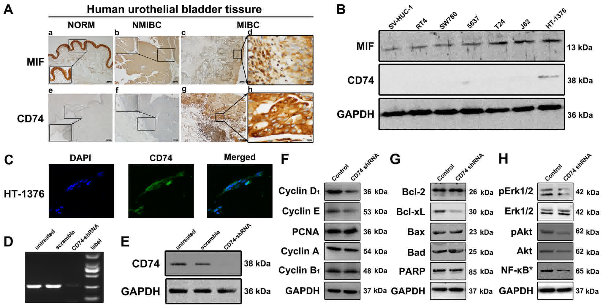 | Figure 1.Expression of MIF and CD74 in tissue
samples and cells, and protein expression levels in HT-1376 cells
following knockdown of CD74. (A) Little or no expression of
immunoreactive CD74 was identified in the urothelial layers of
normal bladder and NMIBC samples, but sections of MIBC samples
exhibited strong immunoreactive signals of CD74. In contrast, MIF
demonstrated strong immunoreactions in the normal and UCB samples.
(B) Western blotting assay results for MIF and CD74 in the cultured
urothelial SV-HUV-1, SW780, 5637, T24, J82 and HT-1376 cell lines.
HT-1376 was the only one identified to express CD74, but all cells
expressed MIF to a certain extent. (C) Immunofluorescence
microscopy (×100, magnification) indicated positive CD74 staining
in HT-1376 cells. (D) CD74 shRNA lentiviral particles abrogated the
RNA expression of CD74 in the HT-1376 cells compared with untreated
and scramble cells. (E) CD74 short hairpin RNA lentiviral particles
abrogated the protein expression of CD74 in the HT-1376 cells when
compared with the untreated and scramble cells. (F) Knockdown of
CD74 modulated the expression levels of not only Cyclin D1, Cyclin
E, but also intranuclear NF-κB p65, pAkt, pErk1/2. (G and H)
However, no significant changes in Bcl-2, Bad, cleaved PARP and
Erk1/2 were observed among all groups. *intranuclear NF-κB. NMIBC,
non-muscle-invasive bladder cancer; MIBC, muscle-invasive bladder
cancer; UCB, urothelial cell carcinoma of the bladder; MIF,
macrophage migration inhibitory factor; CD74, cluster of
differentiation; p65, transcription factor p65; Akt, RAC-alpha
serine/threonine protein kinase; p, phosphorylated; Erk1/2,
Extracellular regulated protein kinase 1/2; Bcl-2, B-cell lymphoma
2; Bcl-xL, Bcl-extra large; Bax, Bcl-2-associated X protein; PARP,
poly(adenosine 5′-diphosphate-ribose) polymerase. |
Expression levels of proteins
associated with proliferation, invasion and angiogenesis are
affected by knockdown of CD74 in HT-1376 cells
RT-PCR and Western blot analysis showed that CD74
shRNA lentiviral particles were reduced CD74 expression in HT-1376
cells (Fig. 1D and E). Western
blotting assays indicated that Cyclin D1, Cyclin E (Fig. 1F), Bcl-xL (Fig. 1G), pErk1/2, pAkt, Akt and intranuclear
NF-κB (Fig. 1H) levels in the
CD74-knockdown HT-1376 cells were downregulated compared with
untreated cells.
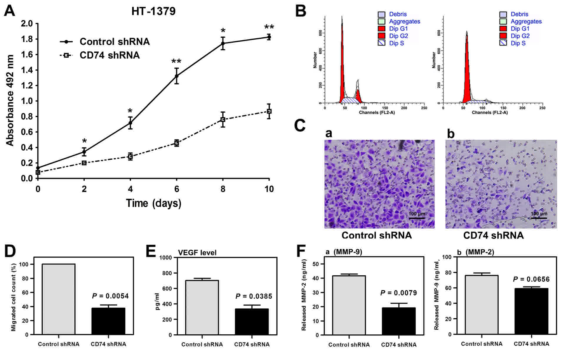
Knockdown of CD74 by CD74 shRNA
lentiviral particles suppresses the proliferative and invasive
abilities of HT-1376 cells
Knockdown of CD74 attenuated cell proliferation in
HT-1376 cells compared with the scramble group (Fig. 2A). The flow cytometry assay indicated
that knockdown of CD74 significantly increased the proportion of
G1-stage cells (P<0.001) and decreased the proportions of G2
stage (P<0.001) and S stage cells (P=0.0149), compared with
scramble shRNA cells (Fig. 2B). The
cell invasion assay showed that knockdown of CD74 significantly
attenuated the invasion ability of HT-1376 cells (Fig. 2C and D). An indicated that the
secretion of VEGF and MMP-9 was significantly reduced in
CD74-knockdown-HT-1376 cells compared with the shRNA control, while
not significantly reduced in MMP-2 (Fig.
2E and F).
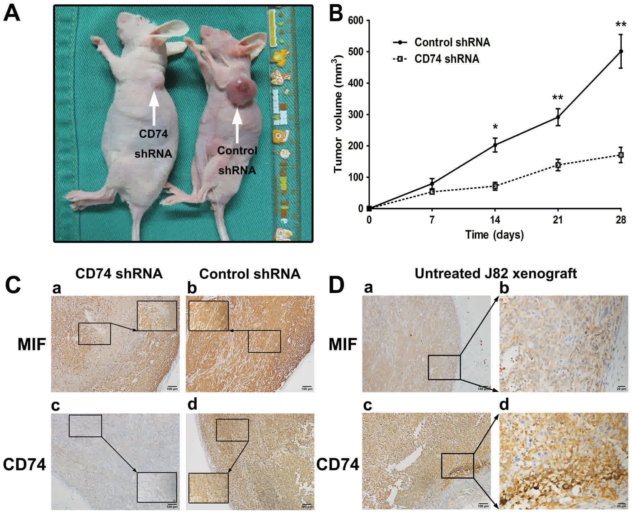
CD74 knockdown inhibits UCB growth and
MVD in xenograft nude mice
CD74 knockdown inhibited the tumorigenesis of
HT-1376 cells in vivo (Fig.
3A-C). The average weight of the tumors in the CD74 shRNA group
was 24.20±2.19 g (%Δ=6.92), and the average weight of the tumors in
the control shRNA group was 22.37±0.98 g (%Δ=−2.32). The MVD values
of 56.8±18.2 and 42.9±14.7 for scramble and CD74-knockdown groups,
respectively, were significantly different (P=0.0114). CD74 in the
wild-type J82 tumor was indicated using immunostaining (Fig. 3D).
Discussion
The present study investigated MIF and CD74
expression patterns in human normal and UCB samples and different
cell lines, and also explored the association between the clinical
characteristics and expression levels of MIF and CD74 in UCB cases,
and the roles of CD74 in urothelial cells. The results suggested
that MIF ubiquitously appeared in benign and malignant bladder
tissues, but CD74 was expressed primarily in malignant UCB samples
of MIBC, but also marginally in normal and NMIBC tissues, which
suggests that CD74 may be a unique and promising marker for
high-grade UCB or MIBC. In addition, knockdown of CD74 in the
HT-1376 cells attenuated the level of proliferation, invasion and
angiogenesis via downregulated Erk1/2 and PI3K/Akt pathways in the
in vitro study, which was concordant with previous studies
(15,16). The tumorigenesis assay indicated that
the knockdown of CD74 in the HT-1376 cells resulted in lower tumor
volumes, and notably induced the expression of CD74 in the
wild-type J82 tumors.
At first, CD74 was identified as a part of the major
histocompatibility complex II and a chaperone of MIF, which was
demonstrated to exert MIF functions within the immune system and
initiate inflammation (21–23). CD74 is an evolutionarily-conserved
type II protein expressed in the cellular membrane, and has various
roles in several key processes including antigen presentation,
B-cell differentiation, inflammatory signaling and carcinogenesis
signaling (14,15). Previous evidence has suggested that
unlike MIF, which was ubiquitous in human tissue, CD74 was
expressed in higher quantities in malignant tissues compared with
benign tissues, and is an essential part of the MIF pathway axis
(24–27).
In the present study, it was identified that the
knockdown of CD74 decreased the level of proliferation, invasion
(MMPs) and angiogenesis (VEGF and MVD) in UCB cells in the in
vitro and in vivo experiments, which may be the result
of the inhibition of Erk1/2 and PI3K/Akt pathways (28). MMPs are a family of proteolytic
enzymes involved in a number of phases of cancer progression,
including angiogenesis, invasiveness and metastasis, and may be
downregulated by ISO-1, a specific inhibitor of MIF (29,30). VEGF
and intranuclear NF-κB also appear to be involved in the MIF-CD74
pathway (31,32), which was also demonstrated in the
present study.
The characteristics of CD74 have been investigated
in a number of types of hematopoietic cancer for a long time
(13,33). The humanized mAb milatuzumab that
targets CD74 has been assessed in clinical trials (34), and additional studies indicated that
milatuzumab may conjugate with doxorubicin (Dox) and enhance the
cytotoxicity of Dox (34,35). Notably, Dox is an important agent in
UCB intravesical chemotherapy, suggesting that CD74-targeted
therapeutic treatment may be a better choice than DOX.
The present study has also demonstrated that UCB J82
cells exhibited no CD74 signals in vitro, but that they were
induced to express CD74 in BALB/c-nu mice in vivo. Due to
the number factors involved in the occurrence of tumor xenografts,
we hypothesized that the pattern of chemokine-receptors expressed
on cultured cells in vitro or individual cells in
vivo was determined by the cell's lineage, state of
differentiation, and micro-environmental factors including
chemokines concentration, the presence of inflammatory cytokines
and hypoxia (31). Additional studies
in this area are required.
In the present study, the expression and potential
roles of CD74 were analyzed via in vitro and in vivo
experiments. It was indicated that the expression of CD74 was
associated with MIBC/high grade of the UCB, while the knockdown of
CD74 attenuated the proliferation, invasion and angiogenesis of
HT-1376.
Acknowledgements
Not applicable.
Funding
This work was supported by the National Natural
Science Foundation of China (grant no. 81302231), the Beijing
Outstanding Talent Training (grant no. 2014000021469G0104), the
Beijing Municipal Administration of Hospitals' Youth Program (grant
no. QML20160303) and the Beijing Chao-Yang Hospital 1351 Talents
Project Funding (grant no. CYXX-2017-11).
Availability of data and materials
The datasets generated or analyzed during this study
are included in this published article.
Authors' contributions
WQ and NX were responsible for the data analysis. JG
and WW were major contributors in writing the manuscript and
statistical analysis. LS, HP, and YN were responsible for the
acquisition of data. MW and FY performed analysis and
interpretation of data. All authors read and approved the final
manuscript.
Ethics approval and consent to
participate
Ethical approval was granted by the Beijing
Chao-Yang Hospital Institutional Research Ethical Board. Informed
consent was obtained from all patients enrolled.
Consent for publication
Informed consent was obtained from all patients
enrolled.
Competing interests
The all authors declare that they have no competing
interests.
Glossary
Abbreviations
Abbreviations:
|
UCB
|
urothelial cell carcinoma of the
bladder
|
|
NMIBC
|
non-muscle-invasive bladder cancer
|
|
MIBC
|
muscle-invasive bladder cancer
|
|
MIF
|
macrophage migration inhibitory
factor
|
|
VEGF
|
vascular endothelial growth factor
|
|
MMP
|
matrix metalloproteinase
|
|
NF-κB
|
Nuclear Factor κB
|
|
PCNA
|
Proliferating Cell Nuclear Antigen
|
|
Erk1/2
|
Extracellular regulated protein kinase
1/2
|
|
TLR4
|
Toll-like receptor 4
|
|
Dox
|
doxorubicin
|
References
|
1
|
Burger M, Catto JW, Dalbagni G, Grossman
HB, Herr H, Karakiewicz P, Kassouf W, Kiemeney LA, La Vecchia C,
Shariat S and Lotan Y: Epidemiology and risk factors of urothelial
bladder cancer. Eur Urol. 63:234–241. 2013. View Article : Google Scholar : PubMed/NCBI
|
|
2
|
McConkey DJ, Lee S, Choi W, Tran M,
Majewski T, Lee S, Siefker-Radtke A, Dinney C and Czerniak B:
Molecular genetics of bladder cancer: Emerging mechanisms of tumor
initiation and progression. Urol Oncol. 28:429–440. 2010.
View Article : Google Scholar : PubMed/NCBI
|
|
3
|
Kaufman DS, Shipley WU and Feldman AS:
Bladder cancer. Lancet. 374:239–249. 2009. View Article : Google Scholar : PubMed/NCBI
|
|
4
|
Serrano C, Morales R, Suárez C, Núñez I,
Valverde C, Rodón J, Humbert J, Padrós O and Carles J: Emerging
therapies for urothelial cancer. Cancer Treat Rev. 38:311–317.
2012. View Article : Google Scholar : PubMed/NCBI
|
|
5
|
Meeks JJ, Bellmunt J, Bochner BH, Clarke
NW, Daneshmand S, Galsky MD, Hahn NM, Lerner SP, Mason M, Powles T,
et al: A systematic review of neoadjuvant and adjuvant chemotherapy
for muscle-invasive bladder cancer. Eur Urol. 62:523–533. 2012.
View Article : Google Scholar : PubMed/NCBI
|
|
6
|
Meyer-Siegler KL, Leifheit EC and Vera PL:
Inhibition of macrophage migration inhibitory factor decreases
proliferation and cytokine expression in bladder cancer cells. BMC
Cancer. 4:342004. View Article : Google Scholar : PubMed/NCBI
|
|
7
|
Taylor JA III, Kuchel GA, Hegde P,
Voznesensky OS, Claffey K, Tsimikas J, Leng L, Bucala R and Pilbeam
C: Null mutation for macrophage migration inhibitory factor (MIF)
is associated with less aggressive bladder cancer in mice. BMC
Cancer. 7:1352007. View Article : Google Scholar : PubMed/NCBI
|
|
8
|
Al-Abed Y and VanPatten S: MIF as a
disease target: ISO-1 as a proof-of-concept therapeutic. Future Med
Chem. 3:45–63. 2011. View Article : Google Scholar : PubMed/NCBI
|
|
9
|
Babu SN, Chetal G and Kumar S: Macrophage
migration inhibitory factor: A potential marker for cancer
diagnosis and therapy. Asian Pac J Cancer Prev. 13:1737–1744. 2012.
View Article : Google Scholar : PubMed/NCBI
|
|
10
|
Calandra T and Roger T: Macrophage
migration inhibitory factor: A regulator of innate immunity. Nat
Rev Immunol. 3:791–800. 2003. View
Article : Google Scholar : PubMed/NCBI
|
|
11
|
Larson DF and Horak K: Macrophage
migration inhibitory factor: Controller of systemic inflammation.
Crit Care. 10:1382006. View
Article : Google Scholar : PubMed/NCBI
|
|
12
|
Becker-Herman S, Arie G, Medvedovsky H,
Kerem A and Shachar I: CD74 is a member of the regulated
intramembrane proteolysis-processed protein family. Mol Biol Cell.
16:5061–5069. 2005. View Article : Google Scholar : PubMed/NCBI
|
|
13
|
Borghese F and Clanchy FI: CD74: An
emerging opportunity as a therapeutic target in cancer and
autoimmune disease. Expert Opin Ther Targets. 15:237–251. 2011.
View Article : Google Scholar : PubMed/NCBI
|
|
14
|
Lue H, Thiele M, Franz J, Dahl E,
Speckgens S, Leng L, Fingerle-Rowson G, Bucala R, Lüscher B and
Bernhagen J: Macrophage migration inhibitory factor (MIF) promotes
cell survival by activation of the Akt pathway and role for
CSN5/JAB1 in the control of autocrine MIF activity. Oncogene.
2635:5046–5059. 2007. View Article : Google Scholar
|
|
15
|
Starlets D, Gore Y, Binsky I, Haran M,
Harpaz N, Shvidel L, Becker-Herman S, Berrebi A and Shachar I:
Cell-surface CD74 initiates a signaling cascade leading to cell
proliferation and survival. Blood. 107:4807–4816. 2006. View Article : Google Scholar : PubMed/NCBI
|
|
16
|
Choi JW, Kim Y, Lee JH and Kim YS: CD74
expression is increased in high-grade, invasive urothelial
carcinoma of the bladder. Int J Urol. 20:251–255. 2013. View Article : Google Scholar : PubMed/NCBI
|
|
17
|
Edge S, Byrd D, Compton C, Fritz A, Greene
F and Trotti A: AJCC cancer staging manual. edition 7. New York,
NY: Springer; 2010
|
|
18
|
Bradford MM: A rapid and sensitive method
for the quantitation of microgram quantities of protein utilizing
the principle of protein-dye binding. Anal Biochem. 72:248–254.
1976. View Article : Google Scholar : PubMed/NCBI
|
|
19
|
Association for assessment and
accreditation of laboratory animal care international. [Internet].
2011. AAALAC International rules of accreditation. Section 2.d.
[Cited April 2012]. http://www.aaalac.org/accreditation/rules.cfm
|
|
20
|
Offersen BV, Pfeiffer P, Hamilton-Dutoit S
and Overgaard J: Patterns of angiogenesis in nonsmall-cell lung
carcinoma. Cancer. 91:1500–1509. 2001. View Article : Google Scholar : PubMed/NCBI
|
|
21
|
Cooke G, Armstrong ME and Donnelly SC:
Macrophage migration inhibitory factor (MIF), enzymatic activity
and the inflammatory response. Biofactors. 35:165–168. 2009.
View Article : Google Scholar : PubMed/NCBI
|
|
22
|
Conroy H, Mawhinney L and Donnelly SC:
Inflammation and cancer: Macrophage migration inhibitory factor
(MIF)-the potential missing link. QJM. 103:831–836. 2010.
View Article : Google Scholar : PubMed/NCBI
|
|
23
|
Bernhagen J, Krohn R, Lue H, Gregory JL,
Zernecke A, Koenen RR, Dewor M, Georgiev I, Schober A, Leng L, et
al: MIF is a noncognate ligand of CXC chemokine receptors in
inflammatory and atherogenic cell recruitment. Nat Med. 13:587–596.
2007. View
Article : Google Scholar : PubMed/NCBI
|
|
24
|
Cheng RJ, Deng WG, Niu CB, Li YY and Fu Y:
Expression of macrophage migration inhibitory factor and CD74 in
cervical squamous cell carcinoma. Int J Gynecol Cancer.
21:1004–1012. 2011. View Article : Google Scholar : PubMed/NCBI
|
|
25
|
Zheng YX, Yang M, Rong TT, Yuan XL, Ma YH,
Wang ZH, Shen LS and Cui L: CD74 and macrophage migration
inhibitory factor as therapeutic targets in gastric cancer. World J
Gastroenterol. 18:2253–2261. 2012. View Article : Google Scholar : PubMed/NCBI
|
|
26
|
Metodieva G, Nogueira-de-Souza NC,
Greenwood C, Al-Janabi K, Leng L, Bucala R and Metodiev MV:
CD74-dependent deregulation of the tumor suppressor scribble in
human epithelial and breast cancer cells. Neoplasia. 15:660–668.
2013. View Article : Google Scholar : PubMed/NCBI
|
|
27
|
Matsuura S, Shinmura K, Kamo T, Igarashi
H, Maruyama K, Tajima M, Ogawa H, Tanahashi M, Niwa H, Funai K, et
al: CD74-ROS1 fusion transcripts in resected non-small cell lung
carcinoma. Oncol Rep. 30:1675–1680. 2013. View Article : Google Scholar : PubMed/NCBI
|
|
28
|
Bifulco C, McDaniel K, Leng L and Bucala
R: Tumor growth-promoting properties of macrophage migration
inhibitory factor. Curr Pharm Des. 14:3790–3801. 2008. View Article : Google Scholar : PubMed/NCBI
|
|
29
|
Hadler-Olsen E, Winberg JO and
Uhlin-Hansen L: Matrix metalloproteinases in cancer: Their value as
diagnostic and prognostic markers and therapeutic targets. Tumour
Biol. 34:2041–2051. 2013. View Article : Google Scholar : PubMed/NCBI
|
|
30
|
Khoufache K, Bazin S, Girard K,
Guillemette J, Roy MC, Verreault JP, Al-Abed Y, Foster W and Akoum
A: Macrophage migration inhibitory factor antagonist blocks the
development of endometriosis in vivo. PLoS One. 7:e372642012.
View Article : Google Scholar : PubMed/NCBI
|
|
31
|
Giannice R, Erreni M, Allavena P,
Buscaglia M and Tozzi R: Chemokines mRNA expression in relation to
the Macrophage migration inhibitory factor (MIF) mRNA and vascular
endothelial growth factor (VEGF) mRNA expression in the
microenvironment of endometrial cancer tissue and normal
endometrium: A pilot study. Cytokine. 64:509–515. 2013. View Article : Google Scholar : PubMed/NCBI
|
|
32
|
Cao WG, Morin M, Sengers V, Metz C, Roger
T, Maheux R and Akoum A: Tumour necrosis factor-alpha up-regulates
macrophage migration inhibitory factor expression in endometrial
stromal cells via the nuclear transcription factor NF-kappaB. Hum
Reprod. 21:421–428. 2006. View Article : Google Scholar : PubMed/NCBI
|
|
33
|
Stein R, Mattes MJ, Cardillo TM, Hansen
HJ, Chang CH, Burton J, Govindan S and Goldenberg DM: CD74: A new
candidate target for the immunotherapy of B-cell neoplasms. Clin
Cancer Res. 13:S5556–S5563. 2007. View Article : Google Scholar
|
|
34
|
Berkova Z, Tao RH and Samaniego F:
Milatuzumab-a promising new immunotherapeutic agent. Expert Opin
Investig Drugs. 19:141–149. 2010. View Article : Google Scholar : PubMed/NCBI
|
|
35
|
Stein R, Smith MR, Chen S, Zalath M and
Goldenberg DM: Combining milatuzumab with bortezomib, doxorubicin,
or dexamethasone improves responses in multiple myeloma cell lines.
Clin Cancer Res. 15:2808–2817. 2009. View Article : Google Scholar : PubMed/NCBI
|