Introduction
Cervical lymph node metastasis of nasopharyngeal
carcinoma (NPC) has an incidence as high as 70% (1), and is a key factor that affects the
clinical staging of NPC, treatment plan and prognosis (2). The current existing international
standards for NPC staging originated from the 7th edition of the
Union for International Cancer Control/American Joint Committee on
Cancer (UICC/AJCC) staging system that is primarily based on
palpation (3,4). Palpation of lymph nodes usually depends
on the subjectivity of doctors, which may interfere with the
accuracy of staging and the development of individualized treatment
plans. In previous years, intensity modulated radiation therapy
(IMRT) has become an important therapy for NPC (5–12). As a
type of precision radiotherapy, IMRT requires precise anatomical
locations; however, the 7th edition of the UICC/AJCC staging system
has included body surface positions that are used for the
determination of cervical lymph node metastasis (13). For example, in this staging system,
supraclavicular fossa (SCF) is defined as a triangle area formed by
the upper edge of the sternal end of the clavicle, the upper edge
of the outer end of the clavicle and the neck-shoulder
intersection; however, this area may include the IV region and the
tail of V region in the international cervical lymph node partition
system (14). Therefore, SCF, a
position that does not have accurate positioning in cross sections
in imaging, cannot satisfy the requirements by IMRT. Thus, the 7th
edition of UICC/AJCC staging system has certain limitations
regarding the outline of NPC target areas, the definition of
treatment plans and the evaluation of prognosis (15).
Initially, the Radiation Therapy Oncology Group
(RTOG) divides cervical lymph nodes into retropharyngeal region, I
(Ia, Ib) region, II (IIa, IIb) region, III region, IV region, V
region and VI region (16).
Subsequently, retrostyloid space and supraclavicular region are
included in a modified version of RTOG division standards (17). In 2013, RTOG division standards
further defined IVb, Vc, VII, VIII, IX and X regions (18). The internationally recognized cervical
lymph node imaging division standards are widely used among the
radiation oncology research community. The present study used RTOG
division standards and magnetic resonance imaging (MRI) to
investigate cervical lymph node metastasis of NPC and to establish
a novel N staging standard for NPC based on IMRT via a prospective
multicenter clinical trial.
Materials and methods
Patients
Between January 2006 and December 2009, 492 patients
with NPC without distant metastasis from six hospitals (The First
Affiliated Hospital of Guangxi Medical University, Nanning; Liuzhou
Worker Hospital, Liuzhou; First People's Hospital of Yulin City,
Yulin; People's Hospital of Guangxi Zhuang Autonomous Region,
Nanning; Affiliated Hospital of Guilin Medical University, Guilin;
Wuzhou Red Cross Hospital, Wuzhou, Guangxi, China) were included in
the present study. Among them, 338 were male and 154 were female.
The median age of the group was 45 years old (range, 18–81 years
old). All patients received IMRT. Patients with a Karnofsky
performance status of ≥70, who met criteria for blood counts and
other tests (i.e., serum creatinine ≤1.6 mg/dl and serum bilirubin
≤1.5 mg/dl; white blood cell ≥3,600/mm3, platelet
≥100,000/mm3 and hemoglobin ≥12.0 g/dl for males, ≥11.0
g/dl for females) were eligible. Prior to treatment, all patients
received detailed physical examination, general situation
appraisal, blood routine examination, nasopharyngeal fiberscope
examination, chest X-ray or computed tomography (CT), abdominal
ultrasound and MRI of areas including the nasopharynx and neck.
Patients with N2-N3 stage received additional bone scanning. All
procedures were approved by the Ethics Committee of Guangxi Medical
University (Nanning, China). Written informed consent was obtained
from all patients or their families prior to enrolment in the
present study.
MRI
MR images were obtained using a 1.5-T MRI scanner
(GE Healthcare Life Sciences, Little Chalfont, UK). All patients
received routing and enhanced scanning. Scanning directions were
cross sectional, sagittal, coronal, T2-weighted (TR 3,000–4,000 ms,
TE 102–110 ms), T1-weighted (TR 2,200–2,400 ms, TE 77–109 ms, TI
750 ms) and enhanced T1-weighted scanning. Head quadrature coil was
adopted, with a thickness of 6 mm, interlayer space of 1 mm and
matrix of 256×192. Cross-sectional scanning ranged from suprasellar
cistern to the lower edge of the clavicle. The contrast medium was
gadopentetate dimeglumine-diethylenetriaminepentacetate, with a
dose of 15 ml.
Clinical staging
All MR images were independently reviewed using a
picture archiving and communication system by two physicians. The
stage of this research group was defined according to the 7th
edition of UICC/AJCC clinical staging standard, taking into account
patient symptoms and physical examination information. Lymph node
metastasis was diagnosed by MRI, but not by palpation, according to
the guidelines of RTOG 2013 edition (19,20).
Therapeutic method
A total of 492 patients with NPC received IMRT
during the whole process. Computed tomography contrast-enhanced
scanning was applied from the skull cap to 3 cm below the clavicle,
with a layer distance of 3 mm and layer thickness of 3 mm. Under
the guidance of Report 50 and Report 62 of International Commission
on Radiation Units and Measurements, gross tumor volume (GTV)
included primary tumor sites, and their invasion range (GTVnx),
retropharyngeal metastatic lymph nodes (GTVrpn) and cervical
metastatic lymph node (GTVnd) (21).
The clinical target volume (CTV) range may be adjusted according to
involvement degrees. For example, CTV1 should include GTVnx,
GTVrpn, the whole nasopharyngeal mucosa and submucosal 5 mm region;
CTV2 should include CTV1, as well as some of the following:
Posterior nasal cavity, pterygopalatine fossa, posterior maxillary
sinus, part of the posterior ethmoid sinus, lateral pharyngeal
space, skull base, part of cervical vertebra and slope. Planning
target volume (PTV) included position errors and organ movements
during treatments, which are usually externally expanded for 3–5 mm
based on GTVs and CTVs. The prescription doses were as follows:
PGTVnx and PTVrpn (68–74 Gy), PTVnd (66–70 Gy), PTV1 (60–66 Gy) and
PTV2 (50–56 Gy; 5 fractions/week for a total of 30–33 fractions).
The setting of restricted dosages for critical organs followed
international consensus (21,22).
All stages were defined according to the 7th edition
of the UICC/AJCC staging standards. Of the 477 patients with Stage
II–IVB disease, 93.70% patients (461/492) received chemotherapy,
including 51.0% (235/461) with concurrent chemotherapy, 37.09%
(171/461) with induction + concurrent chemotherapy, 7.59% (35/461)
with concurrent + adjuvant chemotherapy, 4.12% (19/461) with
induction + concurrent + adjuvant chemotherapy, and 0.22% (1/461)
with induction chemotherapy. The chemotherapy drugs were primarily
platinum-based. All centers used identical chemotherapy
protocols.
Follow-ups
Regular follow-ups commenced from 3 months after the
patients ended their treatment. The follow-up period was defined as
the period starting from the commencement date of treatment to the
last date of regular follow-up or to the time of mortality of
patients. By December 31st 2014, the last date of regular
follow-up, the median follow-up period was 64.1 months (6–92
months). A percentage of 96.3% of patients had complete follow-up
data of 5 years. The main analysis factors included overall
survival (OS), disease-free survival (DFS), relapse-free survival
(RFS) and distant metastasis-free survival (DMFS).
Statistical analysis
All results were analyzed using SPSS v19.0
statistical software (IBM Corp., Armonk, NY, USA). The Kaplan-Meier
method was used to calculate various survival rates. The log-rank
test was used to examine the significance of differences in
survival rate. Analyses of prognosis were performed using
univariate analysis or multivariate analysis. P<0.05 was
considered to indicate a statistically significant difference. Data
are presented as the mean ± standard error of the mean.
Results
Rate of lymph node metastasis of NPC
is high, with retropharyngeal and II regional lymph nodes being the
most likely to have metastasis
To determine the distribution of metastatic lymph
nodes, the locations of the metastasis were recorded for all 492
patients with NPC. Among the 492 patients, 428 (87%) had cervical
lymph node metastasis, including 82 (19.2%) cases with left
cervical metastasis only, 77 (18.0%) cases with right cervical
metastasis only and 269 (62.9%) cases with cervical metastasis on
both sides. The number of patients with retropharyngeal lymph node
metastasis was 339 (79.2%), including 128 cases of bilateral
metastasis (38.1%) and 210 cases of unilateral metastasis (61.9%).
The number of patients with II regional lymph node metastasis was
351 (82.0%), including 189 cases of bilateral metastasis (44.2%;
Table I). Among the 428 patients with
lymph node metastasis, only 4 patients without II regional
metastasis had III regional metastasis (0.9%), including 1 case
that demonstrated IV, Va and Vb regional metastasis. Among patients
without II and III regional metastasis, no IV regional metastasis
was observed. These results suggested that the rate of lymph node
metastasis of NPC was high, with retropharyngeal and II regional
lymph nodes being the most likely to have metastasis.
 | Table I.Percentage and distribution of the
metastatic lymph nodes in 428 patients with NPC. |
Table I.
Percentage and distribution of the
metastatic lymph nodes in 428 patients with NPC.
|
| No. of patients
(%) |
|---|
| Level | Left | Right | Bilateral | Total |
|---|
| Ia | 0 (0.0) | 0 (0.0) | 0 (0.0) | 0 (0.0) |
| Ib | 3 (0.7) | 6 (1.4) | 1 (0.2) | 8 (1.9) |
| II | 254 (59.3) | 286 (66.8) | 189 (44.2) | 351 (82.0) |
| III | 104 (24.3) | 99 (23.1) | 35 (8.2) | 168 (47.4) |
| IVa | 21 (4.9) | 15 (3.5) | 4 (0.9) | 32 (7.5) |
| IVb | 1 (0.2) | 0 (0.0) | 0 (0.0) | 1 (0.2) |
| Va | 10 (2.3) | 8 (1.9) | 1 (0.2) | 17 (4.0) |
| Vb | 4 (0.9) | 5 (1.2) | 0 (0.0) | 9 (2.1) |
| Vc | 1 (0.2) | 1 (0.2) | 0 (0.0) | 2 (0.5) |
| VI | 0 (0.0) | 0 (0.0) | 0 (0.0) | 0 (0.0) |
| VIIa | 239 (55.8) | 228 (53.3) | 129 (38.1) | 339 (79.2) |
| VIIb | 0 (0.0) | 0 (0.0) | 0 (0.0) | 0 (0.0) |
| VIII | 0 (0.0) | 0 (0.0) | 0 (0.0) | 0 (0.0) |
| IX | 0 (0.0) | 0 (0.0) | 0 (0.0) | 0 (0.0) |
| Xa | 0 (0.0) | 0 (0.0) | 0 (0.0) | 0 (0.0) |
| Xb | 0 (0.0) | 0 (0.0) | 0 (0.0) | 0 (0.0) |
Retropharyngeal lymph nodes, cervical
lymph node level and cervical lymph node laterality are associated
with the prognosis of patients
To investigate the association between lymph node
characteristics and prognosis, univariate analysis and multivariate
analysis were performed. According to the 7th edition of the
UICC/AJCC staging system, patients at I, II, III, IVa, and IVb
stages accounted for 3.0% (15/492), 14.4% (71/492), 35.8%
(176/492), 38.0% (187/492) and 8.7% (43/492), respectively. In
addition, patients at T1, T2, T3 and T4 stages accounted for 6.7%
(33/492), 18.5% (91/492), 33.7% (166/492) and 41.1% (202/492),
respectively. Furthermore, patients at N0, N1, N2, N3a and N3b
stages accounted for 13.0% (64/492), 32.3% (159/492), 45.9%
(226/492), 2.2% (11/492) and 6.5% (32/492), respectively (data not
shown). The OS rate after 5 years was 80.5%, the DFS rate was
78.6%, the RFS rate was 94.1% and the DMFS rate was 84.3%.
Univariate analysis of 428 patients with cervical lymph node
metastasis demonstrated that lymph nodal level, sizes [measured as
the maximum diameter (Dmax)] had a statistically
significant effect on OS, DFS and DMFS, cervical lymph node
laterality and retropharyngeal region had a statistically
significant effect on OS, DFS, RFS and DMFS, and extracapsular
spread had a statistically significant effect on OS, whereas
liquefaction necrosis had no significant effect (Table II). In addition, distant metastasis
survival and overall survival in Ib, retropharyngeal (VIIa), III
and Va regions were not significantly different from II region,
whereas those in IVa, Vb and lower regions were significantly
different from the II region. Therefore, lymph node levels were
divided into two groups: Level 1 (L1), retropharyngeal (VIIa), Ib,
II, III and Va regions; and level 2 (L2), IVa, IVb, Vb and Vc
regions (Table III). Multivariate
analysis of all 492 patients revealed that Dmax of
cervical lymph nodes, extracapsular spread and liquefaction
necrosis did not have independent prognostic significance. Of note,
retropharyngeal lymph nodes, cervical lymph node level and cervical
lymph node laterality were independent prognostic factors (Table IV). These results indicated that
retropharyngeal lymph nodes, cervical lymph node level and cervical
lymph node laterality are associated with the prognosis of
patients.
| Table II.Univariate analysis of the
association between various cervical lymph node variables and
patient prognosis. |
Table II.
Univariate analysis of the
association between various cervical lymph node variables and
patient prognosis.
| Variables | OS | χ2 | P-value | DFS | χ2 | P-value | RFS | χ2 | P-value | DMFS | χ2 | P-value |
|---|
| Level |
| 30.003 | <0.001 |
| 22.774 | 0.001 |
| 1.170 | 0.978 |
| 31.184 | <0.001 |
| Level
IB | – |
|
| – |
|
| – |
|
| – |
|
|
| Level
II | 82.7 |
|
| 81.9 |
|
| 94.4 |
|
| 87.7 |
|
|
| Level
III | 75.2 |
|
| 73.6 |
|
| 93.3 |
|
| 79.9 |
|
|
| Level
IVa | 67.0 |
|
| 65.6 |
|
| 93.3 |
|
| 71.9 |
|
|
| Level
Va | 92.3 |
|
| 65.9 |
|
| 91.7 |
|
| 74.0 |
|
|
| Level
Vb | 37.0 |
|
| 44.4 |
|
| 100 |
|
| 44.4 |
|
|
| Level
(IVb+Vc) | 33.3 |
|
| 33.3 |
|
| 100 |
|
| 33.3 |
|
|
| Level
VIIa | 78.9 |
|
| 86.5 |
|
| 91.7 |
|
| 85.9 |
|
|
| Group |
| 12.109 | 0.001 |
| 10.682 | 0.001 |
| 0.062 | 0.803 |
| 13.792 | <0.001 |
| Level
1 | 79.8 |
|
| 78.0 |
|
| 93.6 |
|
| 83.9 |
|
|
| Level
2 | 39.2 |
|
| 59.1 |
|
| 95.0 |
|
| 63.6 |
|
|
| Dmax,
cm |
| 14.566 | 0.001 |
| 15.728 | <0.001 |
| 0.575 | 0.750 |
| 14.068 | 0.001 |
| ≤3 | 81.0 |
|
| 77.3 |
|
| 93.6 |
|
| 83.3 |
|
|
| >3
and ≥6 | 78.1 |
|
| 76.4 |
|
| 94.8 |
|
| 81.3 |
|
|
|
>6 | 45.5 |
|
| 36.4 |
|
| 90.9 |
|
| 45.5 |
|
|
| Laterality |
| 12.640 | <0.001 |
| 11.866 | 0.001 |
| 4.645 | 0.031 |
| 6.682 | 0.010 |
|
Unilateral | 89.2 |
|
| 86.3 |
|
| 97.6 |
|
| 88.5 |
|
|
|
Bilateral | 71.2 |
|
| 70.3 |
|
| 92.0 |
|
| 77.7 |
|
|
| RLN |
| 17.872 | 0.046 |
| 14.199 | <0.001 |
| 4.710 | 0.030 |
| 8.970 | 0.003 |
| No | 96.2 |
|
| 91.9 |
|
| 98.9 |
|
| 93.0 |
|
|
|
Yes | 73.3 |
|
| 71.9 |
|
| 92.3 |
|
| 78.9 |
|
|
| Extracapsular
spread |
| 6.071 | 0.014 |
| 1.700 | 0.192 |
| 0.001 | 0.976 |
| 1.921 | 0.166 |
| No | 86.0 |
|
| 79.4 |
|
| 93.7 |
|
| 85.4 |
|
|
|
Yes | 73.8 |
|
| 74.5 |
|
| 93.9 |
|
| 80.2 |
|
|
| Necrosis |
| 0.426 | 0.514 |
| 1.754 | 0.185 |
| 2.636 | 0.104 |
| 0.315 | 0.574 |
| No | 78.2 |
|
| 77.2 |
|
| 94.7 |
|
| 82.2 |
|
|
|
Yes | 72.7 |
|
| 70.2 |
|
| 89.0 |
|
| 80.2 |
|
|
| Table III.Comparison of distant metastasis
survival and OS of various lymph node levels. |
Table III.
Comparison of distant metastasis
survival and OS of various lymph node levels.
|
|
| Risk ratio (95%
CI) |
|---|
|
|
|
|
|---|
| Nodal
variables | No. | Distant metastasis
survival | OS |
|---|
| Level II | 196 | 1 | 1 |
| Level VIIa | 37 | 0.998
(0.382–2.607) | 1.080
(0.475–2.452) |
| Level III | 138 | 1.565
(0.909–2.697) | 1.520
(0.934–2.474) |
| Level IVa | 32 | 2.357
(1.100–5.050)a | 2.124
(1.041–4.335)a |
| Level Va | 13 | 1.779
(0.537–5.893) | 0.462
(0.063–3.381) |
| Level Vb | 9 | 6.318
(2.415–16.532)a | 5.302
(2.057–13.667)a |
| Level (IVb+Vc) | 3 | 10.559
(2.492–44.750)a | 10.491
(2.483–44.318)a |
| Level Ib | 0 | – | – |
| Table IV.Multivariate analysis of cervical
lymph node variables associated with prognosis. |
Table IV.
Multivariate analysis of cervical
lymph node variables associated with prognosis.
| Variables | OS | DFS | RFS | DMFS |
|---|
| Levels 1 and 2 | 0.007 | 0.003 | 0.006 | 0.003 |
|
Dmax | 0.918 | 0.764 | 0.793 | 0.945 |
| Laterality | 0.006 | 0.006 | 0.004 | 0.007 |
| RLN | <0.001 | <0.001 | <0.001 | 0.001 |
| Extracapsular
spread | 0.909 | 0.999 | 0.962 | 0.925 |
| Necrosis | 0.242 | 0.424 | 0.232 | 0.193 |
Novel N staging system proposed in the
present study is more suitable for IMRT compared with the 7th
edition of the UICC/AJCC staging system
According to univariate and multivariate analysis
results, the 492 patients included in the present study were
classified into six subgroups, including the N0 group (no lymph
node metastasis), retropharyngeal lymph nodes (Nrp) group, L1Lu
group (unilateral upper cervical lymph node metastasis), L1Lb group
(bilateral upper cervical lymph node metastasis), L2Lu group
(unilateral lower cervical lymph node metastasis) and L2Lb group
(bilateral lower cervical lymph node metastasis). Compared with the
L1Lu group (hazard ratio=1), the hazard ratio for the risk of
distant metastasis survival and overall survival in the N0 group
was 0. Thus, N0 group was classified as N0 stage. Compared with the
L1Lu group (hazard ratio=1), the hazard ratio for the risk of
distant metastasis survival in the Nrp group was 1.367 and the risk
of overall survival was 1.857. Statistically, the L1Lu and Nrp
groups had no significant difference. Thus, the L1Lu and Nrp group
were classified as N1 stage. However, the hazard ratio for the risk
of distant metastasis survival and overall survival in the L1Lb
group was 2.142 and 2.755, respectively, significantly compared
with that in the L1Lu group (P<0.05). Thus, the L1Lb group was
classified as N2 stage. Compared with the L1Lu group, the hazard
ratio for the risk of distant metastasis survival and overall
survival in the L2Lu group (3.825 and 3.835, respectively), and in
the L2Lb group (4.785 and 5.415, respectively) was also
significantly higher (P<0.05). Thus, the L2Lu and L2Lb groups
were classified as N3 stage (Table
V). According to the effects of different factors on prognosis,
the present study proposed novel N staging standards: N0 (no lymph
node metastasis), N1 [retropharyngeal or/and unilateral upper
cervical (I, II, III, Va, VIIb, VIII, IX and X regions) lymph node
metastasis], N2 [bilateral upper cervical (I, II, III, Va, VIIb,
VIII, IX and X regions) lymph node metastasis] and N3 (lymph node
metastasis in IVa and Vb regions and their lower regions). To
evaluate the novel N staging, differences in the survival
prediction value, the distribution balance and the risk ratio were
compared between the novel N staging and the 7th edition of the
UICC/AJCC staging system. In the proposed novel N staging system,
the OS curves were significantly different among all stages: N0:N1
(χ2=5.198, P<0.05), N0:N2 (χ2=14.663,
P<0.01), N0:N3 (χ2=29.990, P<0.01), N1:N2
(χ2=9.215, P<0.01), N1:N3 (χ2=22.592,
P<0.01) and N2:N3 (χ2=9.305, P<0.01). In addition,
the DMFS curves were significantly different among all stages:
N0:N1 (χ2=5.528, P<0.05), N0:N2
(χ2=11.748, P<0.01), N0:N3 (χ2=25.172,
P<0.01), N1:N2 (χ2=8.525, P<0.01), N1:N3
(χ2=18.934, P<0.01) and N2:N3 (χ2=7.315,
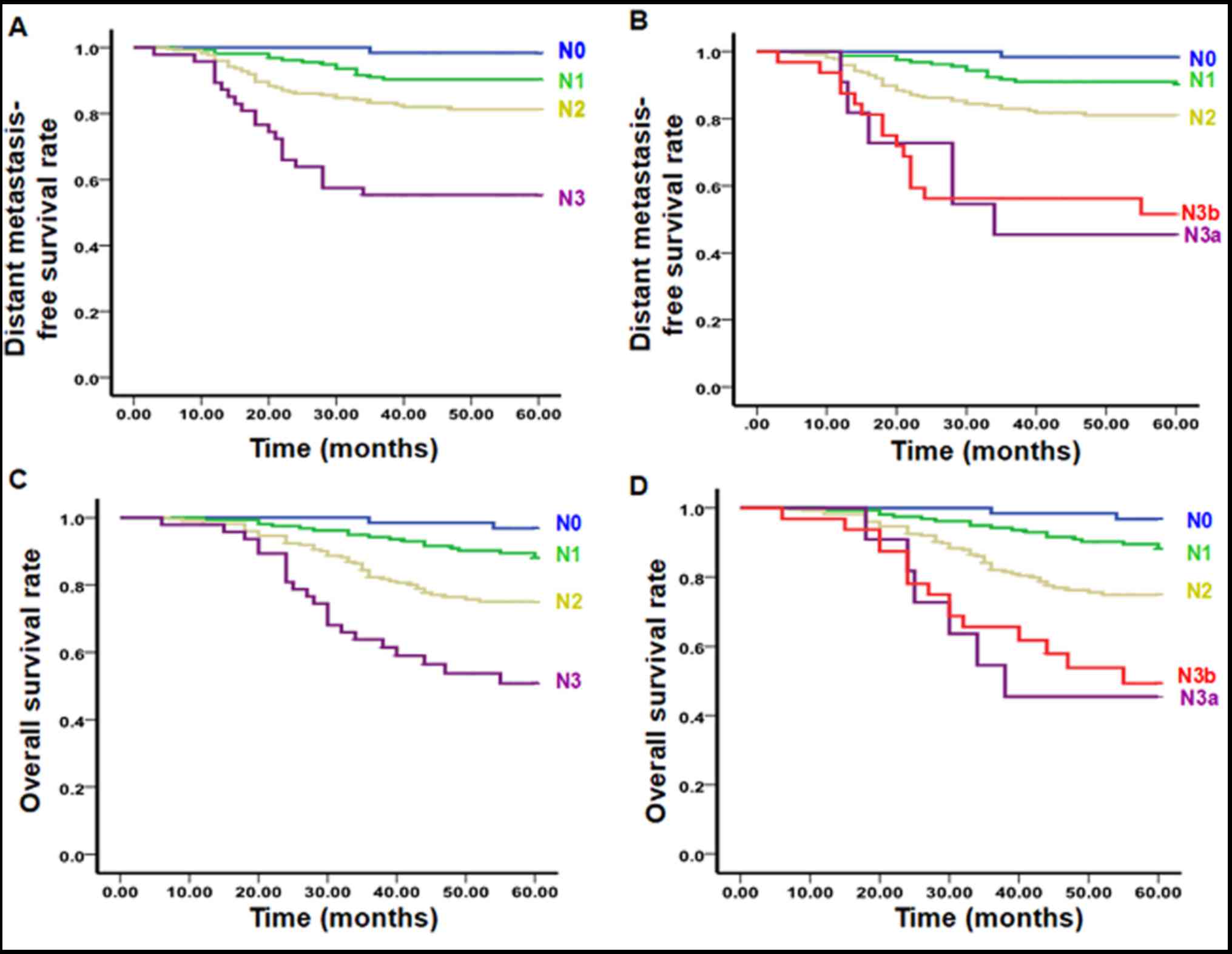
P<0.01; Fig. 1, Table VI). Conversely, OS and DMFS curves
were not significantly different between N3a and N3b stages
according to the 7th edition of the UICC/AJCC staging system
(Fig. 1, Table VI). Furthermore, the ratio of
distribution in N3a and N3b stages in the 7th edition of the
UICC/AJCC staging system accounted for 2.2 and 6.5%, respectively,
whereas the N3 stage in the proposed novel N staging system
accounted for 8.9%, being more balanced compared with the 7th
edition of the UICC/AJCC staging system (Table VII). In addition, the risk ratio for
each stage in the proposed novel N staging system was significantly
different compared with the N0 stage (P<0.05), whereas the risk
ratio for the N1 stage in the 7th edition of the UICC/AJCC staging
system was not significantly different compared with the N0 stage
(P>0.05; Table VII). These
results suggested that the novel N staging system proposed in the
present study was more suitable for IMRT compared with the 7th
edition of the UICC/AJCC staging system.
| Table V.Distribution patterns and failure
hazards of different N subsets. |
Table V.
Distribution patterns and failure
hazards of different N subsets.
|
| Hazard ratio (95%
CI) |
|---|
|
|
|
|---|
| Group | Distant metastasis
survival | OS |
|---|
| L1Lu | 1 | 1 |
| N0 | 0.152
(0.020–1.166) | 0.271
(0.061–1.201) |
| Nrp | 1.367
(0.481–3.879) | 1.857
(0.071–4.657) |
| L1Lb | 2.142
(1.129–4.064)b | 2.755
(1.499–5.065)a |
| L2Lu | 3.825
(1.079–13.556)b | 3.835
(1.092–13.471)a |
| L2Lb | 4.785
(2.181–10.497)b | 5.415
(2.537–11.562)a |
| Table VI.OS rate and distant failure-free
rates of various N stages between the proposed system and the 7th
edition of the UICC/AJCC system. |
Table VI.
OS rate and distant failure-free
rates of various N stages between the proposed system and the 7th
edition of the UICC/AJCC system.
|
| OS | DMFS |
|---|
|
|
|
|
|---|
|
| X2 | P | X2 | P |
|---|
| Proposed
system |
|
|
|
|
| N0:N1 | 5.198 | 0.039 | 5.528 | 0.042 |
| N0:N2 | 14.663 | <0.001 | 11.748 | <0.001 |
| N0:N3 | 29.990 | <0.001 | 25.172 | <0.001 |
| N1:N2 | 9.215 | 0.001 | 8.525 | 0.002 |
| N1:N3 | 22.592 | <0.001 | 18.934 | <0.001 |
| N2:N3 | 9.305 | <0.001 | 7.315 | <0.001 |
| UICC/AJCC
system |
|
|
|
|
| N0:N1 | 4.203 | 0.040 | 4.325 | 0.038 |
| N0:N2 | 14.104 | <0.001 | 11.197 | 0.001 |
| N0:N3a | 39.270 | <0.001 | 39.328 | <0.001 |
| N0:N3b | 34.506 | <0.001 | 31.973 | <0.001 |
| N1:N2 | 11.438 | 0.001 | 6.491 | 0.011 |
| N1:N3a | 35.277 | <0.001 | 28.276 | <0.001 |
| N1:N3b | 34.218 | <0.001 | 31.427 | <0.001 |
| N2:N3a | 6.544 | 0.011 | 9.797 | 0.002 |
| N2:N3b | 9.510 | 0.002 | 12.721 | <0.001 |
| N3a:N3b | 0.121 | 0.728 | 0.070 | 0.792 |
| Table VII.Distribution and differences in risk
ratios between the 7th edition of the UICC/AJCC staging system and
the proposed novel N staging system. |
Table VII.
Distribution and differences in risk
ratios between the 7th edition of the UICC/AJCC staging system and
the proposed novel N staging system.
|
|
| Risk ratio (95%
CI) |
|---|
|
|
|
|
|---|
| N stage | No. | Distant metastasis
survival | OS |
|---|
| Proposed
system |
|
|
|
| N0 | 64 (13.0) | 1 | 1 |
| N1 | 161 (32.7) | 7.557
(1.009–56.613)a | 4.596
(1.077–19.609)a |
| N2 | 223 (45.3) | 13.827
(1.903–100.486)a | 10.000
(2.431–41.132)a |
| N3 | 44 (8.9) | 30.123
(3.993–227.251)a | 18.584
(4.282–80.651)a |
| UICC/AJCC
system |
|
|
|
| N0 | 64 (13.0) | 1 | 1 |
| N1 | 159 (32.3) | 6.316
(0.834–47.812) | 3.955
(0.917–17.050) |
| N2 | 226 (45.9) | 13.288
(1.827–96.612)a | 9.719
(2.361–40.006)a |
| N3a | 11 (2.2) | 51.936
(6.243–432.084)a | 36.384
(7.268–182.153)a |
| N3b | 32 (6.5) | 38.744
(5.091–294.830)a | 23.544
(5.369–103.245)a |
Discussion
NPC has a high rate of cervical lymph node
metastasis and ~70% of patients were diagnosed with cervical lymph
node metastasis at their preliminary diagnosis (1,23). The MRI
data in the present study demonstrated that 87.0% of 492 patients
had lymph node metastasis, which was consistent with the results
reported by Wang et al (24)
and Ho et al (25). The
metastatic rates for retropharyngeal, II, III, IV, Va, Vb and Vc
regions were 79.2, 82.0, 47.4, 7.7, 4.0, 2.1 and 0.5%,
respectively. Consistent with a previous study, the metastatic
rates of each region decreased from the upper region to the lower
region, and the skipping metastatic rate was 0.9% (26). However, it has not been confirmed
whether the retropharyngeal or II region lymph node is the sentinel
lymph node (1,27–30). Lv
et al (31) revealed that the
metastatic rates of retropharyngeal and II region lymph nodes were
74.5 and 75.3%, respectively, suggesting that both are sentinel
lymph nodes. Consistent with this observation, the present study
demonstrated that the metastatic rates of retropharyngeal and II
region lymph nodes in 428 patients were 351 cases (82.0%) and 339
cases (79.2%), respectively. This observation may be associated
with lymph node drainage via retropharyngeal and II regions. In the
7th edition of the UICC/AJCC staging system, retropharyngeal lymph
node metastasis was classified as N1 (32). Tang et al (33) recommended classifying retropharyngeal
lymph node metastasis as N1. The results of the present study
revealed that retropharyngeal lymph node metastasis (regardless of
sides and sizes) is an independent prognostic factor that affects
OS, DFS, RFS and DMFS of patients with NPC. Due to various N
staging standards of NPC that have distinct lymph node parameters
(15,20,29,34,35),
the present study suggested setting a unified standard based on
objective MRI data and international lymph node imaging division
method.
Supraclavicular fossa defined in the 7th edition of
the UICC/AJCC staging system has been demonstrated to have high
risk of distant metastasis. According to the RTOG for Lymph Node
criteria, supraclavicular fossa is located at IV region, the lower
part of V region and the whole supraclavicular region (13). Cervical lymph nodes are an ordered
defense system. Once supraclavicular lymph nodes are affected,
tumor cells may further invade thoracic ducts and possibly the
whole body. Mao et al (34)
suggested that lymph node metastasis regions may be categorized
into retropharyngeal, Ib, II, III, V and IV regions, as well as the
supraclavicular region, when using the N staging based on MRI and
RTOG to evaluate distant metastasis risks. Ng et al
(15) and Yue et al (36) suggested replacing supraclavicular
fossae in UICC/AJCC standards with IV and Vb regions. Li et
al (37) revealed that lower
cervical lymph node metastasis (IV, Vb and supraclavicular regions)
is an independent prognostic factor that affects survival (37). Consistent with these reports, the
present study divided the lymph node level into Level 1
(retropharyngeal, Ib, II, III and Va regions) and Level 2 (IVa,
IVb, Vb and Vc regions), and univariate and multivariate analyses
confirmed the significant differences in prognosis between the
two.
NPC usually metastasizes according to the direction
of lymphatic drainage (38). As it is
different from other malignant tumors in the head and neck, NPC
usually has lymph node metastasis (bilateral or unilateral) during
its early stage, with a 40% rate of bilateral lymph node metastasis
(39). The primary difference between
N1 and N2 stages in the 7th edition of the UICC/AJCC staging system
is unilateral or bilateral lymph node metastasis. The present study
demonstrated that OS, DFS, RFS and DMFS rates were significantly
different between unilateral and bilateral metastases, suggesting
that cervical lymph node laterality is an independent prognostic
factor for NPC.
A previous literature review investigating the data
for the N-staging system for NPC revealed that the prognostic
significance attributed to size was controversial (40–44). Lee
et al (4) demonstrated that
the largest lymph node size was independently significant in
predicting survival. However, certain reports revealed that lymph
node size was not an independent prognostic factor (40–43). The
multivariate analysis of present study indicated that lymph node
size was not an independent prognostic factor. According to the 7th
edition of the UICC/AJCC staging system, the diagnosis of lymph
nodes >6 cm was primarily based on palpation, which is
subjective. Few lymph nodes >6 cm may be diagnosed by MRI or CT.
Therefore, it is still controversial whether lymph node size should
be included in the N staging standards.
The prognostic significance of extracapsular spread
in the treatment of NPC remains unclear. Mao et al (34) suggested that lymph node extracapsular
spread should be classified into N2 stage as a staging factor. The
results of the univariate analysis of the present study
demonstrated that extracapsular spread does not significantly
affect RFS, DMFS and DFS rates. In addition, multivariate analysis
indicated that cervical lymph node extracapsular spread was not an
independent prognostic factor. This may be due to the lack of
pathological evidence and diagnostic standard. Therefore,
extracapsular spread is not included in the proposed novel N
staging system.
The tumor-node-metastasis staging system is the
comprehensive manifestation of all types of prognosis factors
revealed by the investigation of clinical epidemiology, and the
identification of novel prognostic factors depends on the
improvement of diagnosis and therapy. Due to the continuous
improvement of diagnosis and therapy, prognostic factors are also
changing, and staging system should also be continuously improved.
The staging standard of the 7th edition of the UICC/AJCC published
in 2009 is primarily based on the data of regular two-dimensional
radiotherapy. As the progress of accurate radiotherapy, IMRT has
been more frequently applied in the treatment of NPC than regular
two-dimensional radiotherapy (44–46). In
addition, the 7th edition of UICC/AJCC is primarily based on
palpation (3,4). Palpation of lymph nodes usually depends
on the subjectivity of doctors, which may interfere with the
accuracy of staging and the development of individualized treatment
plans (34). Previous studies have
demonstrated that there were no significant differences in DMFS
between N3a and N3b, and suggested that N3a and N3b may be combined
as N3 in the novel N stage (47,48). The
present study used RTOG division standards and MRI to investigate
cervical lymph node metastasis of NPC, and to establish a novel N
staging standard for NPC based on IMRT in a prospective multicenter
clinical trial. It was proposed that the novel N staging system
include: N0 (no lymph node metastasis), N1 [retropharyngeal or/and
unilateral upper cervical (I, II, III, Va, VIIb, VIII, IX and X
regions) lymph node metastasis], N2 [bilateral upper cervical (I,
II, III, Va, VIIb, VIII, IX and X regions) lymph node metastasis]
and N3 (lymph node metastasis in IVa and Vb regions and their lower
regions). Compared with the 7th edition of the UICC/AJCC staging
system, the novel N staging system has improved risk difference and
distribution balance, as well as distinct DMFS rate and OS rate
between stages. In conclusion, the novel N staging system is more
suitable for IMRT and more accurately predicts the prognosis of
patients with NPC.
Acknowledgements
Not applicable.
Funding
The present study was supported by grants from the
National Natural Science Foundation of China (grant nos. 81460460,
81360405 and 81760542), The Research Foundation of the Science and
Technology Department of Guangxi Province, China (grant nos.
2016GXNSFAA380252 and 2014GXNSFBA118114), Guangxi Medical
University Training Program for Distinguished Young Scholars
(2017), The central government guide local science and technology
development projects (ZY18057006). Medical Excellence Award Funded
by the Creative Research Development Grant from the First
Affiliated Hospital of Guangxi Medical University.
Availability of data and materials
All data generated or analyzed during this study are
included in this published article.
Authors' contributions
MK and RW designed the research and assigned the
tasks to teams. PZ, TW, TZ and JL collected and analyzed data. MK
accessed the relevant information. GL and HY helped with MRI
examination. ML and GF participated in image analysis. JZ was
involved in statistical analysis. RW, GL, HY, GF, ML and JZ
critically revised the manuscript for important intellectual
content. RW approved the final version of the manuscript to be
submitted.
Ethics approval and consent to
participate
All procedures were approved by the Ethics Committee
of Guangxi Medical University (Nanning, China). Written informed
consent was obtained from all patients or their families prior to
enrolment in the present study.
Consent for publication
Not applicable.
Competing interests
The authors declared that they have no competing
interests.
References
|
1
|
Tang L, Mao Y, Liu L, Liang S, Chen Y, Sun
Y, Liao X, Lin A, Liu M, Li L and Ma J: The volume to be irradiated
during selective neck irradiation in nasopharyngeal carcinoma:
Analysis of the spread patterns in lymph nodes by magnetic
resonance imaging. Cancer. 115:680–688. 2009. View Article : Google Scholar : PubMed/NCBI
|
|
2
|
Yi J, Gao L, Huang X, Luo J, Xiao J, Li S,
Wang K, Zhang S, Qu Y and Xu G: Nasopharyngeal carcinoma treated by
intensity-modulated radiotherapy: Long-term results of 416
patients. Chin J Radiat Oncol. 21:196–200. 2012.
|
|
3
|
Edge SB and Compton CC: The American Joint
Committee on Cancer: The 7th edition of the AJCC cancer staging
manual and the future of TNM. Ann Surg Oncol. 17:1471–1474. 2010.
View Article : Google Scholar : PubMed/NCBI
|
|
4
|
Lee AW, Foo W, Poon YF, Law CK, Chan
DKOSK, Tung SY and Ho JH: Staging of nasopharyngeal carcinoma:
Evaluation of N-staging by Ho and UICC/AJCC systems. Union
Internationale Contre le Cancer. American Joint Committee for
Cancer. Clin Oncol (R Coll Radiol). 8:146–154. 1996. View Article : Google Scholar : PubMed/NCBI
|
|
5
|
Lee AW, Sze WM, Au JS, Leung SF, Leung TW,
Chua DT, Zee BC, Law SC, Teo PM, Tung SY, et al: Treatment results
for nasopharyngeal carcinoma in the modern era: The Hong Kong
experience. Int J Radiat Oncol Biol Phys. 61:1107–1116. 2005.
View Article : Google Scholar : PubMed/NCBI
|
|
6
|
Lee AW, Lin JC and Ng WT: Current
management of nasopharyngeal cancer. Semin Radiat Oncol.
22:233–244. 2012. View Article : Google Scholar : PubMed/NCBI
|
|
7
|
Wong FC, Ng AW, Lee VH, Lui CM, Yuen KK,
Sze WK, Leung TW and Tung SY: Whole-field simultaneous
integrated-boost intensity-modulated radiotherapy for patients with
nasopharyngeal carcinoma. Int J Radiat Oncol Biol Phys. 76:138–145.
2010. View Article : Google Scholar : PubMed/NCBI
|
|
8
|
Lee N, Harris J, Garden AS, Straube W,
Glisson B, Xia P, Bosch W, Morrison WH, Quivey J, Thorstad W, et
al: Intensity-modulated radiation therapy with or without
chemotherapy for nasopharyngeal carcinoma: Radiation therapy
oncology group phase II trial 0225. J Clin Oncol. 27:3684–3690.
2009. View Article : Google Scholar : PubMed/NCBI
|
|
9
|
Lee N, Xia P, Quivey JM, Sultanem K, Poon
I, Akazawa C, Akazawa P, Weinberg V and Fu KK: Intensity-modulated
radiotherapy in the treatment of nasopharyngeal carcinoma: An
update of the UCSF experience. Int J Radiat Oncol Biol Phys.
53:12–22. 2002. View Article : Google Scholar : PubMed/NCBI
|
|
10
|
Lee AW, Lau WH, Tung SY, Chua DT, Chappell
R, Xu L, Siu L, Sze WM, Leung TW, Sham JS, et al: Preliminary
results of a randomized study on therapeutic gain by concurrent
chemotherapy for regionally-advanced nasopharyngeal carcinoma:
NPC-9901 trial by the Hong Kong nasopharyngeal cancer study group.
J Clin Oncol. 23:6966–6975. 2005. View Article : Google Scholar : PubMed/NCBI
|
|
11
|
Lin S, Pan J, Han L, Zhang X, Liao X and
Lu JJ: Nasopharyngeal carcinoma treated with reduced-volume
intensity-modulated radiation therapy: Report on the 3-yr outcome
of a prospective series. Int J Radiat Oncol Biol Phys.
75:1071–1078. 2009. View Article : Google Scholar : PubMed/NCBI
|
|
12
|
Kam MK, Leung SF, Zee B, Chau RM, Suen JJ,
Mo F, Lai M, Ho R, Cheung KY, Yu BK, et al: Prospective randomized
study of intensity-modulated radiotherapy on salivary gland
function in early-stage nasopharyngeal carcinoma patients. J Clin
Oncol. 25:4873–4879. 2007. View Article : Google Scholar : PubMed/NCBI
|
|
13
|
Edge SB, Byrd DR, Compton CC, Fritz AG,
Greene FL and Trotti A: AJCC cancer staging manual. 7th ed. New
York, NY: Springer; 2010
|
|
14
|
Som PM, Curtin HD and Mancuso AA:
Imaging-based nodal classification for evaluation of neck
metastatic adenopathy. AJR Am J Roentgenol. 174:837–844. 2000.
View Article : Google Scholar : PubMed/NCBI
|
|
15
|
Ng WT, Lee AW, Kan WK, Chan J, Pang ES,
Yau TK and Lau KY: N-staging by magnetic resonance imaging for
patients with nasopharyngeal carcinoma: Pattern of nodal
involvement by radiological levels. Radiother Oncol. 82:70–75.
2007. View Article : Google Scholar : PubMed/NCBI
|
|
16
|
Grégoire V, Levendag P, Ang KK, Bernier J,
Braaksma M, Budach V, Chao C, Coche E, Cooper JS, Cosnard G, et al:
CT-based delineation of lymph node levels and related CTVs in the
node-negative neck: DAHANCA, EORTC, GORTEC, NCIC, RTOG consensus
guidelines. Radiother Oncol. 69:227–236. 2003. View Article : Google Scholar : PubMed/NCBI
|
|
17
|
Grégoire V, Eisbruch A, Hamoir M and
Levendag P: Proposal for the delineation of the nodal CTV in the
node-positive and the post-operative neck. Radiother Oncol.
79:15–20. 2006. View Article : Google Scholar : PubMed/NCBI
|
|
18
|
Grégoire V, Ang K, Budach W, Grau C,
Hamoir M, Langendijk JA, Lee A, Le QT, Maingon P, Nutting C, et al:
Delineation of the neck node levels for head and neck tumors: A
2013 update. DAHANCA, EORTC, HKNPCSG, NCIC CTG, NCRI, RTOG, TROG
consensus guidelines. Radiother Oncol. 110:172–181. 2014.
View Article : Google Scholar : PubMed/NCBI
|
|
19
|
Gao Y, Hu C, Ying H, Guopei Z, Ling K,
Xiayun H, Tingting X, Xiaoshen W, Jing Y, Suqin W, et al: Treatment
results of nasopharyngeal carcinoma: A retrospective analysis of
1837 cases in a single institute. Chin J Radiat Oncol. 17:335–339.
2008.
|
|
20
|
Chinese clinical staging committee of
nasopharyngeal carcinoma: Nasopharyngeal carcinoma' 92 staging
modification work report. Chin J Radiat Oncol. 18:2–6. 2009.
|
|
21
|
2010 consensus guidelines for
intensity-modulated radiation therapy target area and dose design
for the treatment of nasopharyngeal carcinoma. Chin J Radiat Oncol.
20:267–269. 2011.
|
|
22
|
Lee N, Zhang Q, Kim J, Garden AS,
Mechalakos J, Hu K, Le Q, Glisson BS, Chan AT and Pfister DG: Phase
II study of concurrent and adjuvant chemotherapy with intensity
modulated radiation therapy (IMRT) or three-dimensional conformal
radiotherapy (3D-CRT) + Bevacizumab (BV) for locally or regionally
advanced nasopharyngeal cancer (NPC) [RTOG 0615]: Preliminary
toxicity report. Int J Radiat Oncol Biol Phys. 78 Suppl:S103–S104.
2010. View Article : Google Scholar
|
|
23
|
Gu X: Tumor radiation therapy. Beijing:
Peking Union Medical College Press; pp. 443–448. 2008
|
|
24
|
Wang XS, Yan C, Hu CS, Ying HM, He XY,
Zhou ZR and Ding JH: Study of the medial group retropharyngeal node
metastasis from nasopharyngeal carcinoma based on 3100 newly
diagnosed cases. Oral Oncol. 50:1109–1113. 2014. View Article : Google Scholar : PubMed/NCBI
|
|
25
|
Ho FC, Tham IW, Earnest A, Lee KM and Lu
JJ: Patterns of regional lymph node metastasis of nasopharyngeal
carcinoma: A meta-analysis of clinical evidence. BMC Cancer.
12:982012. View Article : Google Scholar : PubMed/NCBI
|
|
26
|
Wang XS, Hu CS, Ying HM, Zhou ZR, Ding JH
and Feng Y: Patterns of retropharyngeal node metastasis in
nasopharyngeal carcinoma. Int J Radiat Oncol Biol Phys. 73:194–201.
2009. View Article : Google Scholar : PubMed/NCBI
|
|
27
|
King AD, Ahuja AT, Leung SF, Lam WW, Teo
P, Chan YL and Metreweli C: Neck node metastases from
nasopharyngeal carcinoma: MR imaging of patterns of disease. Head
Neck. 22:275–281. 2000. View Article : Google Scholar : PubMed/NCBI
|
|
28
|
Wang X, Li L, Hu C, Zhou Z, Ying H, Ding J
and Feng Y: Patterns of level II node metastasis in nasopharyngeal
carcinoma. Radiother Oncol. 89:28–32. 2008. View Article : Google Scholar : PubMed/NCBI
|
|
29
|
Ng SH, Chang JT, Chan SC, Ko SF, Wang HM,
Liao CT, Chang YC and Yen TC: Nodal metastases of nasopharyngeal
carcinoma: Pattern of disease on MRI and FDG PET. Eur J Nucl Med
Mol Imaging. 31:1073–1080. 2004. View Article : Google Scholar : PubMed/NCBI
|
|
30
|
Liu LZ, Zhang GY, Xie CM, Liu XW, Cui CY
and Li L: Magnetic resonance imaging of retropharyngeal lymph node
metastasis in nasopharyngeal carcinoma: Patterns of spread. Int J
Radiat Oncol Biol Phys. 66:721–730. 2006. View Article : Google Scholar : PubMed/NCBI
|
|
31
|
Lv J, Wang R, Qing Y, Du Q and Zhang T:
Magnetic resonance imaging analysis of regional lymph node
metastasis in 1 298 cases of nasopharyngeal carcinoma. Lin Chung Er
Bi Yan Hou Tou Jing Wai Ke Za Zhi. 26:769–772. 2012.(In Chinese).
PubMed/NCBI
|
|
32
|
Edge SB, Byrd DR, Compton CC, Fritz AG,
Greene FL and Trotti A: American joint committee on cancer staging
manual. 7th ed. New York: Springer; 2009
|
|
33
|
Tang LL, Guo R, Zhou G, Sun Y, Liu LZ, Lin
AH, Mai H, Shao J, Li L and Ma J: Prognostic value and staging
classification of retropharyngeal lymph node metastasis in
nasopharyngeal carcinoma patients treated with intensity-modulated
radiotherapy. PLoS One. 9:e1083752014. View Article : Google Scholar : PubMed/NCBI
|
|
34
|
Mao YP, Liang SB, Liu LZ, Chen Y, Sun Y,
Tang LL, Tian L, Lin AH, Liu MZ, Li L and Ma J: The N staging
system in nasopharyngeal carcinoma with radiation therapy oncology
group guidelines for lymph node levels based on magnetic resonance
imaging. Clin Cancer Res. 14:7497–7503. 2008. View Article : Google Scholar : PubMed/NCBI
|
|
35
|
Leslie S, Mary G and Chreitiann W:
International union against cancer TNM Classification of Malignant
Tumours (7th ed). 2009.
|
|
36
|
Yue D, Xu YF, Zhang F, Lin L, Mao YP, Li
WF, Chen L, Sun Y, Liu LZ, Lin AH, et al: Is replacement of the
supraclavicular fossa with the lower level classification based on
magnetic resonance imaging beneficial in nasopharyngeal carcinoma?
Radiother Oncol. 113:108–114. 2014. View Article : Google Scholar : PubMed/NCBI
|
|
37
|
Li WF, Sun Y, Mao YP, Chen L, Chen YY,
Chen M, Liu LZ, Lin AH, Li L and Ma J: Proposed lymph node staging
system using the International Consensus Guidelines for lymph node
levels is predictive for nasopharyngeal carcinoma patients from
endemic areas treated with intensity modulated radiation therapy.
Int J Radiat Oncol Biol Phys. 86:249–256. 2013. View Article : Google Scholar : PubMed/NCBI
|
|
38
|
Wakisaka M, Mori H, Fuwa N and Matsumoto
A: MR analysis of Nasopharyngeal Carcinoma: Correlation of the
pattern of tumour extent at the primary site with the distribution
of metastasized cervically lymph nodes. Preliminay results. Eur
Radiol. 10:970–977. 2000. View Article : Google Scholar : PubMed/NCBI
|
|
39
|
Wang XS, Hu CS, Wu YR, Qiu XX and Feng Y:
Analysis of computed tomography-based distribution of metastatic
cervical nodes in 218 cases of nasopharyngeal carcinoma. Ai Zheng.
23:1056–1059. 2004.(In Chinese). PubMed/NCBI
|
|
40
|
Teo P, Shiu W, Leung SF and Lee WY:
Prognostic factors in nasopharyngeal carcinoma investigated by
computer tomography-an analysis of 659 patients. Radiother Oncol.
23:79–93. 1992. View Article : Google Scholar : PubMed/NCBI
|
|
41
|
Heng DM, Wee J, Fong KW, Lian LG, Sethi
VK, Chua ET, Yang TL, Tan Khoo HS, Lee KS, Lee KM, et al:
Prognostic factors in 677 patients in Singapore with
nondisseminated nasopharyngeal carcinoma. Cancer. 86:1912–1920.
1999. View Article : Google Scholar : PubMed/NCBI
|
|
42
|
Liu MZ, Tang LL, Zong JF, Huang Y, Sun Y,
Mao YP, Liu LZ, Lin AH and Ma J: Evaluation of sixth edition of
AJCC staging system for nasopharyngeal carcinoma and proposed
improvement. Int J Radiat Oncol Biol Phys. 70:1115–1123. 2008.
View Article : Google Scholar : PubMed/NCBI
|
|
43
|
Chen L, Mao YP, Xie FY, Liu LZ, Sun Y,
Tian L, Tang LL, Lin AH, Li L and Ma J: The seventh edition of the
UICC/AJCC staging system for nasopharyngeal carcinoma is
prognostically useful for patients treated with intensity-modulated
radiotherapy from an endemic area in China. Radiother Oncol.
104:331–337. 2012. View Article : Google Scholar : PubMed/NCBI
|
|
44
|
Pow EH, Kwong DL, McMillan AS, Wong MC,
Sham JS, Leung LH and Leung WK: Xerostomia and quality of life
after intensity-modulated radiotherapy vs. conventional
radiotherapy for early stage nasopharyngeal carcinoma: Initial
report on a randomized controlled clinical trial. Int J Radiat
Oncol Biol Phys. 66:981–991. 2006. View Article : Google Scholar : PubMed/NCBI
|
|
45
|
Peng G, Wang T, Yang KY, Zhang S, Zhang T,
Li Q, Han J and Wu G: A prospective, randomized study comparing
outcomes and toxicities of intensity-modulated radiotherapy vs.
conventional two-dimensional radiotherapy for the treatment of
nasopharyngeal carcinoma. Radiother Oncol. 104:286–293. 2012.
View Article : Google Scholar : PubMed/NCBI
|
|
46
|
Lai SZ, Li WF, Chen L, Luo W, Chen YY, Liu
LZ, Sun Y, Lin AH, Liu MZ and Ma J: How does intensity-modulated
radiotherapy versus conventional two-dimensional radiotherapy
influence the treatment results in nasopharyngeal carcinoma
patients? Int J Radiat Oncol Biol Phys. 80:661–668. 2011.
View Article : Google Scholar : PubMed/NCBI
|
|
47
|
Lee AWM, Ng WT, Chan LK, Chan OSH, Hung
WM, Chan CC, Cheng PTC, Sze H, Lam TS and Yau TK: The
strength/weakness ofthe AJCC/UICC staging system (7th edition) for
nasopharyngeal cancer and suggestions for future improvement. Oral
Oncol. 48:1007–1113. 2012. View Article : Google Scholar : PubMed/NCBI
|
|
48
|
Zong J, Lin S, Lin J, Tang L, Chen B,
Zhang M, Zhang Y, Xu L, Chen Y, Xiao Y, et al: Impact of
intensity-modulated radiotherapy on nasopharyngeal carcinoma:
Validation of the 7th edition AJCC staging system. Oral Oncol.
51:254–259. 2015. View Article : Google Scholar : PubMed/NCBI
|