Introduction
Intrahepatic cholangiocarcinoma (IH-CCA) is a
malignant epithelial tumor arising from hepatic parenchyma in the
biliary tracts. It is the second most prevalent primary hepatic
tumor worldwide with a progressive increase in incidence and
mortality rates in recent years (1–3). At
present, complete surgical resection is the only possible curative
therapy for patients with IH-CCA. For unresectable IH-CCA,
palliative chemotherapy with cisplatin and gemcitabine is the
standard first-line treatment. However, the response to such
therapy remains limited (4). Since no
effective drug is available for the treatment of refractory CCA
(5,6),
novel therapeutic drugs require urgent development.
A possible explanation for the multi-drug resistance
observed in patients with IH-CCA is the ‘desmoid-like’ pattern of
IH-CCA in the liver parenchyma. IH-CCA is well known for its
desmoplastic and hypoplastic stroma, which are both predictive
factors for poor drug penetration and response (7). Recently, α-smooth muscle actin (α-SMA)
expressed by cancer-associated fibroblasts (CAFs), was revealed to
have a major role in altering the tumor microenvironment by
increasing the production of extracellular matrix proteins,
cytokines and growth factors that interact with CCA, thereby
enhancing the aggressive behavior and therapeutic resistance of the
tumor (8).
Several CAF-associated liver parenchyma diseases
have been linked to the risk of developing CCA. For example,
primary sclerosing cholangitis, a chronic inflammatory disease of
the bile duct characterized by fibrosis in the biliary tract, has
been identified as a predisposing factor for IH-CCA.
Immunohistochemical staining has revealed the enhanced expression
of α-SMA in the biliary stroma of these patients (7). The presence of desmoplasia is also
indicative of the existence of a close association between
mammographic density and breast cancer (9).
Nab-paclitaxel, also known as nanoparticle
albumin-bound paclitaxel, is designed to deliver the paclitaxel
compound without the complications induced by chemical solvents. A
follow-up study reported stroma disrupting effects of
nab-paclitaxel in pancreatic cancer (10), which were confirmed later in a phase
III MPACT study that suggested that nab-paclitaxel with gemcitabine
is more effective than gemcitabine alone in the treatment of
pancreatic cancer (11). However, the
role of nab-paclitaxel in treating IH-CCA has not been investigated
despite the fact that IH-CCA is also a desmoplastic tumor.
The present study we initially investigated the
inhibitory effects of paclitaxel and nab-paclitaxel in different
CCA cell lines. Subsequently, a toxin-induced IH-CCA rat model
established in our laboratory was used to evaluate the in
vivo antitumor activity of the standard
gemcitabine/oxaliplatin, paclitaxel and nab-paclitaxel treatments.
Compared with paclitaxel, nab-paclitaxel demonstrated increased
effectiveness in reducing in vivo tumor formation by
disrupting the desmoplastic stroma.
Materials and methods
Cell culture
The CCA KKU-M213 and KKU-100 cell lines were
obtained from the Japanese Collection of Research Bioresources Cell
Bank (JCRB; Osaka, Japan). KKU-213 and KKU-100 cells were cultured
in Dulbecco's modified Eagle's medium (DMEM; Gibco; Thermo Fisher
Scientific, Inc., Waltham, MA, USA), supplemented with 10%
heat-inactivated fetal bovine serum (FBS; GE Healthcare Life
Sciences, Little Chalfont, UK), 100 µg/ml streptomycin, 100 µg/ml
penicillin and 2 mM L-glutamine (Invitrogen; Thermo Fisher
Scientific, Inc.) in a humidified atmosphere containing 5%
CO2 at 37°C.
Cell viability measurements
Cell viability was determined using an MTT cell
viability assay kit (Trevigen, Inc., Gaithersburg, MD, USA),
according to manufacturer's protocols. The cells were seeded at a
density of 2,000 cells/100 µl culture medium/well in 96-well
microplates. At 24 h after seeding, the cells were treated with 0,
5, 10, 20, 40, 80, 160 or 320 nM paclitaxel or nab-paclitaxel
dissolved in dimethyl sulfoxide (DMSO) or DMEM medium containing
DMSO and 10% heat-inactivated fetal bovine serum (FBS; GE
Healthcare Life Sciences), 100 µg/ml streptomycin, 100 µg/ml
penicillin and 2 mM L-glutamine (Invitrogen; Thermo Fisher
Scientific, Inc.) for 72 h in a humidified atmosphere containing 5%
CO2 at 37°C. Subsequently, the cells were incubated in
the medium containing MTT for 4 h and were lysed with DMSO, prior
to the optical density of the resulting supernatant being measured
at 570 nm using a microplate reader (Spectral Max 250; Molecular
Devices, LLC, Sunnyvale, CA, USA).
Apoptotic cell death
Apoptosis was measured using a FITC Annexin V
apoptosis detection kit (BD Biosciences, Franklin Lakes, NJ, USA),
according to manufacturer's instructions. After a 24-h incubation,
cells were treated with DMSO or nab-paclitaxel at 0, 5, 10 or 20 nM
for 48 h in a humidified atmosphere containing 5% CO2 at
37°C. Cells were collected and stained with an Annexin V kit (cat
no. 51-65874X; BD Biosciences) and a propidium iodide kit (cat no.
51-66211E; BD Biosciences) according to the manufacturer's protocol
for 15 min at 25°C in the dark and then analyzed using a
FACSCalibur machine. The data were analyzed using CellQuest
software (version 2.0; BD Biosciences). Experiments were performed
in triplicate, and data are expressed as mean ± standard
deviation.
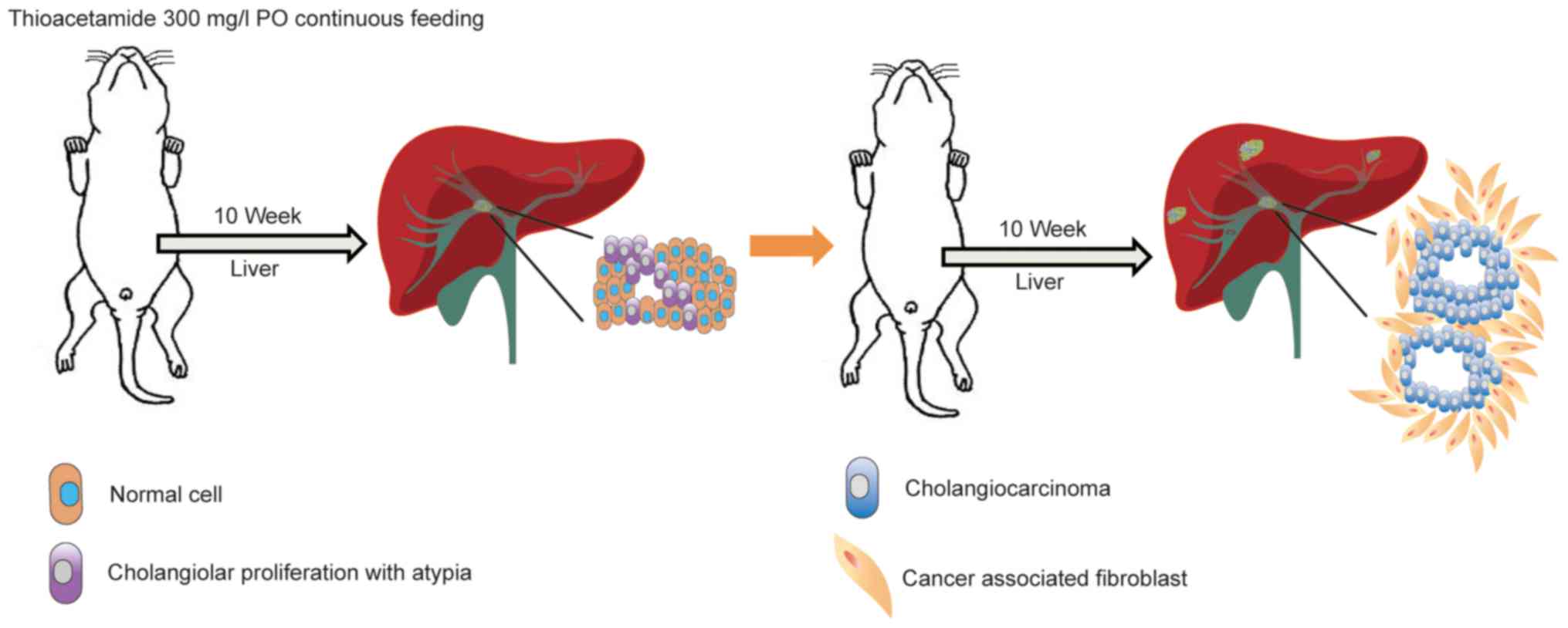
Rat orthotopic tumor graft
A total of 20 adult 8-week old male Sprague-Dawley
(SD) rats (310±14 g; BioLASCO Taiwan Co, Ltd, Taipei, Taiwan) were
used for the experiments. Animals were divided equally (n=5) into
the following four groups: Control (Group 1),
gemcitabine/oxaliplatin treatment (Group 2), paclitaxel treatment
(Group 3) and nab-paclitaxel treatment (Group 4). The rats were
housed in an animal facility room maintained in a 12/12 h
light:dark cycle and at an ambient temperature of 22°C. The animals
had ad libitum access to food and water. The rats were
administered with 300 mg/l thioacetamide (TAA) daily via drinking
water for up to 20 weeks. All drug treatments administered to these
animals were initiated in the 21st week. The
gemcitabine/oxaliplatin group received intraperitoneal injections
of gemcitabine (50 mg/kg) and oxaliplatin (2 mg/kg) once every 2
weeks over a 4-week period. Simultaneously, the paclitaxel group
received an intravenous infusion of paclitaxel (20 mg/kg) for 5
consecutive days. Additionally, the nab-paclitaxel group received
an intravenous slow infusion of nab-paclitaxel (7.5 mg/kg) for 5
consecutive days. The control group received intraperitoneal
injections of phosphate buffered saline (PBS; pH 7.4) (Fig. 1).
Evaluation of treatment efficacy in
rats by positron emission tomography
In order to evaluate the changes in glycolysis in
live animals with liver tumors, 2-deoxy-2-[F-18] fluoro-d-glucose
positron emission tomography (18F-FDG-PET) studies were
performed at the Molecular Imaging Center of Chang Gung Memorial
Hospital (Taoyuan, Taiwan). In brief, 20 rats were treated with
TAA, prior to being subjected to serial PET scanning at 21, 23 and
25 weeks using the Inveon™ system (Siemens AG, Munich, Germany).
Animals were assigned equally to the control and treatment groups
based on their baseline PET results, to ensure similar PET-positive
rates in the two groups. The details of radioligand preparation,
scanning protocols and determination of optimal scanning time have
been previously described by our group (12,13). In
brief, animals were fasted overnight prior to scanning and, 90 min
after 18F-FDG intravenous injection, 30-min static scans
were obtained for all the animals. All imaging studies on animals
were performed at 37°C under anesthesia (2% isoflurane vaporized in
100% oxygen)-controlled imaging bed (Minerve System, Esternay,
France). PET images were reconstructed using the 2-dimensional
ordered subset expectation-maximization method (4 iterations and 16
subsets) without attenuation and scatter corrections. All imaging
data were processed by the PMOD image analysis workstation (PMOD
Technologies Ltd., Zurich, Switzerland). The largest liver tumor
for each animal was identified by careful investigation of the
three tumor image sets obtained for each rat. The uptake of
18F-FDG by the normal and tumor liver tissues was
quantified by calculating the standardized uptake value (SUV). The
SUVs were calculated according to the recommendations of the
European Organization for Research and Treatment of Cancer
(14). Regions of interest (ROIs) in
the tumors were determined by obtaining transverse images of the
selected tumors and measuring the largest diameter. The normal
liver ROIs were also determined similarly by measuring the diameter
of transverse images obtained from the normal liver tissues. The
mean SUV (SUVmean) of the normal and tumor liver tissues
was calculated, and the respective tumor:liver (T/L) radioactivity
ratio were compared for the two tissue types.
Immunofluorescence analysis for
effects of drug treatment on rat tumor stroma
The study samples were obtained from rats treated
with paclitaxel and nab-paclitaxel for 4 weeks. Formalin-fixed,
paraffin-embedded slides (4-µm thickness) were prepared using a
standard protocol. The slides were deparaffinized in 4 5-min
incubations in 100% xylene, then dehydrated in a graded alcohol
serious of 100% for 5 min ×2, 95% for 5 min, 90% for 5 min, 70% for
5 min and distilled H2O for 5 min ×3 at 25°C. Antigen
retrieval took place using pH 6.0 citric buffer at 121°C for 3 min,
washed 3 times in distilled water for 5 min, then twice in
Tris-buffered saline for 5 min. Specimens were mounted using H33258
mounting solution and kept at 4°C until use. The slides were
incubated with a primary antibody against α-SMA (dilution, 1:200;
cat. no. ab5694; Abcam, Cambridge, UK), a marker for activated
fibroblasts, at 4°C overnight. The slides were washed with PBS with
tween 20 following incubation with the primary antibody and were
incubated at room temperature for 1 h with a goat anti-rabbit Alexa
Fluor 568 (IgG H+L) secondary antibody (dilution, 1:200; cat. no.
A11036; Molecular Probes; Thermo Fisher Scientific, Inc.). Nuclei
were stained with Hoechst 33258 [mounted cover slide with 0.1 µg/ml
H33258 mounting solution consisting of 0.1 µg/ml H33258 (cat. no.
H21491; Molecular Probes; Thermo Fisher Scientific, Inc.) in
glycerol (cat no. G7757; Sigma-Aldrich; Merck KGaA) at 25°C and
slides were then kept at 4°C until images were captured following
three days]. Images were captured using the Leica TCS SP8X confocal
system equipped with a LEICA DM6000 CS Microscope (objective lens
63×, subjective lens 10×, digital zoom 1×; final magnification,
×630) (Leica Microsystems, Inc., Buffalo Grove, IL, USA).
Statistical analysis
The data are presented as the mean ± standard
deviation. SPSS software for Windows (Version 21.0; IBM Corp.,
Armonk, NY, USA) was used for statistical analysis. The SUV ratio
between experimental and control animals was calculated using
nonlinear regression analysis. Differences between the data from
experimental and control animals were calculated and verified using
the Mann-Whitney U test or the Kruskal-Wallis test. P<0.05 was
considered to indicate a statistically significant difference.
Results
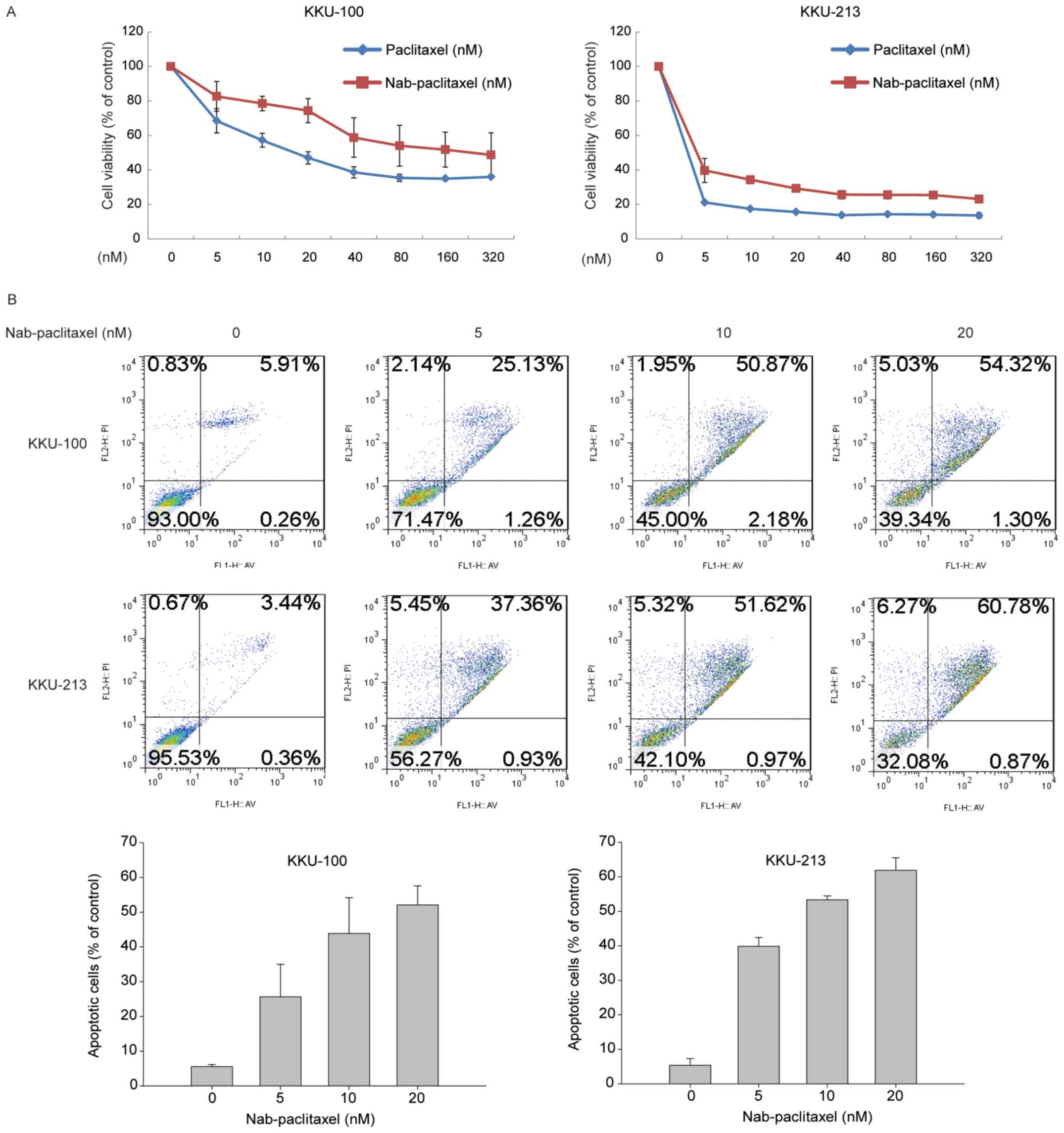
Nab-paclitaxel and paclitaxel induced
similar in vitro anti-proliferative effects
Human CCA KKU-100 and KKU-213 cell lines were
treated with varying doses of paclitaxel and nab-paclitaxel. The
MTT test indicated that the two compounds induced similar
cytotoxicity in the different cell lines (paclitaxel
IC50 values, 17.09 nM for KKU-100 and 3.17 nM for
KKU-213; nab-paclitaxel IC50 values, 25.32 nM for
KKU-100 and 4.15 nM for KKU-213; Fig.
2A). In order to confirm the apoptotic effect of nab-paclitaxel
in CCA cells, the presence of apoptosis and the percentage of
apoptotic cells were determined using a FITC Annexin V apoptosis
detection kit. Nab-paclitaxel treatment increased the percentage of
apoptotic cells within 48 h in a concentration-dependent manner;
54.32 and 60.78% of apoptotic KKU-100 and KKU-213 cells were
observed following treatment with 20 nM nab-paclitaxel (Fig. 2B).
Nab-paclitaxel induced enhanced
antitumor effects compared with paclitaxel in a rat model of
TAA-induced IH-CCA
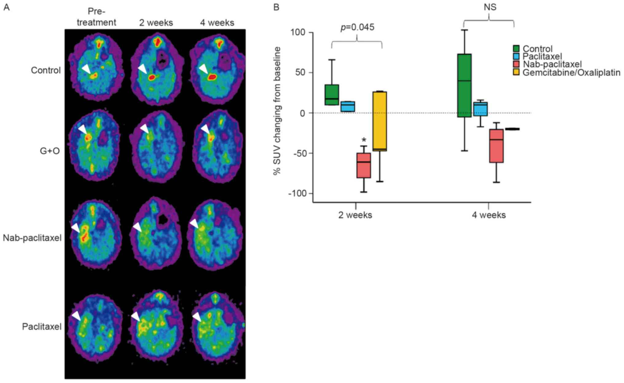
Transverse, sagittal and coronal views of
TAA-induced CCA tumors in control and drug-treated animals were
evaluated by 18F-FDG-PET. Each group exhibited >1
FDG-avid tumor in the liver after 20 weeks of TAA treatment, as
demonstrated in the coronal view of the animal PET-CT (Fig. 3A). In the present study,
nab-paclitaxel, paclitaxel or gemcitabine/oxaliplatin were
administered to rats undergoing TAA treatment. The change in the
T/L ratio of the SUV for each group is presented in Fig. 3B. The T/L ratio of the SUV exhibited
steady elevation from the initial to the final scans in the control
group (7.8–20.6% elevation from 2 to 4 weeks post-experiment). The
T/L ratio of the SUV in the paclitaxel treatment group exhibited a
mild increase until the final scans (5.6 and 5.1% at 2 and 4 weeks
post-experiment, respectively). By contrast, in the nab-paclitaxel
and gemcitabine/oxaliplatin treatment groups, the T/L ratio of the
SUV exhibited a decrease until the final scans (nab-paclitaxel,
−28.5 and −19.1%, respectively; gemcitabine/oxaliplatin, −11.3 and
−5.4%, respectively, at 2 and 4 weeks post-experiment). The results
of the present study indicated that treatment with nab-paclitaxel
induced a significant decrease in the T/L ratio of the SUV,
compared with the values reported for the control group (P<0.05,
control vs. group 3). Furthermore, nab-paclitaxel treatment
resulted in significant suppression of in vivo tumor
viability in the rat CCA model.
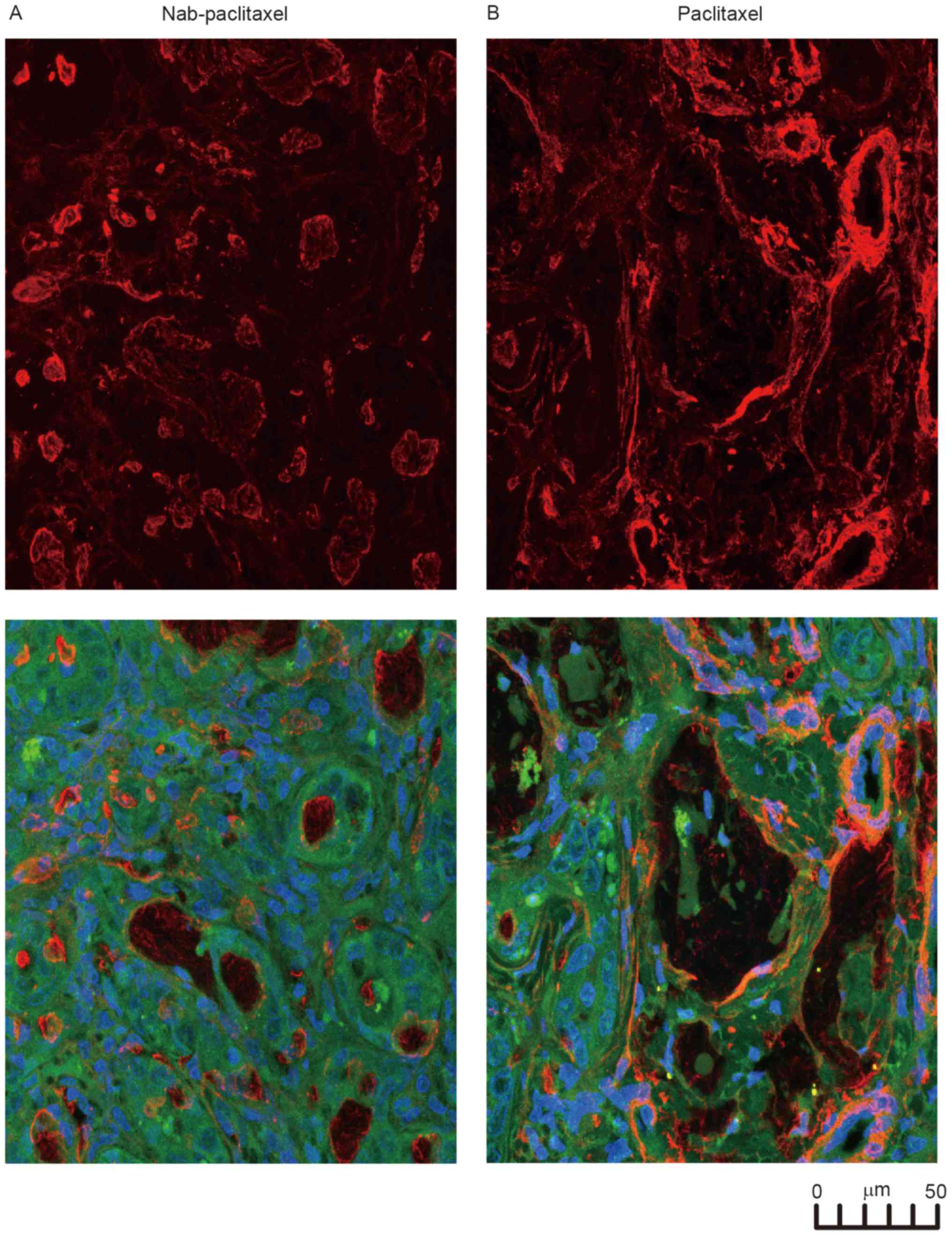
Nab-paclitaxel disrupts
cancer-associated fibroblasts in rat IH-CCA
The immunofluorescence results for the resected
IH-CCA of the rats are presented in Fig.
4. The red staining for α-SMA was stronger and observed over
the entire paclitaxel-treated tumor, compared with the weak,
dispersed staining of the nab-paclitaxel-treated tumor (Fig. 4A). The confocal images in Fig. 4B indicated the anatomical association
among the CAFs (with α-SMA staining), vessels and IH-CCA,
demonstrating that nab-paclitaxel broke the CAFs surrounding the
IH-CCA.
Discussion
The results of the present study demonstrated that
nab-paclitaxel may inhibit IH-CCA by disrupting the stromal CAFs.
In an attempt to study this further, the TAA-induced IH-CCA rat
model was selected for in vivo study. Multifocal bile ductal
proliferation with marked atypia (15) was observed in the liver of the rats
after 9 weeks of TAA administration, contrary to the observations
made in a mouse xenograft model (15). Furthermore, 50% of the rats developed
IH-CCA with intense stromal desmoplasia from the 16th week
(15). From the 16–22th weeks, the
incidence of invasive CCAs increased progressively in 100% of the
rats (Fig. 1) (15). Therefore, this model served as a
useful pre-clinical platform for developing therapeutic strategies
for human CCA, including nab-paclitaxel (12,13).
In the present study, paclitaxel and nab-paclitaxel
induced similar anti-proliferative effects via induction of
apoptosis in two human CCA cell lines (Fig. 2). However, only nab-paclitaxel induces
in vivo antitumor effects in the TAA-induced rat model,
demonstrated by a significant decrease in the T/L ratio of the SUV
(Fig. 3). Paclitaxel did not induce
any in vivo antitumor effects in the TAA-induced rat model.
Furthermore, in the present study, the treatment effects induced by
CAF on desmoplastic stroma were compared between the paclitaxel and
the nab-paclitaxel groups. The disrupting effects on the stroma
were found to be limited to the nab-paclitaxel group (Fig. 4). Therefore, the stromal disrupting
effect may partially explain the effectiveness of nab-paclitaxel
against IH-CCA. The current clinical observations support the
aforementioned result, thereby providing an explanation for the
ineffective use of paclitaxel in the treatment of biliary tract
cancer since 1996 (16). However,
Muc1 and survivin have been suggested to be associated with the
poor prognosis of IH-CCA (17).
Furthermore, whether or not these two proteins changed following
nab-paclitaxel treatment was examined. The immunohistochemistry
stains of Muc1 and survivin exhibited no change prior to and
following all treatments (data not shown), suggesting that
nab-paclitaxel did not target these two proteins.
There are certain limitations to the present study.
To begin with, although the present study investigates an
orthotopic CCA with desmoplastic stroma originating from the rat
liver, such induced CCA may not completely simulate the conditions
observed in human CCA. However, this model overcomes the drawbacks
of a xenograft CCA model that lacks surrounding CAFs. Secondly, the
small number of rats used in the four different treatment groups
may lower the statistical power of the present study. For example,
the animal PET revealed the significant inhibitory effect of
nab-paclitaxel compared with that of paclitaxel, but the
immunofluorescence study was unable to statistically confirm these
observations.
In conclusion, the results of the present study
demonstrated that nab-paclitaxel is effective against IH-CCA via
disruption of the surrounding CAFs. Since there are few treatment
options available for IH-CCA, this result may provide useful
information in designing clinical trials, as well as for the
identification of biomarker in the future.
Acknowledgements
The authors would like to thank Miss Meng-Lun Lu
(Taipei Veterans General Hospital, Tapei, Taiwan) for her
technology support.
Funding
The work was supported by Taiwan Cancer Clinic
Foundation and the Szu-Yuan Research Foundation of International
Medicine. Additional support was provided by the Taipei Veterans
General Hospital (grant nos. V105C-057 and V104E16-003-MY3-2 to
Ming-Huang Chen), the Chang Gung Memorial Hospital (grant nos.
NMRPG5D6032, CMRPG3E1611, CMRPG3E1612, CRRPG3F0031 and NMRPG3F6021
to Chun-Nan Yeh), and the Ministry of Science and Technology (grant
nos. MOST104-2314-B-075-064-MY2 to Ming-Huang Chen,
MOST103-2314-B-182A-081-MY2 and MOST105-2314-B-182A-041-MY2 to
Chun-Nan Yeh).
Availability of data and materials
All data generated or analyzed during this study are
included in this published article.
Authors' contributions
PMC analyzed the data and wrote the manuscript. CTC,
RCW, KCC, CYL and MHaC contributed to study design and data
collection. YHC, TSY helped with the animal experiments. MHuC
helped cell experiments. CNY guided the animal model and associated
tissue IHC staining analysis. All authors read and approved the
final manuscript.
Ethics approval and consent to
participate
The present study was approved by the Institutional
Animal Care and Use Committee of Chang Gung Memorial Hospital
(Taoyuan, Taiwan).
Consent for publication
Not applicable.
Competing interests
The authors declare that they have no competing
interests.
Reference
|
1
|
Ustundag Y and Bayraktar Y:
Cholangiocarcinoma: A compact review of the literature. World J
Gastroenterol. 14:6458–6466. 2008. View Article : Google Scholar : PubMed/NCBI
|
|
2
|
Khan SA, Thomas HC, Davidson BR and
Taylor-Robinson SD: Cholangiocarcinoma. Lancet. 366:1303–1314.
2005. View Article : Google Scholar : PubMed/NCBI
|
|
3
|
Patel T: Increasing incidence and
mortality of primary intrahepatic cholangiocarcinoma in the United
States. Hepatology. 33:1353–1357. 2001. View Article : Google Scholar : PubMed/NCBI
|
|
4
|
Valle J, Wasan H, Palmer DH, Cunningham D,
Anthoney A, Maraveyas A, Madhusudan S, Iveson T, Hughes S, Pereira
SP, et al: Cisplatin plus gemcitabine versus gemcitabine for
biliary tract cancer. N Engl J Med. 362:1273–1281. 2010. View Article : Google Scholar : PubMed/NCBI
|
|
5
|
Hezel AF, Deshpande V and Zhu AX: Genetics
of biliary tract cancers and emerging targeted therapies. J Clin
Oncol. 28:3531–3540. 2010. View Article : Google Scholar : PubMed/NCBI
|
|
6
|
Zhu AX and Hezel AF: Development of
molecularly targeted therapies in biliary tract cancers:
Reassessing the challenges and opportunities. Hepatology.
53:695–704. 2011. View Article : Google Scholar : PubMed/NCBI
|
|
7
|
Sirica AE and Gores GJ: Desmoplastic
stroma and cholangiocarcinoma: Clinical implications and
therapeutic targeting. Hepatology. 59:2397–2402. 2014. View Article : Google Scholar : PubMed/NCBI
|
|
8
|
Sirica AE: The role of cancer-associated
myofibroblasts in intrahepatic cholangiocarcinoma. Nat Rev
Gastroenterol Hepatol. 9:44–54. 2011. View Article : Google Scholar : PubMed/NCBI
|
|
9
|
DeClerck YA: Desmoplasia: A response or a
niche? Cancer Discov. 2:772–724. 2012. View Article : Google Scholar : PubMed/NCBI
|
|
10
|
Alvarez R, Musteanu M, Garcia-Garcia E,
Lopez-Casas PP, Megias D, Guerra C, Muñoz M, Quijano Y, Cubillo A,
Rodriguez-Pascual J, et al: Stromal disrupting effects of
nab-paclitaxel in pancreatic cancer. Br J Cancer. 109:926–933.
2013. View Article : Google Scholar : PubMed/NCBI
|
|
11
|
Von Hoff DD, Ervin T, Arena FP, Chiorean
EG, Infante J, Moore M, Seay T, Tjulandin SA, Ma WW, Saleh MN, et
al: Increased survival in pancreatic cancer with nab-paclitaxel
plus gemcitabine. N Engl J Med. 369:1691–1703. 2013. View Article : Google Scholar : PubMed/NCBI
|
|
12
|
Chen MH, Lin KJ, Yang WL, Kao YW, Chen TW,
Chao SC, Chang PM, Liu CY, Tzeng CH, Chao Y, et al: Gene
expression-based chemical genomics identifies heat-shock protein 90
inhibitors as potential therapeutic drugs in cholangiocarcinoma.
Cancer. 119:293–303. 2013. View Article : Google Scholar : PubMed/NCBI
|
|
13
|
Chen MH, Chiang KC, Cheng CT, Huang SC,
Chen YY, Chen TW, Yeh TS, Jan YY, Wang HM, Weng JJ, et al:
Antitumor activity of the combination of an HSP90 inhibitor and a
PI3K/mTOR dual inhibitor against cholangiocarcinoma. Oncotarget.
5:2372–2389. 2014. View Article : Google Scholar : PubMed/NCBI
|
|
14
|
Young H, Baum R, Cremerius U, Herholz K,
Hoekstra O, Lammertsma AA, Pruim J and Price P: Measurement of
clinical and subclinical tumour response using
[18F]-fluorodeoxyglucose and positron emission tomography: Review
and 1999 EORTC recommendations. European organization for research
and treatment of cancer (EORTC) PET study group. Eur J Cancer.
35:1773–1782. 1999. View Article : Google Scholar : PubMed/NCBI
|
|
15
|
Yeh CN, Maitra A, Lee KF, Jan YY and Chen
MF: Thioacetamide-induced intestinal-type cholangiocarcinoma in
rat: An animal model recapitulating the multi-stage progression of
human cholangiocarcinoma. Carcinogenesis. 25:631–636. 2004.
View Article : Google Scholar : PubMed/NCBI
|
|
16
|
Jones DV Jr, Lozano R, Hoque A, Markowitz
A and Patt YZ: Phase II study of paclitaxel therapy for
unresectable biliary tree carcinomas. J Clin Oncol. 14:2306–2310.
1996. View Article : Google Scholar : PubMed/NCBI
|
|
17
|
Sirica AE, Dumur CI, Campbell DJ, Almenara
JA, Ogunwobi OO and Dewitt JL: Intrahepatic cholangiocarcinoma
progression: Prognostic factors and basic mechanisms. Clin
Gastroenterol Hepatol. 7 11 Suppl:S68–S78. 2009. View Article : Google Scholar : PubMed/NCBI
|