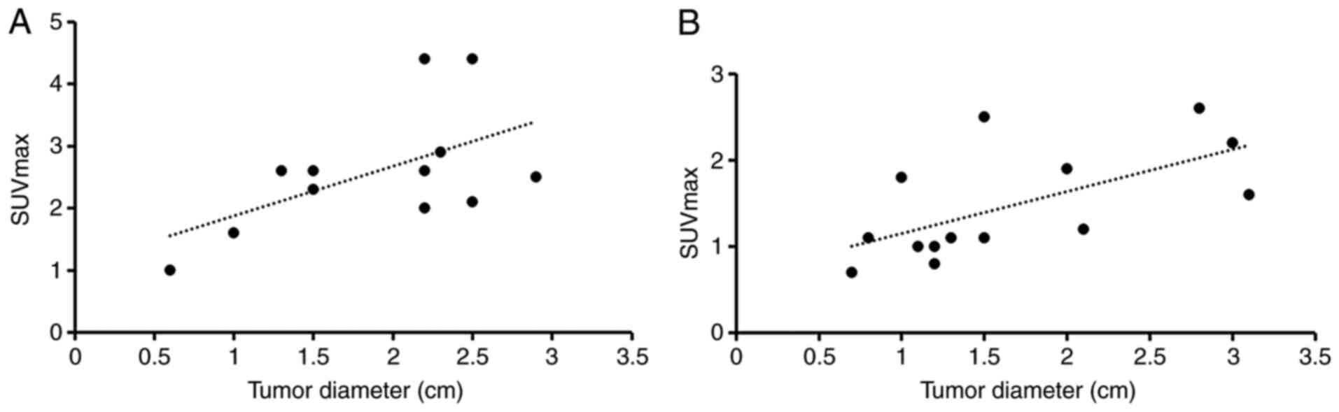
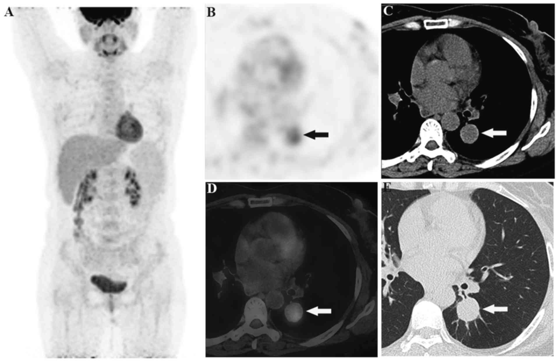
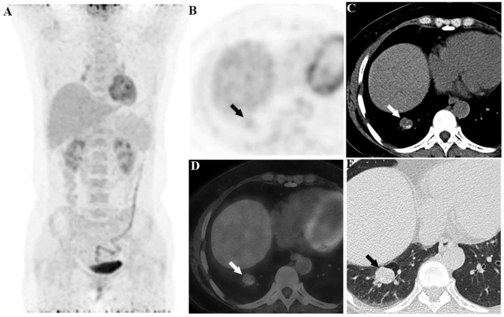
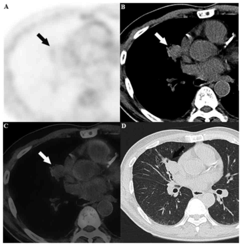
|
1
|
Sugio K, Yokoyama H, Kaneko S, Ishida T
and Sugimachi K: Sclerosing hemangioma of the lung: radiographic
and pathological study. Ann Thorac Surg. 53:295–300. 1992.
View Article : Google Scholar : PubMed/NCBI
|
|
2
|
Iyoda A, Hiroshima K, Shiba M, Haga Y,
Moriya Y, Sekine Y, Shibuya K, Iizasa T and Fujisawa T:
Clinicopathological analysis of pulmonary sclerosing hemangioma.
Ann Thorac Surg. 78:1928–1931. 2004. View Article : Google Scholar : PubMed/NCBI
|
|
3
|
Neuman J, Rosioreanu A, Schuss A, Turi G,
Yung E, Trow TK, Williams L and Katz DS: Radiology-pathology
conference: Sclerosing hemangioma of the lung. Clin Imaging.
30:409–412. 2006. View Article : Google Scholar : PubMed/NCBI
|
|
4
|
Edey AJ and Hansell DM: Incidentally
detected small pulmonary nodules on CT. Clin Radiol. 64:872–884.
2009. View Article : Google Scholar : PubMed/NCBI
|
|
5
|
Lei Y, Yong D, Jun-Zhong R, Zhi Y and
Zi-Tong W: Treatment of 28 patients with sclerosing hemangioma (SH)
of the lung. J Cardiothorac Surg. 7:342012. View Article : Google Scholar : PubMed/NCBI
|
|
6
|
Shin SY, Kim MY, Oh SY, Lee HJ, Hong SA,
Jang SJ and Kim SS: Pulmonary sclerosing pneumocytoma of the lung:
CT characteristics in a large series of a tertiary referral center.
Medicine (Baltimore). 94:e4982015. View Article : Google Scholar : PubMed/NCBI
|
|
7
|
Lin KH, Chang CP, Liu RS and Wang SJ: F-18
FDG PET/CT in evaluation of pulmonary sclerosing hemangioma. Clin
Nucl Med. 36:341–343. 2011. View Article : Google Scholar : PubMed/NCBI
|
|
8
|
Uhlén N, Grundberg O, Jacobsson H, Sundin
A, Dobra K, Sánchez-Crespo A, Axelsson R and Kölbeck KG: 18F-FDG
PET/CT Diagnosis of bronchopulmonary carcinoids versus pulmonary
hamartomas. Clin Nucl Med. 41:263–267. 2016. View Article : Google Scholar : PubMed/NCBI
|
|
9
|
Miyagawa-Hayashino A, Tazelaar HD, Langel
DJ and Colby TV: Pulmonary sclerosing hemangioma with lymph node
metastases: Report of 4 cases. Arch Pathol Lab Med. 127:321–325.
2003.PubMed/NCBI
|
|
10
|
Katakura H, Sato M, Tanaka F, Sakai H,
Bando T, Hasegawa S, Nakashima Y and Wada H: Pulmonary sclerosing
hemangioma with metastasis to the mediastinal lymph node. Ann
Thorac Surg. 80:2351–2353. 2005. View Article : Google Scholar : PubMed/NCBI
|
|
11
|
Chien NC, Lin CW and Tzeng JE: Sclerosing
haemangioma with lymph node metastasis. Respirology. 14:614–616.
2009. View Article : Google Scholar : PubMed/NCBI
|
|
12
|
Komatsu T, Fukuse T, Wada H and Sakurai T:
Pulmonary sclerosing hemangioma with pulmonary metastasis. Thorac
Cardiovasc Surg. 54:348–349. 2006. View Article : Google Scholar : PubMed/NCBI
|
|
13
|
Maeda R, Isowa N, Miura H, Tokuyasu H,
Kawasaki Y and Yamamoto K: Bilateral multiple sclerosing
hemangiomas of the lung. Gen Thorac Cardiovasc Surg. 57:667–670.
2009. View Article : Google Scholar : PubMed/NCBI
|
|
14
|
Kamaleshwaran KK, Rajan F, Mehta S,
Mohanan V and Shinto AS: Multiple pulmonary sclerosing hemangiomas
(pneumocytoma) mimicking lung metastasis detected in fluorine-18
fluorodeoxyglucose positron emission tomography/computed
tomography. Indian J Nucl Med. 29:168–170. 2014. View Article : Google Scholar : PubMed/NCBI
|
|
15
|
Hedlund GL, Bisset GS III and Bove KE:
Malignant neoplasms arising in cystic hamartomas of the lung in
childhood. Radiology. 173:77–79. 1989. View Article : Google Scholar : PubMed/NCBI
|
|
16
|
Rossi G1, Cavazza A, Valli R, Torricelli
P, Richeldi L, Rivasi F and Brambilla E: Atypical lipomatous tumour
(lipoma-like well-differentiated liposarcoma) arising in a
pulmonary hamartoma and clinically presenting with pneumothorax.
Lung Cancer. 39:103–106. 2003. View Article : Google Scholar : PubMed/NCBI
|
|
17
|
Lee BJ, Kim HR, Cheon GJ, Koh JS, Kim CH
and Lee JC: Squamous cell carcinoma arising from pulmonary
hamartoma. Clin Nucl Med. 36:130–131. 2011. View Article : Google Scholar : PubMed/NCBI
|
|
18
|
Jiang L, Tan H, Panje CM, Yu H, Xiu Y and
Shi H: Role of 18F-FDG PET/CT Imaging in Intrahepatic
Cholangiocarcinoma. Clin Nucl Med. 41:1–7. 2016. View Article : Google Scholar : PubMed/NCBI
|
|
19
|
Hara M, Iida A, Tohyama J, Miura N,
Shiraki N, Itoh M, Ohba S and Tateyama H: FDG-PET findings in
sclerosing hemangioma of the lung: A case report. Radiat Med.
19:215–218. 2001.PubMed/NCBI
|
|
20
|
Chen Q, Wu LJ, Hu H, Song J, Wu Y, Yan J
and Shi J: A case of pulmonary sclerosing hemangioma with low
(18)FDG uptake in PET. Oncol Lett. 3:646–648. 2012. View Article : Google Scholar : PubMed/NCBI
|
|
21
|
Lee E, Park CM, Kang KW, Goo JM, Kim MA,
Paeng JC, Lee HJ, Park HS and Chung DH: 18F-FDG PET/CT features of
pulmonary sclerosing hemangioma. Acta Radiol. 54:24–29. 2013.
View Article : Google Scholar : PubMed/NCBI
|
|
22
|
Lowe VJ, Fletcher JW, Gobar L, Lawson M,
Kirchner P, Valk P, Karis J, Hubner K, Delbeke D, Heiberg EV, et
al: Prospective investigation of positron emission tomography in
lung nodules. J Clin Oncol. 16:1075–1084. 1998. View Article : Google Scholar : PubMed/NCBI
|
|
23
|
Chung MJ, Lee KS, Han J, Sung YM, Chong S
and Kwon OJ: Pulmonary sclerosing hemangioma presenting as solitary
pulmonary nodule: Dynamic CT findings and histopathologic
comparisons. AJR Am J Roentgenol. 187:430–437. 2006. View Article : Google Scholar : PubMed/NCBI
|