Introduction
Keloids are a dermatologic condition that affects
1/10 people in Africa (1), and has an
incidence of 0.15% in the Asian population (2). Although it is a benign hyperplasia, it
causes dermatologic dysfunction and esthetic deformity by invading
adjacent normal tissues (3). Frequent
episodes of itching and pain also occur, resulting in physical and
psychological distress (4). The
majority of keloids are initiated by minor skin trauma, such as
folliculitis and/or acne, and grow with chronic inflammation of the
reticular layer of the dermis (5). A
typical pathognomonic characteristic of keloids is the presence of
thickened and hyalinized collagen (6). They are problematic in plastic surgery
due to little being known about their etiology or optimal
treatment.
Keloids are considered to result from prolonged,
aberrant wound healing that involves excessive fibroblast
participation and collagen deposition (7). Fibroblasts are the primary effector
cells of keloid tissue and are characterized by excessive
proliferation, disordered apoptosis, and increased secretion of
extracellular matrix (8). Numerous
previous studies have investigated the pathogenesis of keloids with
respect to genetics, mechanics, endocrinology, immunology and
nutrition (9–11). The skin injury-wound tension theory
was a milestone in our understanding of keloid formation (9). Keloids are frequently observed on the
anterior chest and scapular regions, but rarely on the scalp or
anterior lower legs; this is closely associated with the frequency
of local physical tension or movement in these regions (9). Reducing skin tension around wounds or
scars can, therefore, be beneficial in the prevention and treatment
of keloids (12). The characteristic
shapes of keloid scars are largely determined by the direction of
local mechanical forces on the skin (13). Other influences, besides these local
factors, also promote keloid development, including genetic factors
(single nucleotide polymorphisms) (14), systemic factors (hypertension
(15) and estrogen levels) (16), endocrinal factors (physiological
hyperactivity of the sebaceous gland) (17) and metabolic factors (higher adenosine
triphosphate levels and insufficient oxygen levels) (18). Although keloid development is known to
involve complex pathways, the exact mechanisms by which keloid
formation is initiated and regulated remain to be elucidated.
In our previous study, an investigation of
differential long non-coding RNA (lncRNA) expression between
keloids and normal skin tissue was conducted, and it was revealed
that the lncRNA CANCA1G-AS1 (CAS1) was significantly upregulated in
keloid tissue (19). This suggested
that CAS1 may be involved in the mechanism of keloid formation. In
the present study, an investigation of the biological role of CAS1
in cell proliferation, cell migration, cytokine secretion and
collagen secretion was performed, with controlled calcium channel
protein expression in human keloid fibroblasts using
loss-of-function studies.
Materials and methods
Patient samples
This study was approved by the institutional review
board at Peking Union Medical College (Beijing, China), in
accordance with the principles of the Declaration of Helsinki.
Keloid and corresponding normal skin from 16 patients, who received
surgery at The Peking Union Medical College Hospital (Beijing,
China) (19), was previously obtained
in 2014. Written informed consent was obtained from all patients
prior to surgery. None of the patients in the study received any
local treatment, such as corticosteroid injections, or radiation
therapy prior to surgery.
Cell culture
Cultures were established from keloid specimens of
three patients, processed within 4 h of post-surgical excision,
using conventional methods (20).
Briefly, these specimens were washed three times in 1X PBS and the
epidermis and subdermal fat were removed. The remaining dermis was
dissected and incubated in Dulbecco's modified Eagle's medium
(DMEM; Thermo Fisher Scientific, Inc., Waltham, MA, USA)
supplemented with 10% fetal bovine serum (Thermo Fisher Scientific,
Inc.) at 37°C in a 5% CO2 humidified atmosphere. The
DMEM was replaced every 3 days and keloid fibroblasts were used
during passages 3–5. Detailed information about primary keloid
fibroblast isolation and culture was described in our previous
report (19).
Transfection
Three small interfering RNAs (siRNAs) targeting
different sites of lncRNA CAS1 (GenBank accession no. NR_038439.1)
were designed and synthesized by Suzhou GenePharma Co., Ltd.
(Jiangsu, China) to knock down CAS1 expression, as well as a
control siRNA that did not target CAS1 as the negative control
(NC). The sequences were as follows: Cas1 siRNA-1,
5-CCCUCAACCCAAGGAAGAUTT-3; Cas1 siRNA-2, 5-GCCUUCGCAACUCAUUCAUTT-3;
Cas1 siRNA-3, 5-CCGUGUGAAGGGAGCAAUUTT-3; NC,
5-UUCUCCGAACGUGUCACGUTT-3. Keloid fibroblasts were seeded in 6-well
plates (1×10⁵ cells/well) and incubated overnight in an
atmosphere of 5% CO2 at 37°C to allow their full
extension and adherence prior to transfection. The cells were grown
to 70–90% confluency, and the siRNAs were transfected using
Lipofectamine® 2000 (Thermo Fisher Scientific,
Inc.), according to the manufacturer's instructions. The cells were
collected for further experiments 48 h after transfection.
Reverse transcription-quantitative
polymerase chain reaction (RT-qPCR)
Total RNA was isolated from cells using
TRIzol® reagent (Thermo Fisher Scientific, Inc.) 48 h
after transfection, according to the manufacturer's instructions.
The expression of CAS1 and other target genes was quantified
relative to endogenous GAPDH expression, using RT-qPCR with
the following primer sets: CAS1 forward, 5′-TGTGCTTCACCATGCTCCAT-3′
and reverse, 5′-ATTAGTGCTCCGGCCAACAA-3′; GAPDH forward,
5′-GGTCACCAGGGCTGCTTTTA-3′ and reverse,
5′-GGATCTCGCTCCTGGAAGATG-3′; CACNA1G forward,
5′-CACGGTCATCTCGCCTATCT-3′ and reverse, 5′-TCCTTGTTGCTCTCCTCCAG-3′;
transforming growth factor-β (TGF-β), forward,
5′-GCAACAATTCCTGGCGATAC-3′ and reverse, 5′-CTAAGGCGAAAGCCCTCAAT-3′;
COL1A1, forward 5′-AAGACATCCCACCAATCACC-3′ and reverse,
5′-CGTCATCGCACAACACCTT-3′; and COL3A1 forward,
5′-CTGCCATCCTGAACTCAAGAGTGG-3′ and reverse,
5′-CCATCCTCCAGAACTGTGTAGG-3′. The PCR conditions were as follows:
95°C for 60 sec, followed by 40 cycles of 95°C for 10 sec, and
finally 60°C for 45 sec. After amplification, real-time data
acquisition and analysis were performed. The relative quantitative
results were calculated using the 2−∆∆Cq method
(21).
MTS assay
Cell proliferation was assessed using an MTS assay
(Promega Corporation, Madison, WI, USA). Cells were plated at a
density of 5×10³ cells/well in 96-well plates and
incubated overnight in an atmosphere of 5% CO2 at 37°C.
After transfection, 20 µl MTS was added into each well containing
100 µl DMEM (Thermo Fisher Scientific, Inc.), and the cells were
then incubated at 37°C for 1 h in a humidified 5% CO2
incubator. Absorbance was detected at 490 nm using a microplate
reader (Bio-Rad Laboratories, Inc., Hercules, CA, USA).
Cell cycle analysis by flow
cytometry
Cells were collected 48 h after transfection, washed
with PBS and trypsinized with 0.025% trypsin-EDTA to yield single
cell suspensions. They were then fixed in ice-cold 70% ethanol and
stained with 50 µg/ml propidium iodide solution (Sigma-Aldrich;
Merck KGaA, Darmstadt, Germany) containing 10 µg/ml RNaseA (Tiangen
Biotech Co., Ltd., Beijing, China). A BD Accuri™ C6 flow
cytometer was used for flow cytometric analysis, and the cell cycle
profiles were analyzed using ModFit LT software for Windows Version
3.2 (Verity Software House, Inc., Topsham, ME, USA).
Scratch wound migration assay
Keloid fibroblasts were seeded uniformly
(1×10⁴ cells/well) into 6-well plates and grown
overnight. A total of 48 h after transfection, a scratch wound was
made across the center of each confluent cell culture monolayer
using a sterile 200 µl pipette tip, and any non-adherent cells were
washed off with 1X PBS. Conditioned DMEM was then reapplied to the
cells according to the previous treatment regimen, and the plates
were incubated for a further 72 h in at atmosphere of 5%
CO2 at 37°C). Each well was imaged every 24 h using
objective inverted microscopy (Olympus Corporation, Tokyo,
Japan).
Statistical analysis
Statistical analysis was performed using the SPSS
software version 13.0 (SPSS, Inc., Chicago, IL, USA). All results
are presented as the mean ± standard deviation and were analyzed
using a Student's t-test and one-way analysis of variance (ANOVA)
to determine the levels of significance. Bonferroni correction was
used to perform the post-hoc test following ANOVA. P<0.05 was
considered to indicate a statistically significant difference.
Results
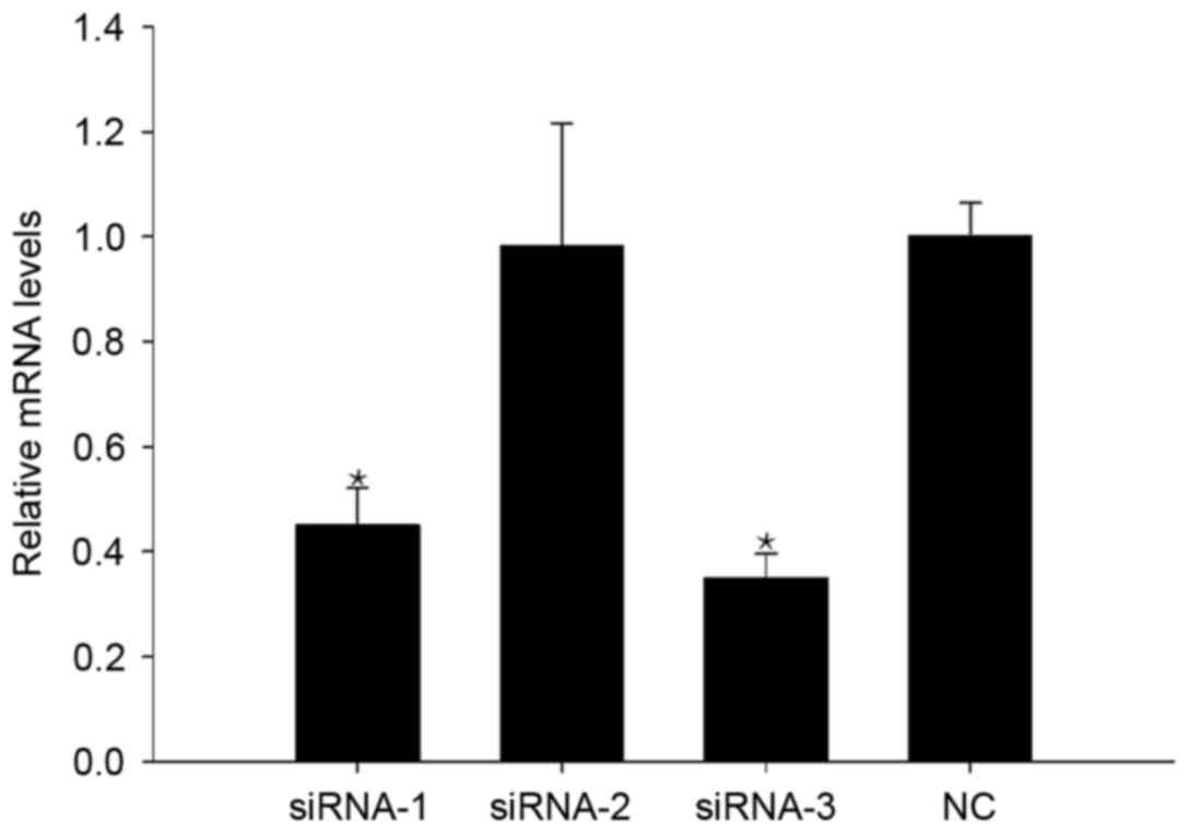
siRNA-3 downregulates CAS1 expression
in keloid fibroblasts
A total of 48 h after transfection, RT-PCR was used
to detect CAS1 mRNA expression in keloid fibroblasts. Of the three
siRNA sequences, siRNA-3 had the greatest effect on reducing CAS1
expression (Fig. 1), and thus was
selected as an effective means of interference for use in further
experiments.
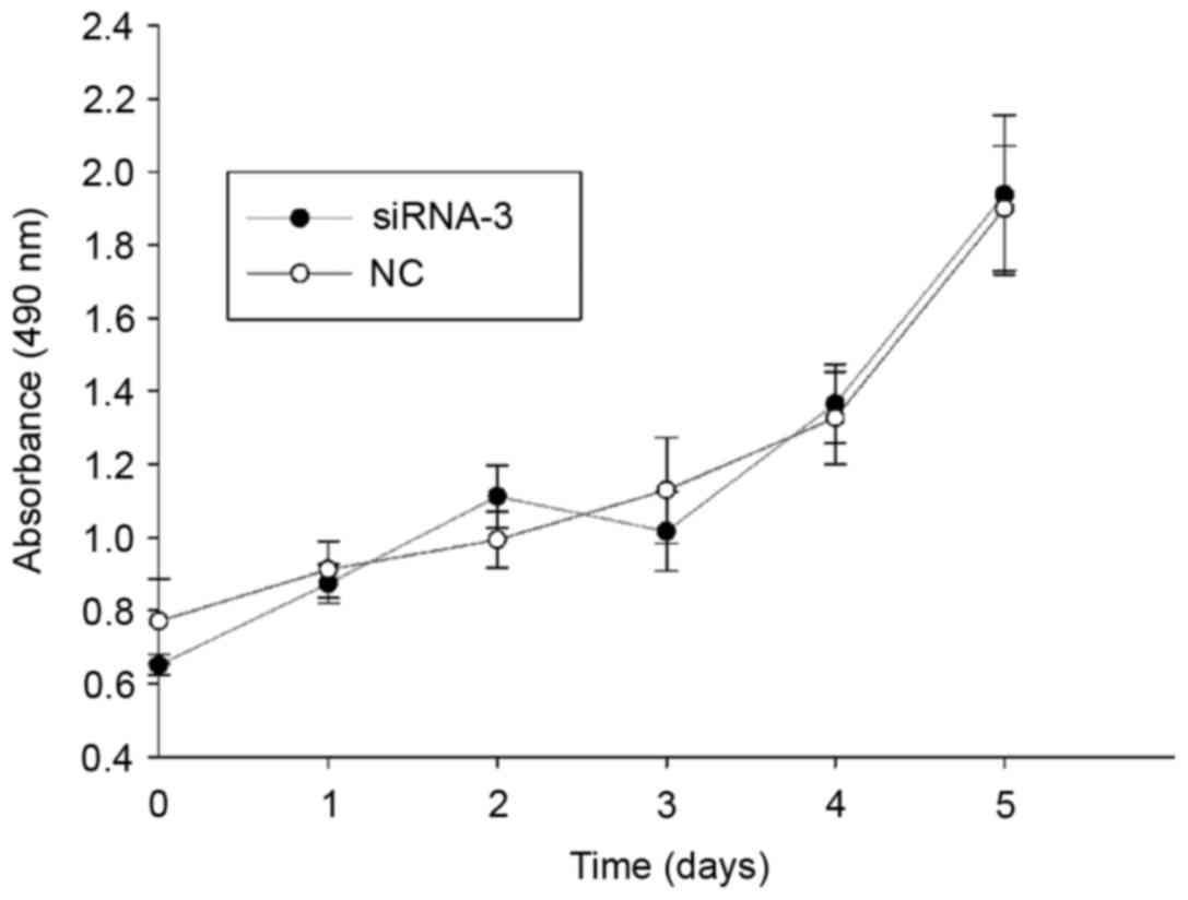
CAS1 knockdown does not inhibit cell
proliferation
To examine the effects of CAS1 on cell
proliferation, an MTS assay, using keloid fibroblasts, was
performed and cell viability was measured every 24 h (Fig. 2). The data revealed that CAS1
knockdown did not change the cell proliferative rate compared with
NC-treated cells. This indicated that CAS1 may not be involved in
cell proliferation.
CAS1 knockdown does not change the
cell mitotic index
To assess the effects of CAS1 on the cell cycle,
analysis of the cell cycle distribution 48 h after siRNA-3
transfection was conducted; this revealed no change in the cell
cycle compared with NC-treated cells (Fig. 3). This suggested that downregulation
of CAS1 expression does not alter the cell cycle in keloid
fibroblasts, which implied that CAS1 does not participate in cell
division.
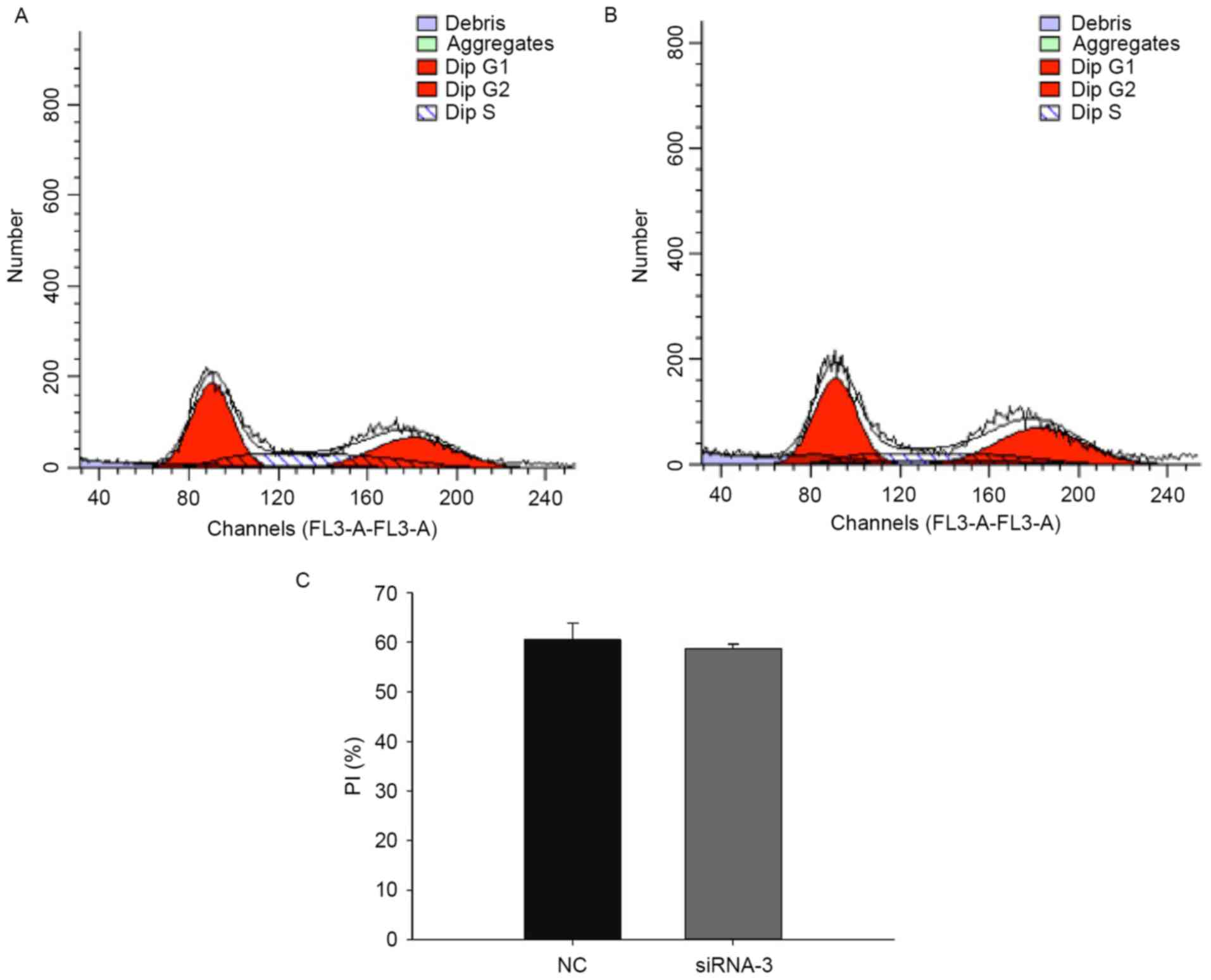 | Figure 3.Effects of CAS1 knockdown on the cell
cycle in keloid fibroblasts. (A) Cell cycle distribution in keloid
fibroblasts 48 h after transfection with the control siRNA.
G1, 39.49%; G2, 33.28%; S, 27.23%. (B) Cell
cycle distribution in keloid fibroblasts 48 h after transfection
with siRNA-3. G1, 41.31%; G2, 38.4%; S,
20.29%. (C) The PI (n=3) equals the sum of the G2 and S
phases of the cell cycle, and is expressed as a percentage. Results
are presented as the mean ± standard deviation of three independent
experiments. P>0.05, compared with NC-treated cells. CAS1,
CACNA1G-AS1; siRNA, small interfering RNA; PI, proliferation index;
FL3, flavagline; NC, negative control. |
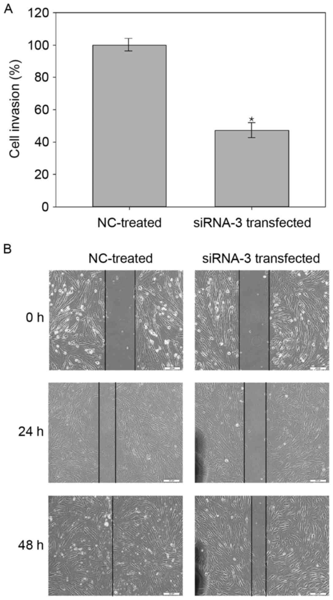
CAS1 knockdown decreases the rate of
wound closure
The scratch wound assay demonstrated that
transfected cells had a slower closure rate than NC-treated cells
(Fig. 4), suggesting that the
downregulation of CAS1 expression reduced the fibroblast migration
rate. This indicated that CAS1 may serve a role in keloid
fibroblast migration.
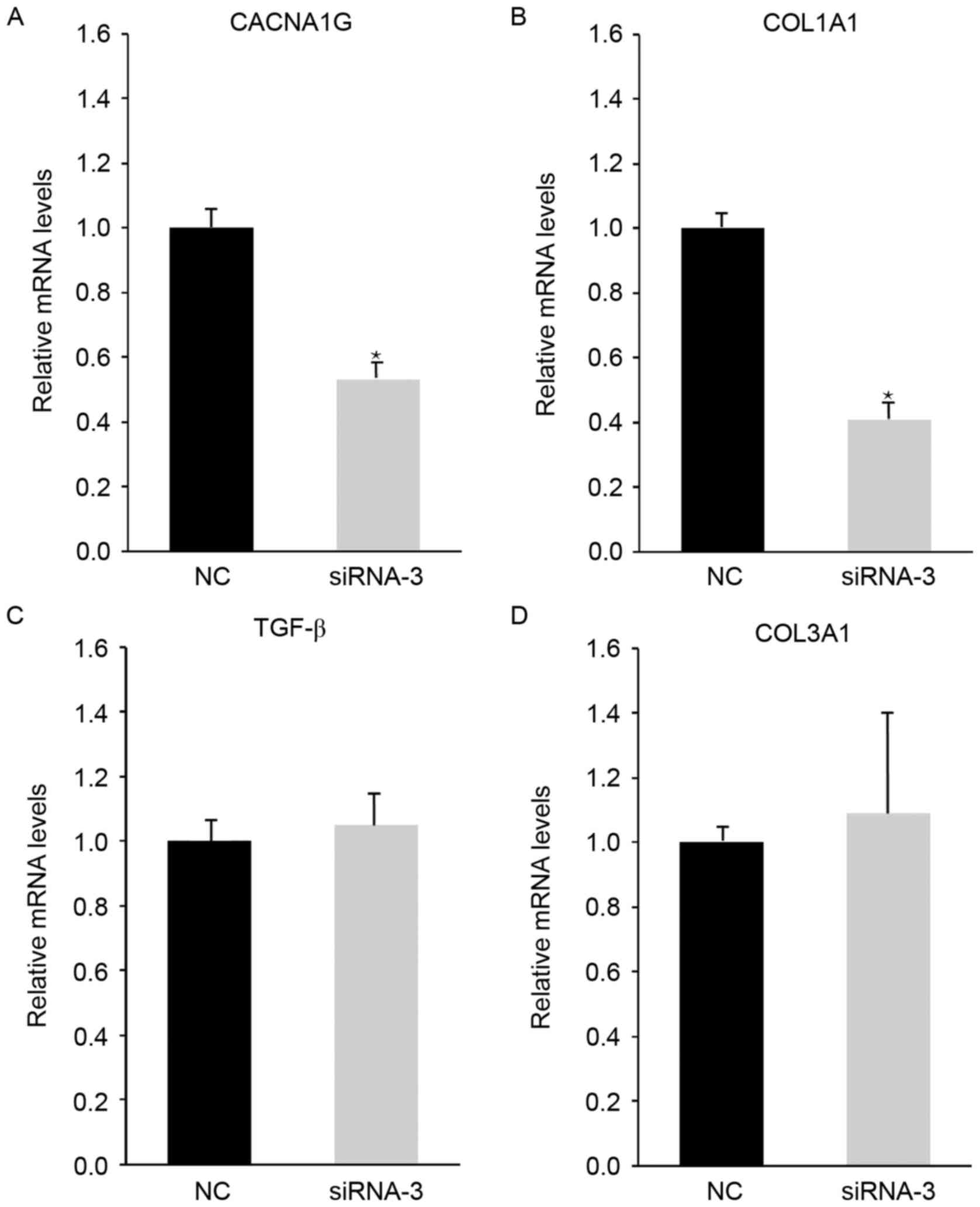
CAS1 knockdown alters CACNA1G and
COL1A1 expression
To explore the interaction between CAS1 and the
biological properties of keloid fibroblasts, analysis of
CACNA1G, TGF-β, COL1A1 and COL3A1 expression using
RT-PCR was conducted. Post-siRNA-3 transfection, CACNA1G and
COL1A1 expression decreased, while TGF-β and
COL3A1 expression was unchanged, compared with in the
NC-treated cells (Fig. 5). These
results suggested that CAS1 knockdown affects calcium channels and
collagen secretion. The absence of a notable effect on TGF-β
expression following CAS1 knockdown supports these findings, which
suggests that CAS1 does not affect the cell cycle.
Discussion
In the present study, it was demonstrated that CAS1
knockdown significantly downregulated CACNA1G and type I
collagen expression, but that it had little effect on TGF-β
and type III collagen expression. The wound healing assay also
demonstrated that CAS1 knockdown inhibited keloid fibroblast
invasion, but the MTS assay and cell cycle analysis revealed that
it had a limited impact on cell proliferation.
lncRNAs are a group of RNA molecules that range in
length from 200–100,000 nucleotides and do not encode proteins
(22). Numerous studies have
demonstrated that lncRNAs participate in various regulatory
processes, including transcriptional activation, transcriptional
interference and intranuclear transport (22–24).
lncRNAs have also been identified to serve an important role in the
development, growth and progression of human carcinomas, acting as
oncogenic drivers through diverse mechanisms, including cell
proliferation, invasion, apoptosis and the secretion of essential
proteins (25). For example, several
lncRNAs are differentially expressed in melanoma cell lines
compared with their controls. One of these lncRNAs, SPRY4-IT1, is
derived from an intron of the SPRY4 gene, and is
predominantly localized in the cytoplasm of melanoma cells, as
identified by RNA fluorescence in situ hybridization
analysis (26). SPRY4-IT1 RNA
interference impairs cell growth and differentiation, and leads to
higher rates of apoptosis in melanoma cell lines. Thus, higher
SPRY4-IT1 expression may be important to the molecular etiology of
human melanoma, and could be used as an early biomarker for
melanoma detection (26).
In an earlier study (19), microarray analysis was used to
identify differential lncRNA expression between three pairs of
keloid and normal skin tissues. A total of 1,731 lncRNAs were
identified to be consistently upregulated and 782 were
downregulated in keloids (fold-change ≥2.0). Validation of our
findings using quantitative RT-PCR revealed consistency with these
microarray results. It was also demonstrated that 11 signaling
pathways were upregulated and 44 were downregulated in keloid
tissues compared with the healthy controls. Within the
co-expression network, one lncRNA was connected with numerous
mRNAs, and vice versa. Bioinformatic analysis indicated that lncRNA
CACNA1G-AS1 may be crucial for keloid formation (19).
Numerous studies (27–29) have
demonstrated that lncRNAs may serve important roles in carcinomas
and fibrotic diseases by regulating the process of cell
proliferation, invasion, apoptosis, and the secretion of essential
proteins.
One of the metastasis-associated lncRNAs, HOTAIR,
was previously observed to be highly expressed in primary melanoma
lymph node metastases (27).
Knockdown of HOTAIR using siRNAs reduced motility and invasion of
the human melanoma cell line A375. siHOTAIR also suppressed gelatin
matrix degradation, suggesting that HOTAIR promotes gelatinase
activity. These data indicate that lncRNAs may be involved in
melanoma metastasis. In the present study, it was revealed that
CAS1 may be involved in keloid fibroblast invasion, which provided
new insights into the keloid pathology of invasion into adjacent
normal tissues.
Mounting evidence exists for the deregulation of
lncRNAs in fibrotic diseases, indicating that these molecules are
differentially expressed during fibrotic remodeling (28,29). For
example, it was recently demonstrated that Meg3 inhibits hepatic
stellate cell activation and liver fibrogenesis. In vitro,
Meg3 overexpression limits the TGF-β1-induced proliferation of
hepatic stellate cells and activates p53-dependent apoptosis in
fibrotic livers (28). Additionally,
depletion of the lncRNA ZEB2NAT was demonstrated to decrease
epithelial-mesenchymal transition (EMT)-associated gene expression
and cancer cell invasion in urinary bladder cancer-associated
fibroblasts. These results provide support for fibroblast induction
of EMT and the invasion of urinary bladder cancer cells through the
TGF-β1-ZEB2NAT axis (29). However,
no significant association was identified between CAS1 and cell
proliferation or TGF-β expression in the present study.
CAS1 is the antisense RNA of CACNA1G, which is the
mRNA of the T-type channel protein Cav3.1 (30). Numerous antisense lncRNAs have been
identified to be involved in the regulatory gene-net of diseases.
For example, proliferating cell nuclear antigen (PCNA)-AS1, the
antisense of PCNA, is significantly upregulated in hepatocellular
carcinoma. It promotes tumor growth in vitro and in
vivo through RNA hybridization, which increases PCNA mRNA
stability (31).
Clinical trials (32–34) have
demonstrated that verapamil, a calcium channel blocker that targets
L-type and T-type channels, was effective at preventing and
treating keloids with no major side effects. Calcium antagonists
are reported to promote a change in cell shape from bipolar to
spherical, which may reflect their calmodulin inhibitor-like
behavior involving calcium-independent alteration or rearrangement
of the actin cytoskeleton (35).
Alternatively, the effects of calcium antagonists may be similar to
that of cytochalasin B, which alters the cell shape by disrupting
stress fibers, and inducing the expression of collagenase and
protease (36). Further studies
(37,38) revealed that verapamil induces
procollagenase expression and increases collagenase; it also
inhibits the synthesis of extracellular matrix molecules, including
collagen, fibronectin and glycosaminoglycans. Fibroblasts in
keloids exhibit elevated levels of interleukin 6 and vascular
endothelial growth factor, which are decreased by verapamil,
reducing cell proliferation and increasing apoptosis (39). Verapamil was also indicated to prevent
keloid formation by inhibiting proliferation and TGF-β1 expression
in fibroblasts (40). The finding
that CACNA1G expression was downregulated by CAS1 knockdown
suggests that CAS1 affects calcium channel expression, leading to a
reduction in collagen levels and cell invasion.
In conclusion, CAS1 may promote the expression of
the calcium channel protein CACNA1G and type I collagen, and also
have a positive effect on cell migration in human keloid
fibroblasts, rendering it a potential new therapeutic target for
keloids.
Acknowledgements
The authors would like to thank Mr. Chongkai Li from
Tongji University (Shanghai, China), the Center of Excellence in
Tissue Engineering (Institute of Basic Medical Sciences) and the
School of Basic Medicine (Chinese Academy of Medical Sciences) for
technical support.
Funding
The present study was supported by Integrative
Medicine Talents Program of Shanghai Municipal Commission of Health
and Family Planning (grant no. ZY3-RCPY-4-2029).
Availability of data and materials
The datasets used and/or analyzed during the current
study are available from the corresponding author on reasonable
request.
Authors' contributions
YL made substantial contributions to acquisition of
data, and was involved in drafting the manuscript. XBL made
substantial contributions to conception and design. PW and XJW made
substatial contributions to interpretation of data and revised the
manuscript critically for important intellectual content. XL made
substantial contributions to analysis and interpretation of data.
ZQM made substantial contributions to conception and design, gave
final approval of the version to be published and agreed to be
accountable for all aspects of the work. All authors read and
approved the final manuscript.
Ethics approval and consent to
participate
The present study was approved by the institutional
review board at Peking Union Medical College (Beijing, China;
approval no. ZS-1301), in accordance with the principles of the
Declaration of Helsinki. Written informed consent was obtained from
all patients for the use of their keloids and normal skin.
Consent for publication
Written informed consent was obtained from all
patients for the publication of any associated data and
accompanying images.
Competing interests
The authors declare that they have no competing
interests.
Glossary
Abbreviations
Abbreviations:
|
CAS1
|
CACNA1G-AS1
|
|
lncRNA
|
long non-coding RNA
|
|
PCNA
|
proliferating cell nuclear antigen
|
|
siRNA
|
small interfering RNA
|
|
TGF-β
|
transforming growth factor-β
|
References
|
1
|
Robles DT and Berg D: Abnormal wound
healing: Keloids. Clin Dermatol. 25:26–32. 2007. View Article : Google Scholar : PubMed/NCBI
|
|
2
|
Sun LM, Wang KH and Lee YC: Keloid
incidence in Asian people and its comorbidity with other
fibrosis-related diseases: A nationwide population-based study.
Arch Dermatol Res. 306:803–808. 2014. View Article : Google Scholar : PubMed/NCBI
|
|
3
|
Huang C, Murphy GF, Akaishi S and Ogawa R:
Keloids and hypertrophic scars: Update and future directions. Plast
Reconstr Surg Glob Open. 1:e252013. View Article : Google Scholar : PubMed/NCBI
|
|
4
|
Bock O, Schmid-Ott G, Malewski P and
Mrowietz U: Quality of life of patients with keloid and
hypertrophic scarring. Arch Dermatol Res. 297:433–438. 2006.
View Article : Google Scholar : PubMed/NCBI
|
|
5
|
Ud-Din S, Volk SW and Bayat A:
Regenerative healing, scar-free healing and scar formation across
the species: Current concepts and future perspectives. Exp
Dermatol. 23:615–619. 2014. View Article : Google Scholar : PubMed/NCBI
|
|
6
|
Lee JY, Yang CC, Chao SC and Wong TW:
Histopathological differential diagnosis of keloid and hypertrophic
scar. Am J Dermatopathol. 26:379–384. 2004. View Article : Google Scholar : PubMed/NCBI
|
|
7
|
Huang C, Akaishi S, Hyakusoku H and Ogawa
R: Are keloid and hypertrophic scar different forms of the same
disorder? A fibroproliferative skin disorder hypothesis based on
keloid findings. Int Wound J. 11:517–522. 2014. View Article : Google Scholar : PubMed/NCBI
|
|
8
|
Mofikoya BO, Adeyemo WL and Abdus-salam
AA: Keloid and hypertrophic scars: A review of recent developments
in pathogenesis and management. Nig Q J Hosp Med. 17:134–139.
2007.PubMed/NCBI
|
|
9
|
Ogawa R, Okai K, Tokumura F, Mori K,
Ohmori Y, Huang C, Hyakusoku H and Akaishi S: The relationship
between skin stretching/contraction and pathologic scarring: The
important role of mechanical forces in keloid generation. Wound
Repair Regen. 20:149–157. 2012. View Article : Google Scholar : PubMed/NCBI
|
|
10
|
Naylor MC and Brissett AE: Current
concepts in the etiology and treatment of keloids. Facial Plast
Surg. 28:504–512. 2012. View Article : Google Scholar : PubMed/NCBI
|
|
11
|
Jin Z: Increased c-Met phosphorylation is
related to keloid pathogenesis: Implications for the biological
behaviour of keloid fibroblasts. Pathology. 46:25–31. 2014.
View Article : Google Scholar : PubMed/NCBI
|
|
12
|
Ogawa R, Akaishi S, Huang C, Dohi T, Aoki
M, Omori Y, Koike S, Kobe K, Akimoto M and Hyakusoku H: Clinical
applications of basic research that shows reducing skin tension
could prevent and treat abnormal scarring: The importance of
fascial/subcutaneous tensile reduction sutures and flap surgery for
keloid and hypertrophic scar reconstruction. J Nippon Med Sch.
78:68–76. 2011. View Article : Google Scholar : PubMed/NCBI
|
|
13
|
Akaishi S, Akimoto M, Ogawa R and
Hyakusoku H: The relationship between keloid growth pattern and
stretching tension: Visual analysis using the finite element
method. Ann Plast Surg. 60:445–451. 2008. View Article : Google Scholar : PubMed/NCBI
|
|
14
|
Nakashima M, Chung S, Takahashi A,
Kamatani N, Kawaguchi T, Tsunoda T, Hosono N, Kubo M, Nakamura Y
and Zembutsu H: A genome-wide association study identifies four
susceptibility loci for keloid in the Japanese population. Nat
Genet. 42:768–771. 2010. View
Article : Google Scholar : PubMed/NCBI
|
|
15
|
Arima J, Huang C, Rosner B, Akaishi S and
Ogawa R: Hypertension: A systemic key to understanding local keloid
severity. Wound Repair Regen. 23:213–221. 2015. View Article : Google Scholar : PubMed/NCBI
|
|
16
|
Park TH and Chang CH: Keloid recurrence in
pregnancy. Aesthetic Plast Surg. 36:1271–1272. 2012. View Article : Google Scholar : PubMed/NCBI
|
|
17
|
Fong EP and Bay BH: Keloids-the sebum
hypothesis revisited. Med Hypotheses. 58:264–269. 2002. View Article : Google Scholar : PubMed/NCBI
|
|
18
|
Ichioka S, Ando T, Shibata M, Sekiya N and
Nakatsuka T: Oxygen consumption of keloids and hypertrophic scars.
Ann Plast Surg. 60:194–197. 2008. View Article : Google Scholar : PubMed/NCBI
|
|
19
|
Liang X, Ma L, Long X and Wang X: LncRNA
expression profiles and validation in keloid and normal skin
tissue. Int J Oncol. 47:1829–1838. 2015. View Article : Google Scholar : PubMed/NCBI
|
|
20
|
Russell SB, Russell JD, Trupin KM, Gayden
AE, Opalenik SR, Nanney LB, Broquist AH, Raju L and WIlliams SM:
Epigenetically altered wound healing in keloid fibroblasts. J
Invest Dermatol. 130:24892010. View Article : Google Scholar : PubMed/NCBI
|
|
21
|
Livak KJ and Schmittgen TD: Analysis of
relative gene expression data using real-time quantitative PCR and
the 2(-Delta Delta C(T)) method. Methods. 25:402–408. 2001.
View Article : Google Scholar : PubMed/NCBI
|
|
22
|
Gibb EA, Brown CJ and Lam WL: The
functional role of long non-coding RNA in human carcinomas. Mol
Cancer. 10:382011. View Article : Google Scholar : PubMed/NCBI
|
|
23
|
Vance KW and Ponting CP: Transcriptional
regulatory functions of nuclear long noncoding RNAs. Trends Genet.
30:348–355. 2014. View Article : Google Scholar : PubMed/NCBI
|
|
24
|
Kung JT, Colognori D and Lee JT: Long
noncoding RNAs: Past, present, and future. Genetics. 193:651–669.
2013. View Article : Google Scholar : PubMed/NCBI
|
|
25
|
Prensner JR and Chinnaiyan AM: The
emergence of lncRNAs in cancer biology. Cancer Discov. 1:391–407.
2011. View Article : Google Scholar : PubMed/NCBI
|
|
26
|
Khaitan D, Dinger ME, Mazar J, Crawford J,
Smith MA, Mattick JS and Perera RJ: The melanoma-upregulated long
noncoding RNA SPRY4-IT1 modulates apoptosis and invasion. Cancer
Res. 71:3852–3862. 2011. View Article : Google Scholar : PubMed/NCBI
|
|
27
|
Tang L, Zhang W, Su B and Yu B: Long
noncoding RNA HOTAIR is associated with motility, invasion, and
metastatic potential of metastatic melanoma. Biomed Res Int.
2013:2510982013. View Article : Google Scholar : PubMed/NCBI
|
|
28
|
He Y, Wu YT, Huang C, Meng XM, Ma TT, Wu
BM, Xu FY, Zhang L, Lv XW and Li J: Inhibitory effects of long
noncoding RNA MEG3 on hepatic stellate cells activation and liver
fibrogenesis. Biochim Biophys Acta. 1842:2204–2215. 2014.
View Article : Google Scholar : PubMed/NCBI
|
|
29
|
Zhuang J, Lu Q, Shen B, Huang X, Shen L,
Zheng X, Huang R, Yan J and Guo H: TGFβ1 secreted by
cancer-associated fibroblasts induces epithelial-mesenchymal
transition of bladder cancer cells through lncRNA-ZEB2NAT. Sci Rep.
5:119242015. View Article : Google Scholar : PubMed/NCBI
|
|
30
|
Fukunaga K: Cognitive function and
calcium. Cognitive improvement through T type calcium channel
stimulation. Clin Calcium. 25:247–254. 2015.(In Japanese).
PubMed/NCBI
|
|
31
|
Yuan SX, Tao QF, Wang J, Yang F, Liu L,
Wang LL, Zhang J, Yang Y, Liu H, Wang F, et al: Antisense long
non-coding RNA PCNA-AS1 promotes tumor growth by regulating
proliferating cell nuclear antigen in hepatocellular carcinoma.
Cancer Lett. 349:87–94. 2014. View Article : Google Scholar : PubMed/NCBI
|
|
32
|
D'Andrea F, Brongo S, Ferraro G and Baroni
A: Prevention and treatment of keloids with intralesional
verapamil. Dermatology. 204:60–62. 2002. View Article : Google Scholar : PubMed/NCBI
|
|
33
|
Wang R, Mao Y, Zhang Z, Li Z, Chen J and
Cen Y: Role of verapamil in preventing and treating hypertrophic
scars and keloids. Int Wound J. 13:461–468. 2016. View Article : Google Scholar : PubMed/NCBI
|
|
34
|
Alexandrescu D, Fabi S, Yeh LC,
Fitzpatrick RE and Goldman MP: Comparative results in treatment of
keloids with intralesional 5-FU/Kenalog, 5-FU/Verapamil, Enalapril
Alone, verapamil alone, and laser: A case report and review of the
literature. J Drugs Dermatol. 15:1442–1447. 2016.PubMed/NCBI
|
|
35
|
Grossman E and Messerli FH: Calcium
antagonists. Prog Cardiovasc Dis. 47:34–57. 2004. View Article : Google Scholar : PubMed/NCBI
|
|
36
|
Lee RC, Doong H and Jellema AF: The
response of burn scars to intralesional verapamil. Report of five
cases. Arch Surg. 129:107–111. 1994. View Article : Google Scholar : PubMed/NCBI
|
|
37
|
Doong H, Dissanayake S, Gowrishankar TR,
LaBarbera MC and Lee RC: The 1996 Lindberg Award. Calcium
antagonists alter cell shape and induce procollagenase synthesis in
keloid and normal human dermal fibroblasts. J Burn Care Rehabil.
17:497–514. 1996. View Article : Google Scholar : PubMed/NCBI
|
|
38
|
Lee RC and Ping JA: Calcium antagonists
retard extracellular matrix production in connective tissue
equivalent. J Surg Res. 49:463–466. 1990. View Article : Google Scholar : PubMed/NCBI
|
|
39
|
Giugliano G, Pasquali D, Notaro A, Brongo
S, Nicoletti G, D'Andrea F, Bellastella A and Sinisi AA: Verapamil
inhibits interleukin-6 and vascular endothelial growth factor
production in primary cultures of keloid fibroblasts. Br J Plast
Surg. 56:804–809. 2003. View Article : Google Scholar : PubMed/NCBI
|
|
40
|
Xu SJ, Teng JY, Xie J, Shen MQ and Chen
DM: Comparison of the mechanisms of intralesional steroid,
interferon or verapamil injection in the treatment of proliferative
scars. Zhonghua Zheng Xing Wai Ke Za Zhi. 25:37–40. 2009.(In
Chinese). PubMed/NCBI
|