Introduction
According to Chinese cancer statistics in 2015
(1), thyroid cancer is the most
commonly diagnosed cancer type in women aged <30 years, followed
by breast cancer at age 30–59 years. Invasion and metastasis are
the leading causes of mortality in patients with breast cancer
(2). Therefore, in addition to
surgery, chemotherapy, radiotherapy and targeted therapy
treatments, finding effective ways to improve the treatment of
patients with cancer is of primary concern.
Vasodilator-stimulated phosphoprotein (VASP), a
member of the Enabled (Ena)/VASP family, is an actin-associated
protein. VASP is mainly distributed at locations associated with
adhesion and migration, including the anterior border of the
lamellar parapodium, filopodia cacumen and adhesion plaque, and has
a highly dynamic position in the cellular membrane (3). It is involved in regulating actin
filament rearrangement in the process of cell adhesion and
expansion, and is associated with cell adhesion, cytomorphosis and
mobility (4,5). According to Krause et al
(3), an increase in VASP expression
promotes the movement of the Listeria bacteria in
vitro. VASP induces the assembling of actin via binding of
ActA, which accelerates the movement of the Listeria
bacterium inside cells. Furthermore, Sechi et al (6) reported that deleting the domain of ActA
that directly combines with VASP, effectively suppresses the
movement of the Listeria bacterium inside cells. In
eukaryotic cells, the expression levels of VASP are positively
correlated with the formation speed of lamellipodia in mouse
melanoma B16-F1 cells (6). Notably,
overexpression of VASP in wild-type NIH 3T3 cells resulted in
malignant transformation of the cells (7). It has also been revealed that increased
expression levels of VASP are correlated with decreased cellular
differentiation and an increased pathological stage of lung tumors
(8). These data suggested that VASP
may serve a crucial role in regulating cellular migration, in
addition to tumorigenesis and development.
Rho GTPases [Rho, Ras-related C3 botulinum toxin
substrate (Rac)1 and cell division control protein 42 homolog
(Cdc42)] are important signaling molecules that are involved in
regulating cell migration through cellular cytoskeletal
rearrangement (9,10). The activation of RAC1, one member of
the family, promotes the recruitment of the integrin family,
participates in the assembling of the cytoskeleton, and induces the
accumulation and polarization of tumor invasion (11). However, as a key factor in the
regulation of cell migration, the relative contribution of RAC1 to
cytoskeletal rearrangement depends on specific conditions and cell
type. It was revealed that there were no significant changes in
cellular migratory abilities in RAC1-deficient rat fibroblasts and
macrophages (12). However,
downregulation of RAC1 was demonstrated to result in the inhibition
of migration in various types of cells, including gastric,
testicular and colorectal carcinoma cells (13). Notably, regarding the association
between RAC1 and VASP, Fryer et al (14) reported that in human umbilical vein
endothelial cells and HeLa cells, p21-activated kinase (PAK; serine
21), a downstream molecule of RAC1 phosphorylated by cGMP-dependent
protein kinase (PKG), promoted the formation of Pak/VASP and
thereby affected cell motility.
Therefore, the present study focused on examining
the role of RAC1 and VASP in the development of breast cancer, by
analyzing data in Kaplan, Oncomine and The Cancer Genome Atlas
(TCGA), and using in vitro experiments in breast cancer
MCF-7 cells.
Materials and methods
Online databases
Patients with breast cancer were divided into a low
expression level group and a high expression level group, according
to the median RAC1 or VASP mRNA expression levels (15). Data from the Kaplan (http://kmplot.com/analysis/index.php?p=service&cancer=breast)
(Data set no. Affy ID, 202205_at VASP; Affy ID, 208640_at p21-RAC1,
RAC1 and TC-2), Oncomine (https://www.oncomine.com/resource/login.html) and TCGA
(http://xena.ucsc.edu/public-hubs/)
databases were used for correlation analysis of different gene
expression levels and patient clinical information.
Analysis of the association between
VASP, RAC1 and molecular subtypes of breast cancer based on data
from the TCGA database
The clinical information data of patients with
breast cancer were collected from TCGA and analyzed using UCSC Xena
(http://xena.ucsc.edu/#analyze;
University of California, Santa Cruz, CA, USA). The mRNA expression
level of RAC1 and VASP in normal breast cancer tissue was the
lowest, increasing gradually in the Luminal A, Luminal B, BLBC and
Her-2 enrichment subtype groups.
Cell culture
The breast carcinoma MCF-7 cell line and the 293T
cell line were purchased from the Department of Pathophysiology,
Medical College of Wuhan University (Wuhan, China). Cells were
cultured in Dulbecco's modified Eagle's medium (DMEM; Hyclone; GE
Healthcare, Logan, UT, USA) supplemented with 10% newborn calf
serum (Gibco; Thermo Fisher Scientific, Inc., Waltham, MA, USA) and
1% antibiotics (100 U/ml penicillin G and 100 U/ml streptomycin
sulfate) at 37°C in an environment containing 5% CO2.
Cells were passaged at 70–80% confluence using 0.02% EDTA in PBS
and 0.25% trypsin.
Construction of short hairpin (shRNA)
MCF-7 stable cell lines
RAC1 and VASP stable knockdown in the MCF-7 cell
line was established via lentiviral-based stable shRNA
overexpression. The target sequences for RAC1,
5′-GATCCGGAAGGAGATTGGTGCTGTAAAACTCGAGAAAATGTCGTGGTTAGAGGAATTTTTG-3′;
VASP,
5′-CCGGAGGAATTGCAGAAAGTGAAAGCTCGAGCTTTCACTTTCTGCAATTCCTTTTTTG-3′;
and shRNA control, 5′-CAACAAGATGAAGAGCACCAA-3′ were designed by
Sigma-Aldrich; Merck KGaA (Darmstadt, Germany) and constructed into
lentiviral-based green fluorescent protein-shRNA expression
plasmids, pLVshRNA-EGFP-Puro (Beijing Inovogen Tech Co., Ltd.,
Beijing, China). The lentiviruses (4 µg) were packaged into the
293T cell line. The cell culture supernatant was collected 48 and
72 h after transfection, centrifuged at 3,000 × g for 15 min,
filtered through a 0.45-µm polyvinylidene difluoride filter
(Acrodisc) to remove cellular debris, aliquoted into Eppendorf
tubes and stored at−70°C as the viral solution for use. MCF-7 cells
were plated in a 96-well plate at a density of 1×10⁴
cells/well. After 24 h, the viral solution was added to the MCF-7
cells and 10 µg/ml Polybrene (Sigma Aldrich; Merck KGaA) was added
to increase the efficiency of infection. The medium containing the
virus was removed 12 h later and replaced with 200 µl DMEM
supplemented with 10% fetal bovine serum. The cell lines were
screened using cell medium with 2 µg/ml puromycin. The infection
efficiency of the virus was further confirmed by fluorescence
microscopy (magnification, ×200). The interference efficiency of
the cells was detected by reverse transcription-polymerase chain
reaction (RT-PCR) and western blotting.
RT-PCR
To determine the mRNA expression level of RAC1 and
VASP in MCF-7 cells, total RNA was purified using a total RNA
isolation system (Promega Corporation, Madison, WI, USA) and RT-PCR
was performed using the Access RT-PCR system, according to the
manufacturer's protocol (Promega Corporation). The primers used
were as follows: RAC1 forward, 5′ATGTCCGTGCAAAGTGGTATC3′ and
reverse, 5′CTCGGATCGCTTCGTCAAACA3′; VASP forward,
5′-CTGGGAGAAGAACAGCACAACC-3′ and reverse,
5′-AGGTCCGAGTAATCACTGGAGC-3′; GAPDH forward,
5′-TGATGACATCAAGAAGGTGGTGAAG-3′ and reverse,
5′-TCCTTGGAGGCCATGTGGGCCAT-3′. The thermocycling conditions were as
follows: RAC1, 94°C for 40 sec; 54°C for 30 sec and 72°C for 30 sec
for 30 cycles; VASP, 94°C for 40 sec; 60°C for 30 sec and 72°C for
30 sec for 30 cycles. GAPDH was employed as an internal control for
mRNA and protein analyses.
Western blot analysis
The MCF-7 cells were washed twice with PBS and lysed
in ice-cold radioimmunoprecipitation assay buffer (1.5 mM
MgCl2, 1 mM EGTA, 1 mM PMSF, 10 mM NaVO3 and
10 mM PIC), using 100 µl lysis buffer per well. Following 10 min on
ice, the lysates were transferred into microcentrifuge tubes and
centrifuged at 12,000 × g for 15 min at 4°C. Following 12% SDS-PAGE
separation, proteins were electrotransferred to a nitrocellulose
membrane for 90 min for RAC1 and GAPDH, and 120 min for VASP.
Subsequent to blocking with 5% BSA in Tris-buffered saline (TBS) at
37°C for 1 h, the bands were excised according to the protein
marker (PageRuler Prestained Protein Ladder; cat. no. 00575411;
Thermo Fisher Scientific, Inc.) protocol, and membrane strips were
incubated with the following primary antibodies: Anti-RAC1 (cat.
no. 610651; dilution, 1:1,000; BD Biosciences, Franklin Lakes, NJ,
USA), anti-VASP (cat. no. 0010-10; dilution, 1:1,000; ImmunoGlobe
GmbH, Himmelstadt, Germany) and anti-GAPDH (cat. no. SC-69778;
dilution, 1:5,000; Santa Cruz Biotechnology, Inc., Dallas, TX, USA)
overnight at 4°C. Subsequent to 5 washes in TBS with Tween-20
(TBST) for 10 min, the membrane strips were incubated with
dilutions (1:10,000) of horseradish peroxidase-conjugated goat
anti-mouse IgG (cat. no. 16473-1-AP; ProteinTech Group, Inc.,
Chicago, IL, USA) and anti-mouse IgG (cat. no. 10285-1-AP,
ProteinTech Group, Inc.) at 37°C for 2 h, and washed with 5 times
in TBST for 10 min. Following a further washing step, an enhanced
chemiluminescence immune enzymatic reaction (Beijing Zoman
Biotechnology Co., Ltd., Beijing, China) was performed to view the
bands.
Scratch wound healing motility assays
(2D-cell migration assays)
Cells were cultured as mentioned earlier. Cells were
seeded on 6-well plates and allowed to grow to 100% confluency.
Confluent monolayers were scratched with a pipette tip and
maintained in serum-free DMEM medium for 12 h and maintained in an
incubator at 37°C. The plates were washed once with fresh medium to
remove non-adherent cells and images were captured using an
inverted phase-contrast microscope (magnification, ×200; Olympus
Corporation, Tokyo, Japan). The cell migration rate was evaluated
by calculating the average cell migration distance in 48 h.
Transwell assays (3D-cell migration
assays)
A migration assay was performed using the Transwell
system, which allows cells to migrate through a polycarbonate
membrane with 8-µm pores. Cells were trypsinized, washed and
resuspended in serum-free DMEM at 1×10⁶ cells/ml. This
suspension (100 µl) was added to the upper chamber of the
Transwell. After an 8-h incubation at 37°C in the presence of 5%
CO2, the lower chamber was filled with 500 µl DMEM
supplemented with 20% calf serum. Following incubation for 18 h at
37°C in the presence of 5% CO2, the cells were fixed for
30 min in 4% formaldehyde and stained at room temperature for 15
min with 5% crystal violet. The filters were then rinsed thoroughly
in distilled water and checked by bright-field microscopy
(magnification, ×200) to ensure that the cells were adherent and
had migrated. The non-migrating cells were then carefully removed
from the upper surface (inside) of the Transwell with a wet cotton
swab. To quantify cell motility, cells that had migrated to the
bottom surface of the filter were counted. A total of nine evenly
spaced fields of cells were counted in each well, using an inverted
phase-contrast microscope at magnification, ×200.
Cell Counting kit-8 (CCK-8) assay to
detect apoptosis
Cells in the logarithmic growth phase were seeded
onto 96-well cell culture plates at a density of ~4×10³
cells/l, and 100 µl complete DMEM was added to each well. A total
of 6 parallel wells were set up and the cells were incubated in a
37°C-incubator for 24 h. A total of 10 µl CCK-8 solution (Beijing
Zoman Biotechnology Co., Ltd.) diluted in 90 µl medium was added to
each well. The cells were incubated at 37°C for 2 h, and the
absorbance nm was measured at a wavelength of 450 using a
microplate reader.
Flow cytometry to detect
apoptosis
The RAC1-shRNA and negative control (NC)-shRNA MCF-7
cells were cultured in a 6-well plate until the cells reached
60–70% confluency. A total of 1–5×10⁵ cells were
digested with EDTA-free trypsin and 500 µl binding buffer (Beyotime
Institute of Biotechnology, Haimen, China) to resuspend the cells.
A total of 5 µl Annexin V-fluorescein isothiocyanate and 10 µl
propidium iodide (both Beyotime Institute of Biotechnology) was
added to each tube, and these were incubated at room temperature
for 5 min prior to flow cytometry.
Statistical analysis
Data are expressed as the mean ± standard error of
the mean. Differences between groups were analyzed using one-way
analysis of variance followed by a Bonferroni's post-hoc test.
Kaplan-Meier analysis, followed by a Log rank test was performed to
determine survival rates (http://kmplot.com/analysis/index.php?p=service&cancer).
The data of RAC1 and VASP mRNA expression in breast cancer tissues,
and the clinical information of patients with breast cancer were
collected from TCGA and analyzed using the UCSC Xena database
(http://xena.ucsc.edu/#analyze;
University of California, Santa Cruz, CA, USA). The mRNA expression
levels of RAC1 and VASP were quantified. P<0.05 was considered
to indicate a statistically significant difference.
Results
Expression of RAC1 and VASP mRNA is
increased in breast cancer tissue data obtained from online
databases
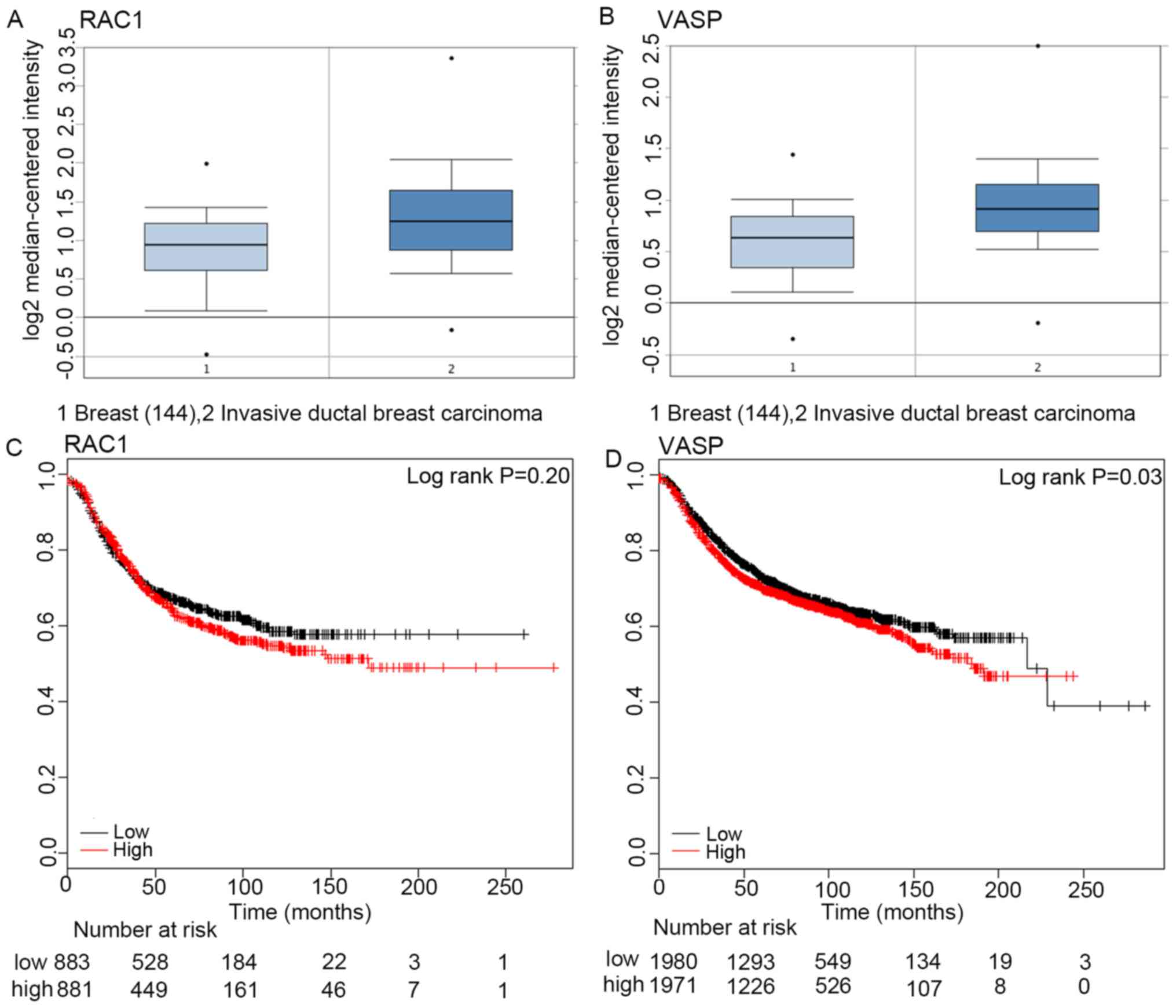
According to the data from Oncomine, the expression
of RAC1 and VASP at the mRNA level in invasive ductal carcinoma
tissues (1,556 cases) was significantly increased compared with
that in control tissues (144 cases; P<0.05, Fig. 1A and B). Furthermore, the patients
with breast cancer were divided into a low expression level group
and a high expression level group, according to the median RAC1 or
VASP mRNA expression levels (15).
The mortality rate of patients with breast cancer at 50 months in
the low RAC1 expression group (883 cases) was higher than that in
the high expression group (881 cases; 40.2% vs. 49%; log rank,
P>0.05; Fig. 1C). Correspondingly,
the mortality rate of patients with breast cancer at 50 months in
the in the high VASP expression group (1,971 cases) was
significantly increased compared with the low expression level
group (1,980 cases) (34.7% vs. 37.8%, log rank P<0.05; Fig. 1D). This indicated that increased VASP
mRNA expression levels were positively associated with poor
prognosis in patients with breast cancer.
Association between the mRNA
expression level of RAC1 or VASP in breast cancer tissues, and the
subtype and staging of breast cancer
The mRNA expression levels of RAC1 and VASP in
breast cancer tissues, and the clinical information of patients
with breast cancer, were collected from TCGA and analyzed using
UCSC Xena (http://xena.ucsc.edu/#analyze; University of
California, Santa Cruz, CA, USA). The mRNA expression levels of
RAC1 and VASP were quantified.
The Prosigna breast Cancer Prognstic Gene Signature
Assay measures the expression level of 50 genes in surgically
resected breast cancer samples to classify a tumor as one of four
intrinsic subtypes (Luminal A, Luminal B, HER2-enriched, and
Basal-like) (16). According to the
Prosigna breast Cancer Prognstic Gene Signature Assay
classification criteria, breast cancer is divided into Luminal A,
Luminal B, Her-2 enrichment and Basal-like breast cancer (BLBC)
subtypes. The control was the Normal group, in which the RAC1 mRNA
expression level was the lowest, increasing gradually in the
Luminal A, Luminal B, BLBC and Her-2 enrichment subtype groups.
Furthermore, the expression in the Luminal A subtype group was
significantly decreased compared with the BLBC subtype and Her-2
subtype groups (P<0.05; Fig. 2A).
The expression in the Luminal B subtype group was significantly
decreased compared with the Her-2 enrichment subtype group
(Fig. 2A; P<0.05).
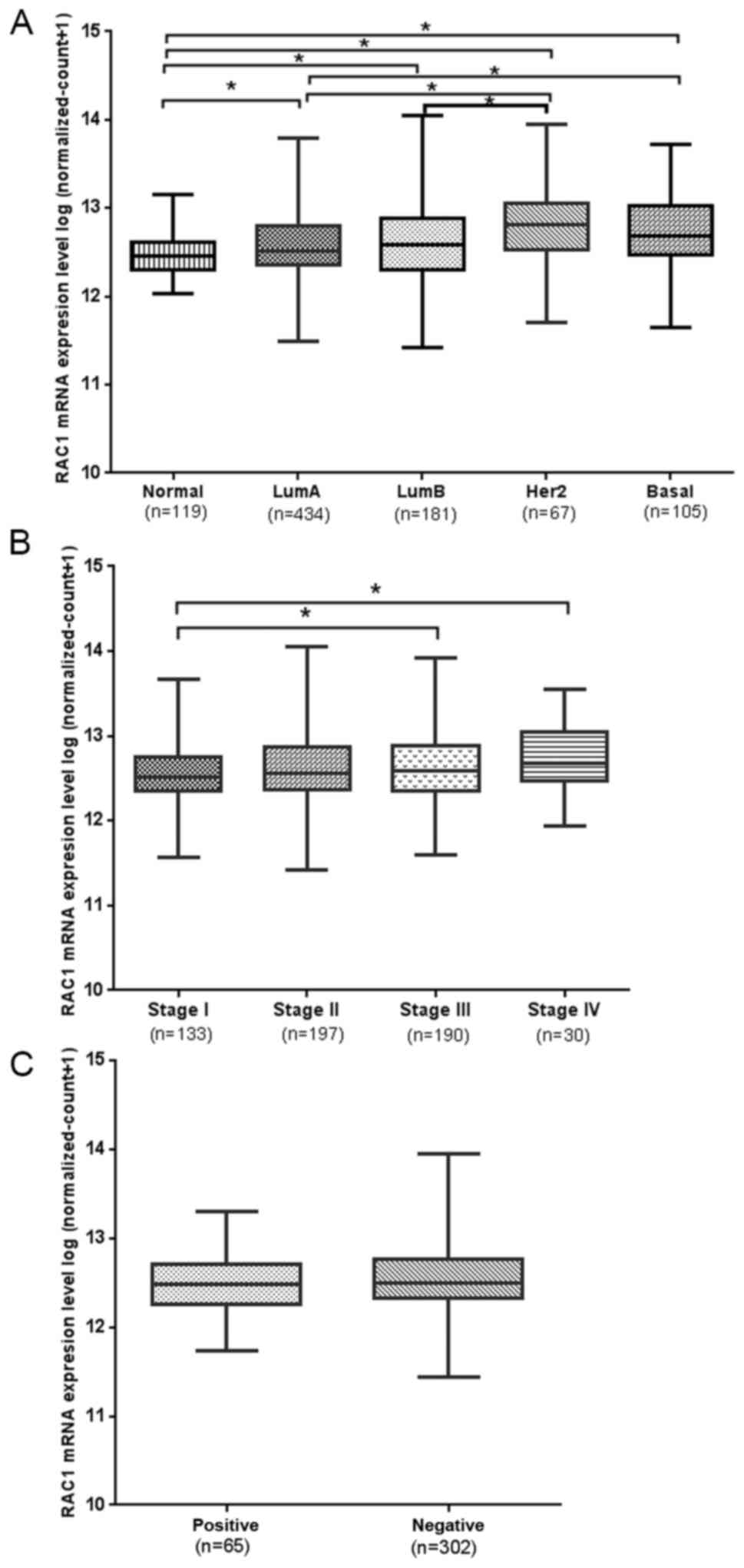 | Figure 2.RAC1 mRNA expression in tumor tissue,
and its association with the classification, staging and lymph node
metastasis of breast cancer. (A) RAC1 mRNA expression in LumA,
LumB, Her2, Basal and Normal tissue. *P<0.05. (B) American Joint
Committee on Cancer staging and RAC1 mRNA expression. *P<0.05.
(C) RAC1 mRNA expression in lymph node metastasis-positive and
-negative breast cancer tissues. RAC1, Ras-related C3 botulinum
toxin substrate 1; LumA, luminal A; LumB, luminal B; Her2, Her-2
enrichment; Basal, basal-like breast cancer. |
According to the American Joint Committee on Cancer
(AJCC) classification criteria (17,18), the
pathological staging of breast cancer was divided into 4 stages,
and the mRNA expression level of RAC1 gradually increased from
stage I–IV. The mRNA expression level of RAC1 in stage I was
decreased compared with stages III and IV, with a significant
difference (P<0.05; Fig. 2B).
However, elevated RAC1 mRNA expression was not associated with
lymph node metastasis (P>0.05; Fig.
2C).
The mRNA expression level of VASP in the Normal
group was the lowest, and its expression level gradually increased
in the Luminal B, Luminal A, BLBC and Her-2 subtype groups. The
mRNA expression level of VASP in the Luminal A subtype was
significantly decreased compared with the Her-2-enriched and BLBC
subtypes. However, it was increased compared with the Luminal B
group (P<0.05; Fig. 3A). The mRNA
expression level of VASP in the Luminal B subtype was significantly
decreased compared with the Her-2 enriched subtype (P<0.05;
Fig. 3A). The expression of VASP mRNA
gradually increased from stage I–IV (P>0.05; Fig. 3B), however, elevated VASP mRNA
expression was not associated with AJCC staging or lymph node
metastasis (P>0.05; Fig. 3C).
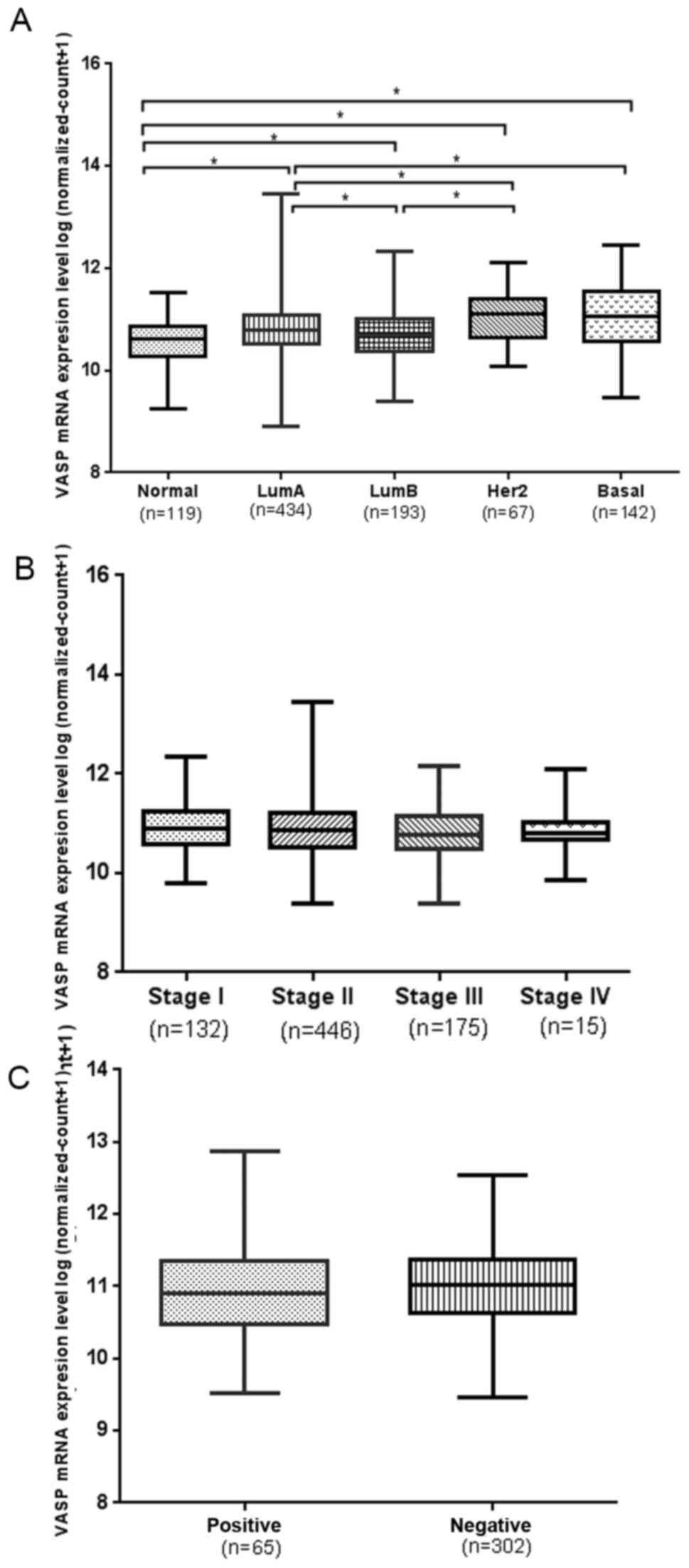 | Figure 3.VASP mRNA expression in tumor tissue,
and its association with the classification, staging and lymph node
metastasis of breast cancer. (A) VASP mRNA expression in LumA,
LumB, Her2, Basal and Normal tissue. *P<0.05. (B) American Joint
Committee on Cancer staging and VASP mRNA expression. P>0.05.
(C) VASP mRNA expression in lymph node metastasis-positive and
-negative breast cancer tissues. VASP, vasodilator-stimulated
phosphoprotein; LumA, luminal A; LumB, luminal B; Her2, Her-2
enrichment; Basal, basal-like breast cancer. |
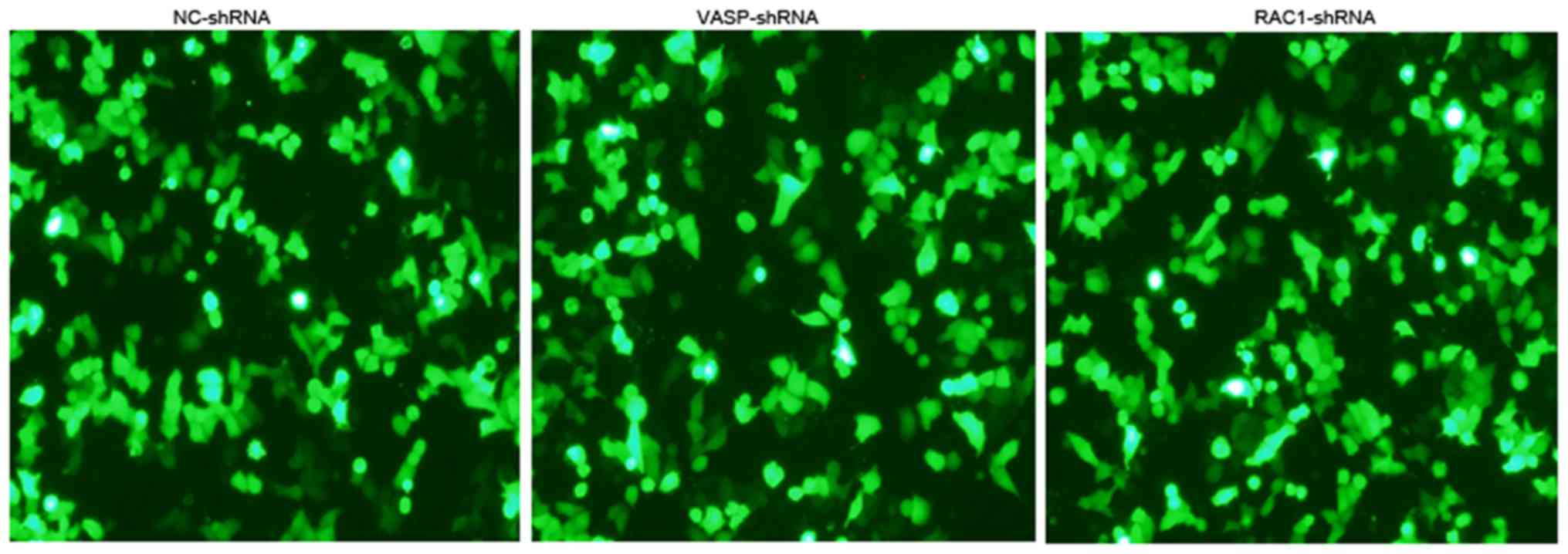
VASP-shRNA interference in MCF-7
cells
The shRNA against the human VASP gene, or NC-shRNA
were stably transfected into MCF-7 cells. Fluorescence microscopy
revealed green fluorescence in the cytoplasm and nuclei, indicating
successful transfection (Fig. 4). The
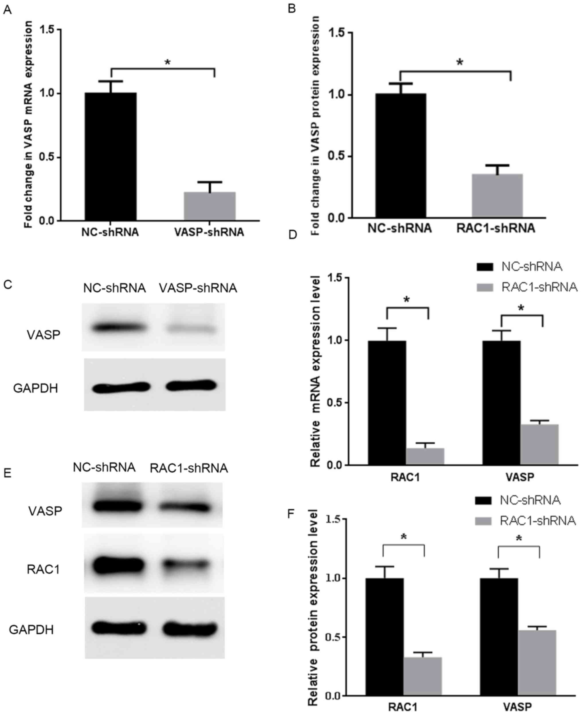
expression level of VASP was detected on the mRNA and protein
levels by RT-qPCR and western blotting, as presented in (Fig. 5A-C). Compared with the stable NC-shRNA
MCF-7 cells, the inhibition rates of mRNA and protein expression
levels of VASP in the stable VASP-shRNA MCF-7 cells were 78 and
65%, respectively (P<0.05). These data indicated the
establishment of stable VASP-shRNA MCF-7 cells.
RAC1-shRNA interference in MCF-7
cells
The shRNA against RAC1 was stably transfected into
MCF-7 cells. Fluorescence microscopy revealed the presence of green
fluorescence in the cytoplasm and nuclei (Fig. 4). The expression of RAC1 and VASP in
the cells was detected on the mRNA and protein levels by RT-qPCR
and western blotting (Fig. 5D-F).
Compared with the stable NC-shRNA MCF-7 cells, the of the RAC1 and
VASP mRNA expression-inhibition rate in the stable RAC1-shRNA MCF-7
cells was 86% (P<0.05) and 67% (P<0.05), respectively. The
RAC1 and VASP protein expression-inhibition rate of the stable
RAC1-shRNA MCF-7 cells was 67% (P<0.05) and 44% (P<0.05),
respectively. These data indicated the establishment of stable
RAC1-shRNA MCF-7 cells, and that RAC1-shRNA transfection interfered
with the expression levels of VASP.
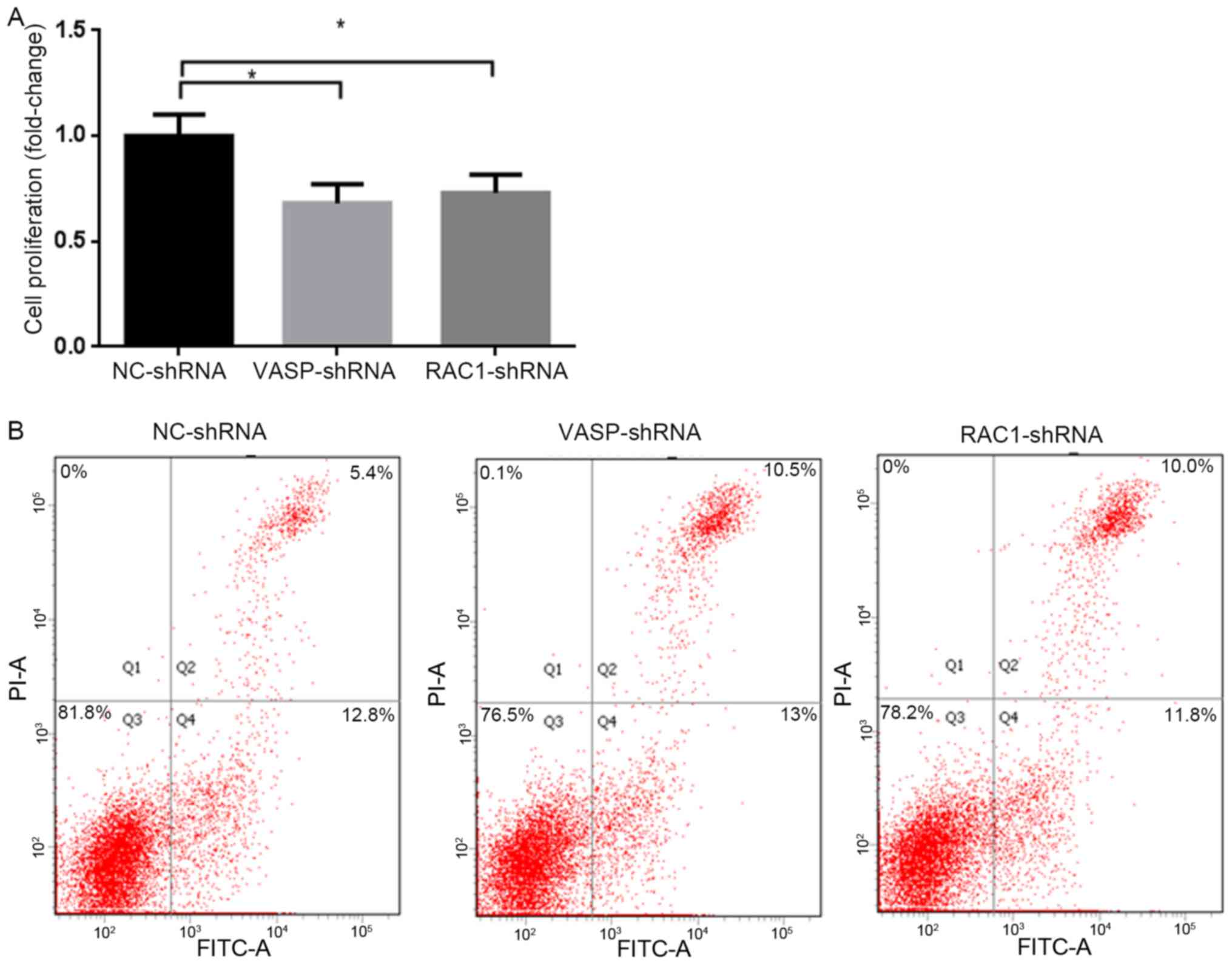
Effect of RAC1 and VASP-shRNA
interference on MCF-7 cell proliferation
In order to study the effect of downregulation of
RAC1 and VASP expression on the proliferation of MCF-7 cells,
stable RAC1-shRNA, VASP-shRNA and NC-shRNA MCF-7 cells were
cultured for 48 h, and the proliferation rate of the cells was
analyzed using a CCK-8 assay. The results revealed that the
proliferation rate of MCF-7 cells in the RAC1-shRNA and VASP-shRNA
groups was significantly decreased compared with the NC-shRNA
group, by 27 and 32%, respectively (P<0.05; Fig. 6A). Compared with the NC-shRNA group,
the apoptotic rates of MCF-7 cells in the RAC1-shRNA and VASP-shRNA
groups were increased significantly, by 4.6 and 5.1%, respectively
(P<0.05; Fig. 6B).
Effect of RAC1 and VASP-shRNA
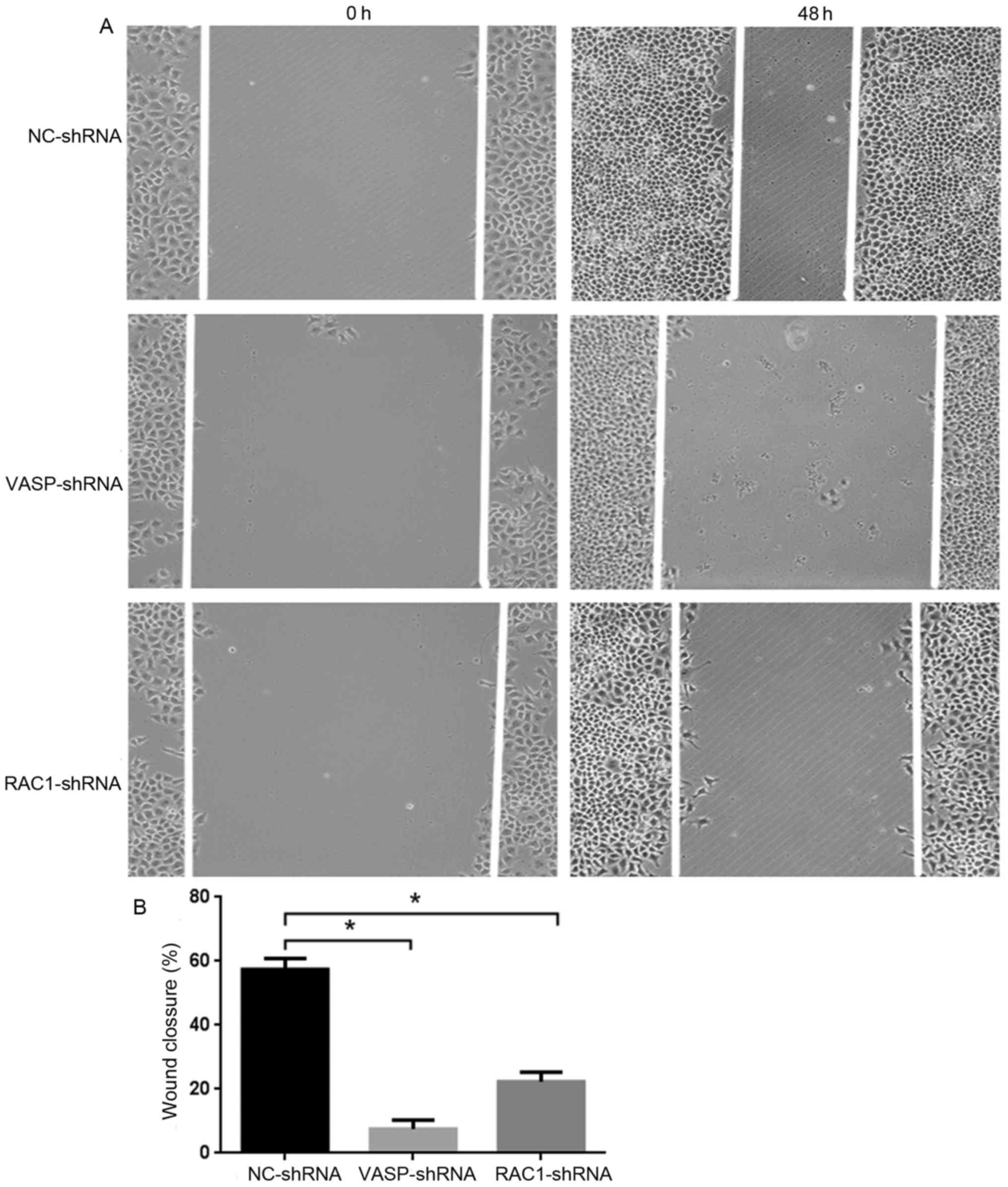
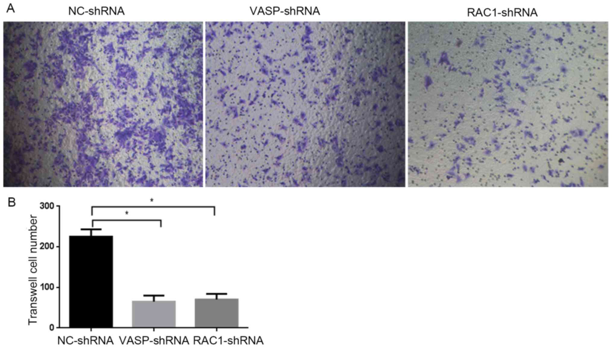
interference on MCF-7 cell migration
In order to study the effect of downregulation of
RAC1 and VASP expression on the migratory ability of MCF-7 cells,
stable RAC1-shRNA, VASP-shRNA and NC-shRNA MCF-7 cells were
cultured for 48 h, and the 2D and 3D migration ability of the cells
was analyzed using wound healing and Transwell assays. As presented
in Fig. 7A, compared with MCF-7 cells
in the NC-shRNA group, the 2D migratory ability of the cells in the
RAC1-shRNA and VASP-shRNA groups was decreased, by 35 and 50%,
respectively (P<0.05; Fig. 7B).
Furthermore, the 3D migratory ability of the cells in the
RAC1-shRNA and VASP-shRNA groups was decreased (Fig. 8A, by 68.9% (P<0.05) and 71.1%,
respectively (P<0.05; Fig. 8B).
These data indicate that downregulation of RAC1 and VASP
effectively inhibited the 2D and 3D migratory ability of MCF-7
cells.
Discussion
Rho GTPases are important downstream signaling
molecules for a number of membrane surface receptors, including G
protein-coupled, tyrosine kinase, cytokine and adhesion molecule
receptors (19,20). Rho GTPases act as molecular switches
that regulate numerous features of cell behavior, including cell
polarity, cytokinesis, particle movement, membrane trafficking,
membrane transport and translocation, cell growth and
transformation, and cell adhesion and motility (21,22).
Members of the Rho family affect cell morphology by controlling the
formation of actin-dependent structures (23). RhoA is involved in the assembly of
tonofibrils, RAC1 regulates the formation of lamellipodia and
membrane wrinkling, and Cdc42 promotes the formation of filopodia
in vascular endothelial cells (23).
Furthermore, it has been demonstrated that Rho GTPases regulate
carcinogenesis and tumor development (24). The mRNA and protein expression levels
of RhoE have been demonstrated to be markedly increased in lung
cancer tissues compared with adjacent non-tumor lung tissues, and
the overexpression of RhoE in non-small cell lung cancer has been
associated with smoking (25). This
suggests that RhoE may serve as a molecular marker to identify
high-risk individuals for lung carcinogenesis. Increased RAC1
activity has been observed, in addition to the overexpression of
PAK1, in urothelial carcinomas of the upper urinary tract, tumor
tissue and metastatic lymph node tissue (26). This was associated with poor
differentiation, local infiltration and lymph node metastasis. In
view of this, the purpose of the present study was to further
examine the association between RAC1 and the development of breast
cancer.
Downstream effectors of activated RAC1 include a
variety of actin-associated proteins, which enable actin filament
polymerization at the edge of the cell lamellipodia (27). The formation of the microfilament
network includes nucleation, the extension of the filament at the
positive end, and branching and dissociation at the negative end.
RAC1 interacts with insulin receptor substrate p53 and the wave
protein to activate the actin-related protein 2/3 complex to induce
actin branch formation (28). In
addition, RAC1 activates PAK1 to allow it to form a complex with,
and phosphorylate, LIM domain kinase (29). LIM-kinase catalyzes phosphorylation of
an N-terminal serine residue of cofilin, thereby inactivating its
F-actin-depolymerizing activity, and leading to the accumulation of
actin filaments and aggregates. These results suggest that
RAC1/Cdc42 promotes the formation of pseudopodia and promotes cell
migration by promoting actin filament polymerization and inhibition
of actin filament depolymerization (30). However, as an actin-associated
protein, the role of VASP in regulating the cytoskeleton is to
promote the extension of actin filaments at the positive end, which
is a crucial step for the formation of actin networks. Notably,
there exists evidence for the presence of Ena/VASP family members
(Mena, VASP and Ena-VASP-like) in the development of solid cancer
(31). The intensity of Mena
expression increases from premalignant to malignant lesions in
various organs, including the large bowel, stomach, cervix and
salivary glands (8,32). Aggressive and migratory Mg-63
osteosarcoma cells contain comparatively higher expression levels
of VASP at the transcriptional and translational levels, compared
with the less aggressive Saos-2 osteosarcoma cells (33). Overexpression of VASP in wild-type NIH
3T3 cells results in metaplasia to cancer cells (34), and the increased expression level of
VASP in lung adenocarcinoma correlates with lower cellular
differentiation and an increased pathological stage of the tumor
(8). Therefore, we hypothesized that
RAC1/VASP participates in the development of breast cancer, and
research was initiated to provide clinical evidence.
The present study used information from multiple
databases to comprehensively analyze the association between RAC1
and VASP mRNA expression levels in breast cancer tissues and the
pathological features of the patients. According to data from
Oncomine, the mRNA expression levels of RAC1 and VASP in invasive
ductal carcinoma tissues were significantly increased compared with
control tissues. Furthermore, according to the Kaplan-Meier data,
the mortality rate of patients with breast cancer at 50 months in
the RAC1 higher expression level group (49%) was increased compared
with the lower expression group (40.2%). The mortality rate of
patients with breast cancer at 50 months in the high VASP
expression level group was significantly increased compared with
the low expression group. Furthermore, according to the data from
TCGA, mRNA expression levels of RAC1 and VASP in the Normal group
were the lowest, and increased gradually in the Luminal A, Luminal
B, BLBC and Her-2 enrichment subtype groups. The mRNA expression
levels of RAC1 and VASP gradually increased in breast cancer
tissues of stages I, II, III and IV. These data indicate that the
high expression levels of RAC1 and VASP in breast cancer are
associated with low cellular differentiation, high pathological
stage and more aggressive tumor subtypes. High VASP mRNA expression
levels were positively associated with a poor overall survival time
in patients with breast cancer. However, the mechanism of RAC1/VASP
function in the progression of breast cancer development requires
further investigation and remains to be fully elucidated.
For further examination of the role of RAC1 and VASP
in the proliferation and migration of breast cancer cells, RAC1 or
VASP-knockdown cell lines established. In the RAC1-shRNA or
VASP-shRNA MCF-7 cells, the protein expression levels of RAC1 and
VASP were downregulated, the proliferation rates were decreased and
the migratory abilities decreased. These data indicated that RAC1
and VASP promoted breast cancer progression and development.
Notably, it was observed that the protein expression level of VASP
was decreased in RAC1-shRNA MCF-7 cells. However, the mechanism
through which RAC1 regulates VASP remains unclear. PAK is a RAC1
downstream target molecule, and when the its serine 21 site is
phosphorylated by PKG, PAK/NCK binding is inhibited and PAK/VASP
binding is enhanced, in order to promote human umbilical vein
endothelial cell motility (30). The
binding takes place between the proline-rich region of PAK and the
EVH1 domain of VASP (3,35). These data may act as a foundation on
which further investigations may be based.
In conclusion, RAC1 and VASP may act as
proto-oncogenes in the development of breast cancer, and VASP may
act as a downstream target of RAC1 to promote the proliferation and
migration of breast cancer cells.
Acknowledgements
Not applicable.
Funding
National Natural Science Foundation of China (grant
no. 81572943).
Availability of data and materials
The datasets used and/or analyzed during the current
study are available from the corresponding author on reasonable
request.
Authors' contributions
YT, LX and LW designed the study and wrote the
manuscript, XX, KL and YM analyzed the data, and YG, DW and YH
performed the cell experiments.
Ethics approval and consent to
participate
Not applicable.
Consent for publication
Not applicable.
Competing interests
The authors declare that they have no competing
interests.
References
|
1
|
Chen W, Zheng R, Baade PD, Zhang S, Zeng
H, Bray F, Jemal A, Yu XQ and He J: Cancer statistics in China,
2015. CA Cancer J Clin. 66:115–132. 2016. View Article : Google Scholar : PubMed/NCBI
|
|
2
|
Lopez-Marure R, Contreras PG and Dillon
JS: Effects of dehydroepiandrosterone on proliferation, migration,
and death of breast cancer cells. Eur J Pharmacol. 660:268–274.
2011. View Article : Google Scholar : PubMed/NCBI
|
|
3
|
Krause M, Dent EW, Bear JE, Loureiro JJ
and Gertler FB: Ena/VASP proteins: Regulators of the actin
cytoskeleton and cell migration. Annu Rev Cell Dev Biol.
19:541–564. 2003. View Article : Google Scholar : PubMed/NCBI
|
|
4
|
Ali M, Rogers LK and Pitari GM: Serine
phosphorylation of vasodilator-stimulated phosphoprotein (VASP)
regulates colon cancer cell survival and apoptosis. Life Sci.
123:1–8. 2015. View Article : Google Scholar : PubMed/NCBI
|
|
5
|
Pula G and Krause M: Role of Ena/VASP
proteins in homeostasis and disease. Handb Exp Pharmacol. 39–65.
2008. View Article : Google Scholar : PubMed/NCBI
|
|
6
|
Sechi AS and Wehland J: ENA/VASP proteins:
Multifunctional regulators of actin cytoskeleton dynamics. Front
Biosci. 9:1294–1310. 2004. View
Article : Google Scholar : PubMed/NCBI
|
|
7
|
Jayakumar T, Lin KC, Lu WJ, Lin CY,
Pitchairaj G, Li JY and Sheu JR: Nobiletin, a citrus flavonoid,
activates vasodilator-stimulated phosphoprotein in human platelets
through non-cyclic nucleotide-related mechanisms. Int J Mol Med.
39:174–182. 2017. View Article : Google Scholar : PubMed/NCBI
|
|
8
|
Dertsiz L, Ozbilim G, Kayisli Y, Gokhan
GA, Demircan A and Kayisli UA: Differential expression of VASP in
normal lung tissue and lung adenocarcinomas. Thorax. 60:576–581.
2005. View Article : Google Scholar : PubMed/NCBI
|
|
9
|
Cardama GA, Gonzalez N, Maggio J, Menna PL
and Gomez DE: Rho GTPases as therapeutic targets in cancer
(Review). Int J Oncol. 51:1025–1034. 2017. View Article : Google Scholar : PubMed/NCBI
|
|
10
|
Orgaz JL, Herraiz C and Sanz-Moreno V: Rho
GTPases modulate malignant transformation of tumor cells. Small
GTPases. 5:e290192014. View Article : Google Scholar : PubMed/NCBI
|
|
11
|
Kazanietz MG and Caloca MJ: The Rac GTPase
in cancer: From old concepts to new paradigms. Cancer Res.
77:5445–5451. 2017. View Article : Google Scholar : PubMed/NCBI
|
|
12
|
Zuzga DS, Pelta-Heller J, Li P, Bombonati
A, Waldman SA and Pitari GM: Phosphorylation of
vasodilator-stimulated phosphoprotein Ser239 suppresses filopodia
and invadopodia in colon cancer. Int J Cancer. 130:2539–2548. 2012.
View Article : Google Scholar : PubMed/NCBI
|
|
13
|
Espina C, Céspedes MV, García-Cabezas MA,
del Pulgar Gómez MT, Boluda A, Oroz LG, Benitah SA, Cejas P, Nistal
M, Mangues R and Lacal JC: A critical role for Rac1 in tumor
progression of human colorectal adenocarcinoma cells. Am J Pathol.
172:156–166. 2008. View Article : Google Scholar : PubMed/NCBI
|
|
14
|
Fryer BH, Wang C, Vedantam S, Zhou GL, Jin
S, Fletcher L, Simon MC and Field J: cGMP-dependent protein kinase
phosphorylates p21-activated kinase (Pak) 1, inhibiting Pak/Nck
binding and stimulating Pak/vasodilator-stimulated phosphoprotein
association. J Biol Chem. 281:11487–11495. 2006. View Article : Google Scholar : PubMed/NCBI
|
|
15
|
Györffy B, Lanczky A, Eklund AC, Denkert
C, Budczies J, Li Q and Szallasi Z: An online survival analysis
tool to rapidly assess the effect of 22,277 genes on breast cancer
prognosis using microarray data of 1,809 patients. Breast Cancer
Res Treat. 123:725–731. 2010. View Article : Google Scholar : PubMed/NCBI
|
|
16
|
Parker JS, Mullins M, Cheang MC, Leung S,
Voduc D, Vickery T, Davies S, Fauron C, He X, Hu Z, et al:
Supervised risk predictor of breast cancer based on intrinsic
subtypes. J Clin Oncol. 27:1160–1167. 2009. View Article : Google Scholar : PubMed/NCBI
|
|
17
|
Ye J, Wang W, Xu L, Duan X, Cheng Y, Xin
L, Zhang H, Zhang S, Li T and Liu Y: A retrospective prognostic
evaluation analysis using the 8th edition of American Joint
Committee on Cancer (AJCC) cancer staging system for luminal A
breast cancer. Chin J Cancer Res. 29:351–360. 2017. View Article : Google Scholar : PubMed/NCBI
|
|
18
|
Penault-Llorca F: Comments on the new
American Joint Committee on Cancer TNM staging for breast cancer.
What's new for the pathologist? Ann Pathol. 23:492–495. 2003.(In
French). PubMed/NCBI
|
|
19
|
Aznar S and Lacal JC: Rho signals to cell
growth and apoptosis. Cancer Lett. 165:1–10. 2001. View Article : Google Scholar : PubMed/NCBI
|
|
20
|
Bar-Sagi D and Hall A: Ras and Rho
GTPases: A family reunion. Cell. 103:227–238. 2000. View Article : Google Scholar : PubMed/NCBI
|
|
21
|
Schaefer A, Reinhard NR and Hordijk PL:
Toward understanding RhoGTPase specificity: Structure, function and
local activation. Small GTPases. 5:62014. View Article : Google Scholar : PubMed/NCBI
|
|
22
|
Zandvakili I, Lin Y, Morris JC and Zheng
Y: Rho GTPases: Anti- or pro-neoplastic targets? Oncogene.
36:3213–3222. 2017. View Article : Google Scholar : PubMed/NCBI
|
|
23
|
Hall A: Rho GTPases and the actin
cytoskeleton. Science. 279:509–514. 1998. View Article : Google Scholar : PubMed/NCBI
|
|
24
|
Roux P, Gauthier-Rouviere C, Doucet-Brutin
S and Fort P: The small GTPases Cdc42Hs, Rac1 and RhoG delineate
Raf-independent pathways that cooperate to transform NIH3T3 cells.
Curr Biol. 7:629–637. 1997. View Article : Google Scholar : PubMed/NCBI
|
|
25
|
Cuiyan Z, Jie H, Fang Z, Kezhi Z, Junting
W, Susheng S, Xiaoli F, Ning L, Xinhua M, Zhaoli C, et al:
Overexpression of RhoE in non-small cell lung cancer (NSCLC) is
associated with smoking and correlates with DNA copy number
changes. Cancer Biol Ther. 6:335–342. 2007. View Article : Google Scholar : PubMed/NCBI
|
|
26
|
Kamai T, Shirataki H, Nakanishi K, Furuya
N, Kambara T, Abe H, Oyama T and Yoshida K: Increased Rac1 activity
and Pak1 overexpression are associated with lymphovascular invasion
and lymph node metastasis of upper urinary tract cancer. BMC
Cancer. 10:1642010. View Article : Google Scholar : PubMed/NCBI
|
|
27
|
Li YL, Shao M and Shi DL: Rac1 signalling
coordinates epiboly movement by differential regulation of actin
cytoskeleton in zebrafish. Biochem Biophys Res Commun.
490:1059–1065. 2017. View Article : Google Scholar : PubMed/NCBI
|
|
28
|
Lim KB, Bu W, Goh WI, Koh E, Ong SH,
Pawson T, Sudhaharan T and Ahmed S: The Cdc42 effector IRSp53
generates filopodia by coupling membrane protrusion with actin
dynamics. J Biol Chem. 283:20454–20472. 2008. View Article : Google Scholar : PubMed/NCBI
|
|
29
|
Dawid IB, Breen JJ and Toyama R: LIM
domains: Multiple roles as adapters and functional modifiers in
protein interactions. Trends Genet. 14:156–162. 1998. View Article : Google Scholar : PubMed/NCBI
|
|
30
|
Edwards DC, Sanders LC, Bokoch GM and Gill
GN: Activation of LIM-kinase by Pak1 couples Rac/Cdc42 GTPase
signalling to actin cytoskeletal dynamics. Nat Cell Biol.
1:253–259. 1999. View
Article : Google Scholar : PubMed/NCBI
|
|
31
|
Zhang YT, Xu LH, Lu Q, Liu KP, Liu PY, Ji
F, Liu XM, Ouyang DY and He XH: VASP activation via the
Gα13/RhoA/PKA pathway mediates cucurbitacin-B-induced actin
aggregation and cofilin-actin rod formation. PLoS One.
9:e935472014. View Article : Google Scholar : PubMed/NCBI
|
|
32
|
Gurzu S, Ciortea D, Ember I and Jung I:
The possible role of Mena protein and its splicing-derived variants
in embryogenesis, carcinogenesis, and tumor invasion: A systematic
review of the literature. Biomed Res Int. 2013:3651922013.
View Article : Google Scholar : PubMed/NCBI
|
|
33
|
Wu G, Wei L, Yu A, Zhang M, Qi B, Su K, Hu
X and Wang J: Vasodilator-stimulated phosphoprotein regulates
osteosarcoma cell migration. Oncol Rep. 26:1609–1615.
2011.PubMed/NCBI
|
|
34
|
Liu K, Li L, Nisson PE, Gruber C, Jessee J
and Cohen SN: Reversible tumorigenesis induced by deficiency of
vasodilator-stimulated phosphoprotein. Mol Cell Biol. 19:3696–3703.
1999. View Article : Google Scholar : PubMed/NCBI
|
|
35
|
Greenwood AI, Kwon J and Nicholson LK:
Isomerase-catalyzed binding of interleukin-1 receptor-associated
kinase 1 to the EVH1 domain of vasodilator-stimulated
phosphoprotein. Biochemistry. 53:3593–3607. 2014. View Article : Google Scholar : PubMed/NCBI
|