Introduction
Cervical cancer is the most common gynecological
malignancy, which causes harm to the health of women globally
(1). The mortality rate of cervical
cancer is the fourth highest among all cancer types (2). In China, there is a significant regional
difference in the incidence of cervical cancer. Cervical cancer is
prominent in poorer provinces, such as Hubei and Shanxi, due to
unsatisfactory sanitary conditions. High-risk human papillomavirus
infection is a major risk factor for cervical cancer, with multiple
sexual partners being a secondary risk factor for the disease.
Other risk factors for cervical cancer also include smoking,
malnutrition and poor health conditions (3). An appropriate individual treatment
program is developed according to the clinical stage, the patient's
age, fertility requirements, general condition and access to
medical technology, and other comprehensive considerations. The
current treatment mainly includes surgery, radiotherapy and
chemotherapy (4,5). Hysterectomy and bilateral
lymphadenectomy are standard treatments for cervical stage I and
stage IIA cancer patients (6).
Radical trachelectomy has been demonstrated to be an effective
treatment for early cervical cancer and is associated with an
acceptable live birth rate (7–10). The
majority of cervical cancer cases can be successfully treated if
diagnosed at an early stage (11).
However, the 5-year survival rate of patients with cervical cancer
is remains low, particularly in advanced cervical cancer. There are
a number of clear clinical signs in the early stage of some
patients with cervical cancer, but they can be easily ignored by
patients. So some patients lose better treatment opportunities.
Therefore, there is a requirement to elucidate the molecular
mechanisms underlying cervical cancer development and progression,
providing a basis for finding potential drug targets and diagnosing
biomarkers of cervical cancer (12,13).
Gene expression microarrays are a frontier
biotechnology. In the current data age, they combine a high
throughput with simultaneous detection of thousands of genes. The
features of gene expression microarrays are automated, integrated
and miniaturized. In the present study, the original GSE26511
dataset was downloaded from the Gene Expression Omnibus (GEO)
database to identify downregulated or upregulated genes in cervical
cancer compared with non-malignant controls. Using gene chip
technology, the analysis found a number of important key roles of
differentially expressed genes (DEGs), which are important in the
initiation and development of cervical cancer. DEGs are the first
choice for the study of molecular targets and diagnostic markers.
In the present study, key DEGs associated with cervical cancer in
tumors and normal samples were identified, and then these genes
were screened according to statistical methods. The study benefited
from current powerful analysis software and statistical methods,
including Gene Ontology (GO) terminology, enrichment analysis,
Kyoto Encyclopedia of Genes and Genomes (KEGG) pathway analysis,
protein-protein interaction (PPI) network analysis and weighted
gene co-expression network analysis (WGCNA).
Materials and methods
Microarray data
The gene expression profiles of the GSE26511 dataset
(14) were downloaded from the GEO
database. The GPL570 [HG-U133 Plus 2] Affymetrix Human Genome U133
Plus 2.0 Array platform was used. The GSE26511 dataset contained 39
samples, including 19 cervical cancer samples and 20 normal
samples.
Screening for DEGs
The statistical software R and packages from
Bioconductor (http://www.bioconductor.org/biocLite.R) were used to
analyze the DEGs between the cervical cancer and normal samples.
The AffyPLM (http://www.bioconductor.org/packages/release/bioc/html/affyPLM.html)
package was used to fit the original data of the chip, obtaining
the weights and residuals diagram, the relative log expression and
the relative standard deviation (normalized unscaled standard
errors) box diagram. Prior to analysis of the data, the microarray
data was quality tested. In this process, R packets, including
packages of affyPLM, packages of Affy and packages of RColorBrewer
(http://www.bioconductor.org/packages/release/bioc/html/affy.html),
were used. Subsequent to removing unqualified samples, a reasonable
and useful sample was obtained. According to the Limma (http://www.bioconductor.org/packages/release/bioc/html/limma.html)
package of Bioconductor, key DEGs were determined. P<0.05 was
considered to indicate a statistically significant difference.
Functional and pathway enrichment
analysis
GO (http://www.geneontology.org/) analysis was extensively
used for a short list of genes with statistically significant
differences in expression. GO terms are divided into three
categories: Biological process, cellular component and molecular
function. The KEGG (http://www.kegg.jp/) website is an online database of
genomes, enzymatic pathways and biochemical that acts as a freely
accessible gene database. Different input data methods are based on
different analyses. Compared with other databases, KEGG has a
powerful graphics function, using graphics rather than harassment
of the text to introduce a large number of metabolic pathways and
the association between the various pathways. P<0.05 was
considered to indicate a statistically significant difference.
PPI network analysis
PPI information of DEGs was acquired from Search
Tool for the Retrieval of Interacting Genes (database (http://www.stringdb.org/). The PPI network assisted
with identifying the key genes and important gene modules, and then
used the network visualization tools, such as Cytoscape (http://www.cytoscape.org/), drawing network diagram.
The PPI network is an experimental network for biological network
visualization. The gene co-expression network attempted to
reconstruct the biological network through the expression of genes.
In the PPI network, nodes and edges (lines) represent proteins and
their interactions (P<0.05).
WGCNA
As a system biology method, gene co-expression
network analysis was performed by the WGCNA package (http://www.genetics.ucla.edu/labs/horvath/CoexpressionNetwork/Rpackages/WGCNA)
to investigate the association between gene expression patterns
(15). The clustering criteria of
WGCNA were biologically significant, and the consequence of this
method is a higher degree of credibility. These results were
obtained in order to complete further investigations, such as those
into association traits, metabolic pathway modeling and the
establishment of gene interaction networks. In a co-expression
network, the expression of each gene at a particular time or space
was treated as a node. WGCNA uses a soft threshold based on the
determination method. Commonly used weighting functions included
sigmoid functions and power functions. The similarity and proximity
of gene co-expression were calculated using soft threshold power.
Analysis of network topology confirmed the final structure of soft
threshold power.
Patient samples
A total of 5 cervical cancer specimens and 5
non-tumor cervical cancer epithelial tissues were collected from
the Cancer Hospital of Hunan Province, Central South University
(Changsha, China). The average age of the patients was 45±5.5
years. Biopsy sample were collected from March 17, 2017 to May 17,
2017. Written informed consent was obtained from each patient, and
the experimental protocols were approved by the Institutional
Review Board of the Cancer Hospital of Hunan Province. Each biopsy
sample was divided into two sections: One section was submitted for
routine histological diagnosis.
The biopsy sample was fixed with 4% paraformaldehyde
for 24 h at 4°C and then was placed in processing cassettes. The
samples were dehydrated through a serial alcohol gradient at room
temperature (50% alcohol for 1 h, 70% alcohol overnight and then
80% alcohol for 1 h, then 95% alcohol for 1 h, and then dehydrated
twice in 100% alcohol for 30 min), and embedded in paraffin wax
blocks. Prior to H&E staining, the 4-µm thick tissue sections
were heated at 60°C for 1 h, dewaxed twice in 100% xylene for 10
min, and then rehydrated through decreasing concentrations of
ethanol (twice in 100% alcohol for 5 min, followed by 95% alcohol
for 5 min and then 80% alcohol for 5 min, then 70% alcohol for 5
min, and then in distilled water for 5 min), and washed in 1X PBS
for 3 min twice at room temperature. The sections were then stained
with hematoxylin for 20 min, washed in distilled water for 5 min
and then stained with eosin for 3 min. The sections were dried in a
fume cupboard at room temperature. The thickness of the sections
was 4 µm. A light microscope was used to observe the sections at
×200 magnification while the other was freshly stored in RNALater
reagent (Qiagen GmbH, Hilden, Germany) at −80°C for the following
experiments.
RNA extraction and reverse
transcription-quantitative polymerase chain reaction (RT-qPCR)
analysis
Total RNA was extracted from the biopsy samples with
the RNeasy® kit (Qiagen GmbH) according to the
manufacturer's protocols. A total of 1 µg of RNA samples were
reverse transcribed into cDNA using TransScript One-Step gDNA
Removal and cDNA Synthesis SuperMix (Vazyme Biotech Co., Ltd.,
Nanjing, China) with oligo-dT primer (R223-01, Vazyme Biotech Co.,
Ltd.; primer sequences unavailable), according to the
manufacturer's protocols. RT-qPCR was performed using TransStart
Top Green qPCRSuperMix (Vazyme Biotech Co., Ltd.) on a
Mastercyclerep realplex4 (Eppendorf, Hamburg, Germany). The PCR
conditions included an initial step at 95°C for 30 sec, followed by
40 cycles of amplification and quantification (95°C for 15 sec,
60°C for 15 sec, and 68°C for 20 sec). GAPDH was used as an
endogenous control for normalization. The sequences of the primers
used for RT-qPCR were as follows: Mucin 1 cell surface associated
(MUC1) forward, 5′-agacgtcagcgtgagtgatg-3′ and reverse,
5′-cagctgcccgtagttctttc-3′; fucosyltransferase 3 (FUT3; Lewis blood
group) forward, 5′-gcaaggcttagaccagttcg-3′ and reverse,
5′-caccagcagctgaaatagca-3′; notch 3 (NOTCH3) forward,
5′-gtcgtggctacactggacct-3′ and reverse, 5′-aatgtccacctcgcaatagg-3′;
fibroblast growth factor 2 (FGF2) forward,
5′-ggtgaaaccccgtctctaca-3′ and reverse, 5′-tctgttgcctaggctggact-3′;
insulin-like growth factor 1 (IGF1) forward,
5′-ggctgaccaagctgaaactc-3′ and reverse, 5′-atcgcttaaacccaggaggt-3′;
hepatocyte growth factor (HGF) forward, 5′-ctggttccccttcaatagca-3′
and reverse, 5′-ctccagggctgacatttgat-3′; and GAPDH forward,
5′-accacagtccatgccatcac-3′ and reverse, 5′-tccaccaccctgttgctgta-3′.
The expression of mRNA was assessed by evaluated quantitation cycle
(Cq) values. The Cq values were normalized with the expression
levels of GAPDH and the relative amount of mRNA specific to each of
the target genes was calculated using the 2−ΔΔCq method
(16).
Statistical analysis
All results were presented as the mean ± standard
error of three independent experiments. Unpaired t-test was used
for statistical analysis. All statistical tests were performed with
GraphPad Prism 5.0 (GraphPad Software Inc., La Jolla, CA, USA). All
statistical tests were two tailed, and P<0.05 was considered to
indicate a statistically significant difference.
Results
Identification of DEGs
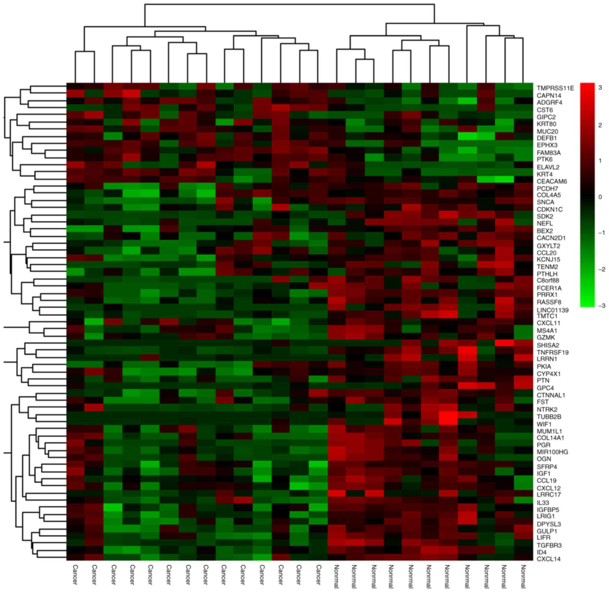
The GSE26511 expression profile shows clear
distinction between the tumor and normal healthy epithelium.
Isolation and identification of DEGs was not only used to assess
the function of the genes, but also assisted in revealing the
pathogenesis of the disease. Gene chip raw data quality detection
lead to 8 low-quality samples being removed from the 20 normal
samples and 6 low-quality samples being removed from the 19
cervical cancer samples. The remaining samples required integration
and processing. Preprocessing with statistical analysis software R
was necessary and important prior to analyzing the microarray data
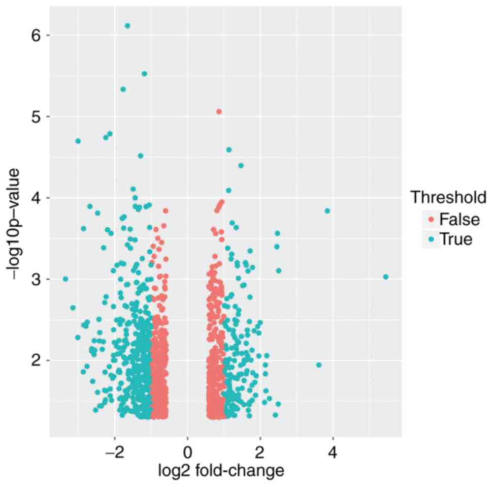
of gene differential expression. Altogether there were 1,263 DEGs,
of which 568 DEGs were selected (fold change >2) (Fig. 1), consisting of 402 significantly
downregulated and 166 significantly upregulated DEGs, for the
subsequent bioinformatics analysis (fold change >2) (Fig. 2).
GO term enrichment analysis of
DEGs
Noordhuis et al (14) performed research mainly concerned with
the molecular mechanism of lymph node metastasis in cervical
cancer. Pathway analysis of microarray expression profiles suggests
that transforming growth factor-β (TGF-β) and p120-associated
non-canonical β-catenin pathways are important in pelvic lymph node
metastasis of early cervical cancer (14). However, the present results showed
that the acquired clean reads were of a sufficiently high quality
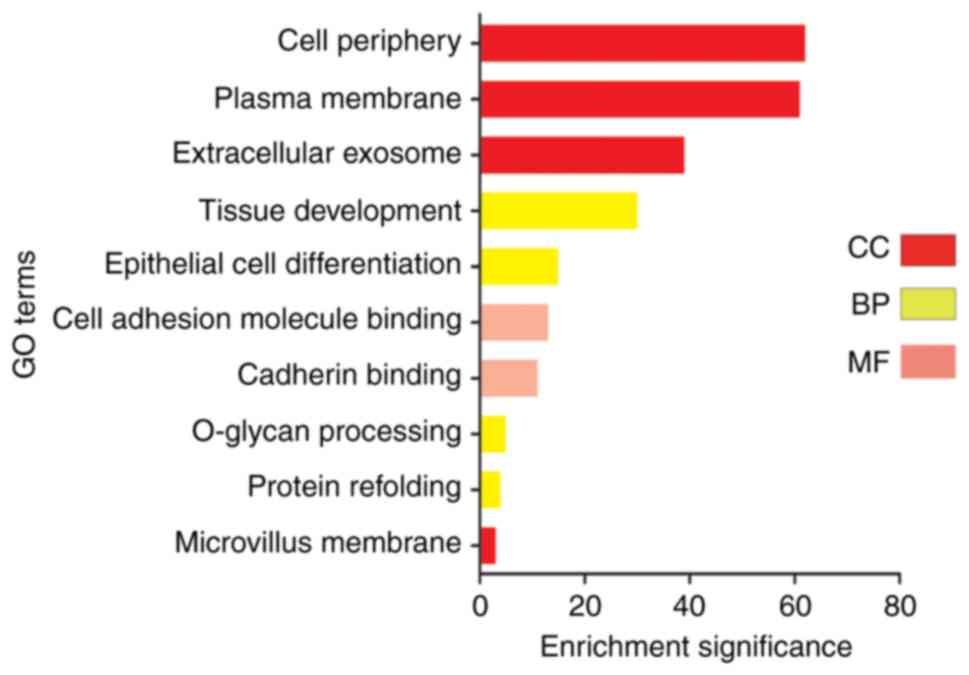
for subsequent analysis. The upregulated DEGs were primarily
enriched in ‘cell adhesion molecule binding’, ‘plasma membrane’,
‘tissue development’, ‘DNA repairing’ and ‘epithelial cell
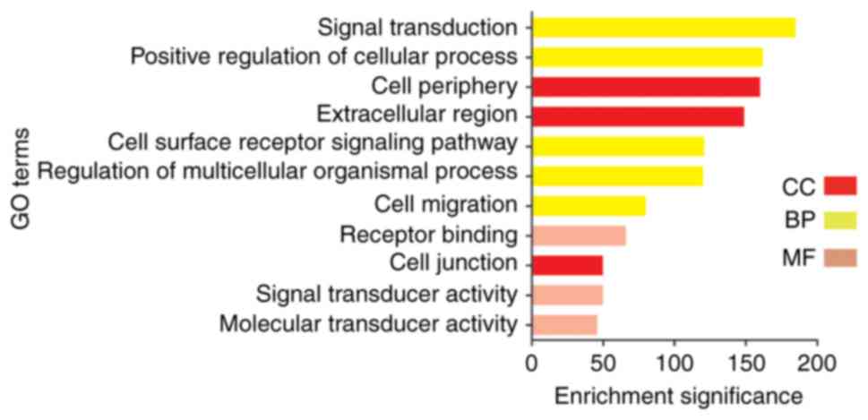
differentiation’. Downregulation of the DEGs was mainly enriched by
‘signal transduction’, ‘cell surface receptor signaling pathway’,
‘positive regulation of biological process’, ‘receptor binding’ and
‘signal transducer activity’. For cellular component, to analyze
the biological functions, biological processes and subcellular
localization of these DEGs, the upregulated DEGs were enriched in
‘cell periphery’, ‘plasma membrane’ and ‘extracellular exosome’.
Subsequent to the scientific analysis, it was found that
downregulated genes were mainly found in ‘cell periphery’,
‘extracellular region’ and ‘cell junction’. For biological cell
molecular function, the DEGs that were upregulated were
significantly enriched in ‘cell adhesion molecule binding’ and
‘cadherin binding’. The DEGs of cervical cancer that were
downregulated were significantly enriched in ‘receptor binding’,
‘signal transducer activity’ and ‘molecular transducer activity’.
With regard to biological process, it was found that downregulated
genes were mainly found in ‘signal transduction’, ‘positive
regulation of cellular process’, ‘cell surface receptor signaling
pathway’, ‘regulation of multicellular organismal process’ and
‘cell migration’. The upregulated DEGs were primarily enriched in
‘epidermal cell differentiation’, ‘tissue development’ and
‘O-glycan processing’. The analysis clearly and intuitively
observed the changes in the enrichment of the DEGs (Figs. 3 and 4).
KEGG pathway analysis of cervical
DEGs
By analyzing the KEGG and Biocarta databases
(https://cgap.nci.nih.gov/Pathways/BioCarta_Pathways),
Noordhuis et al (14) proved
that the five pathways are the TGF-β, nuclear factor of activated T
cells (NFAT), anaplastic lymphoma kinase (ALK), Bcl-2-associated
death promoter (BAD) and protease-activated receptor-1 (PAR1)
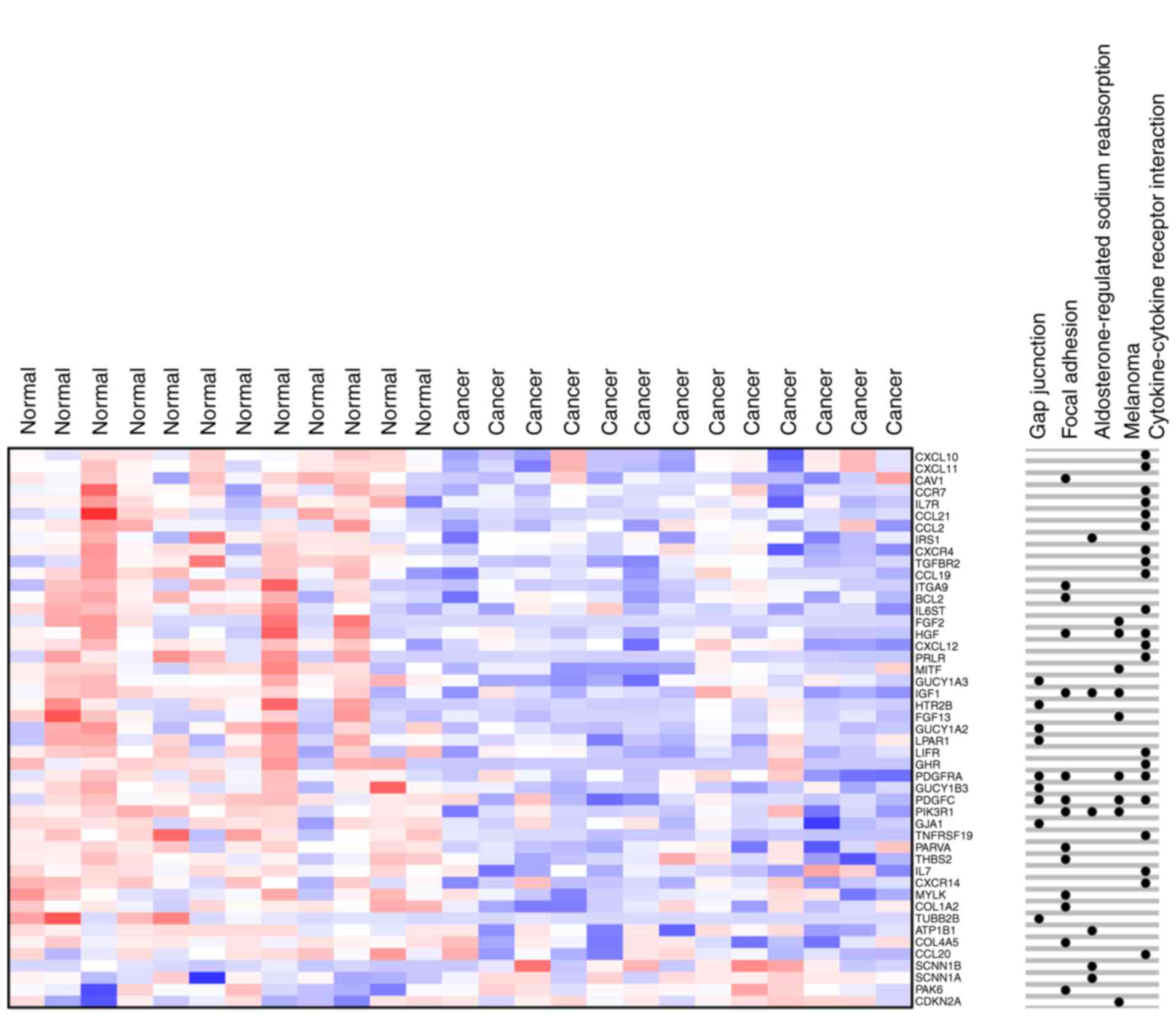
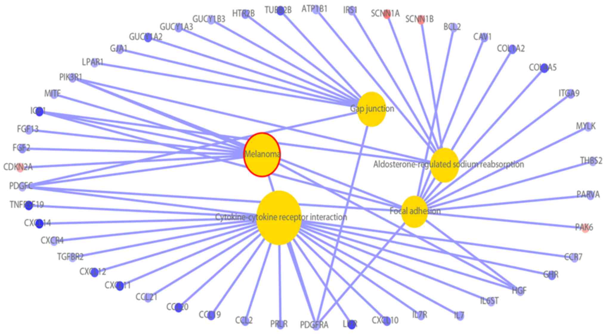
pathways. The present study found five significantly enriched
pathways by KEGG pathway analysis (Fig.
5). First, the enrichment aspect of ‘aldosterone-regulated
sodium reabsorption’ included 4 DEGs that were downregulated and 2
that were upregulated. Second, the enrichment aspect of ‘melanoma’
included 1 upregulated DEG and 8 that were downregulated. Third,
the enrichment aspect of ‘gap junction’ included 9 downregulated
DEGs. Fourth, the enrichment aspect of ‘cytokine-cytokine receptor
interaction’ included 21 downregulated DEGs. Fifth, the enrichment
aspect of ‘focal adhesion’ included 1 upregulated DEG and 13
downregulated DEGs. Through the analysis, the molecular mechanism
of DEGs was observed in more detail (Fig.
6).
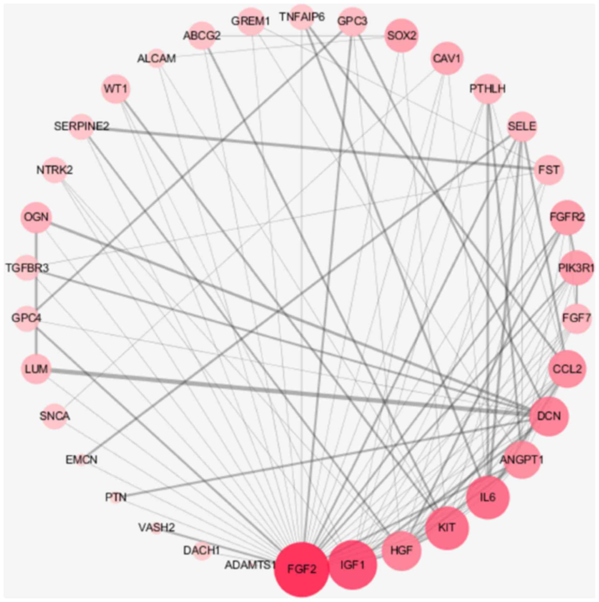
Analysis of DEGs by PPI network
Gene co-expression and the PPI network have a
guiding role in the study of cervical cancer target genes and
proteins, and have a prominent function in the future study of
cervical cancer. Based on the Search Tool for the Retrieval of
Interacting Genes/Proteins database, upregulated and downregulated
genes were selected to observe the association between protein
molecules. The associations between various protein molecules were
highlighted, aiding in the observation and understanding of the
molecular mechanism of cervical cancer. The upregulation of genes
such as FGF2 is associated with tumor formation, and IL6 is
associated with immunity (Fig. 7).
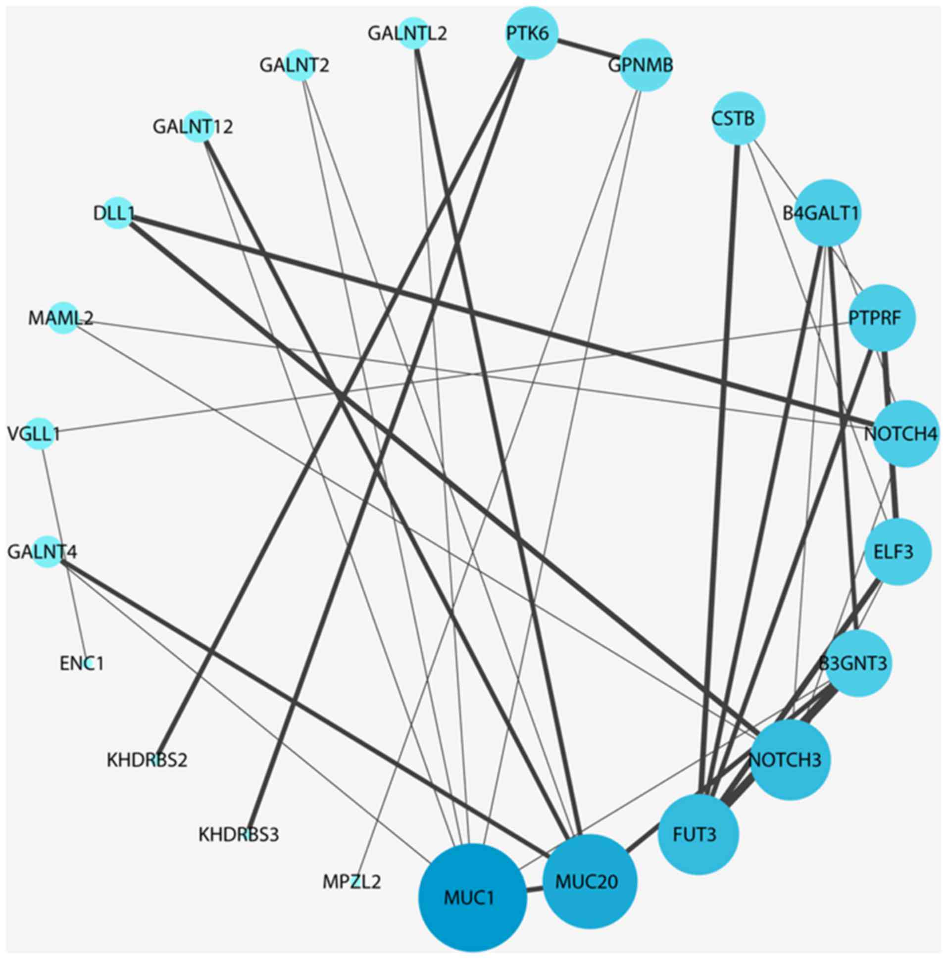
Downregulated genes such as MUC1 are associated with tumor
migration, and FUT3 is associated with the degree of tissue
differentiation (Fig. 8).
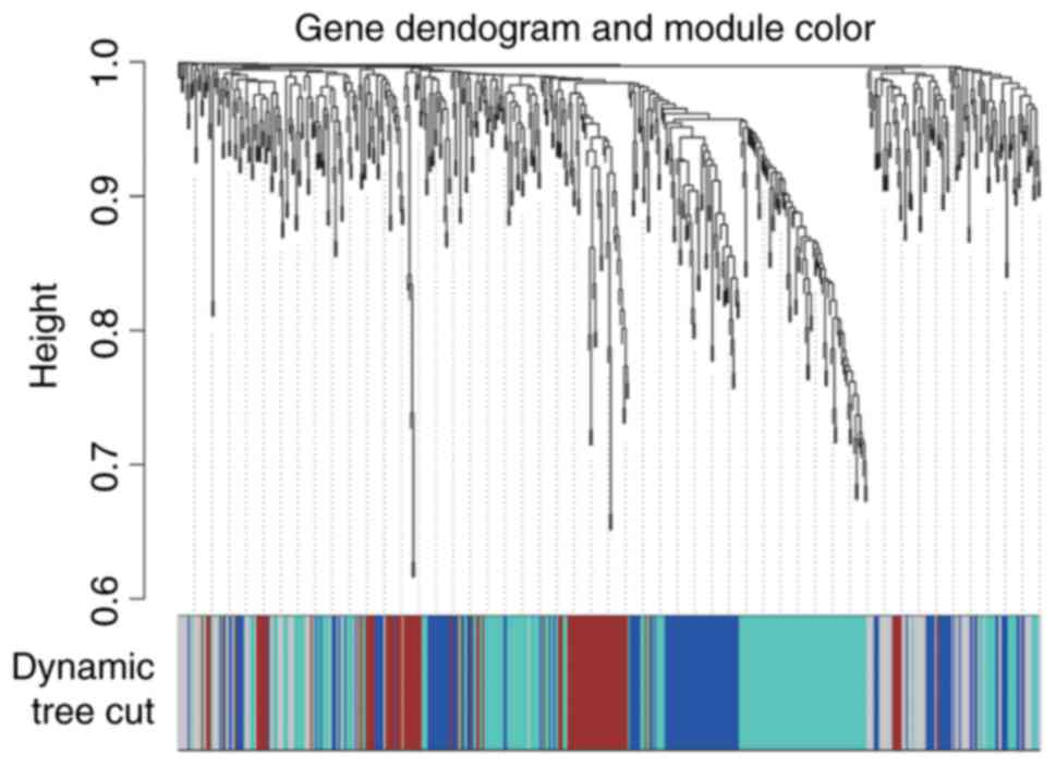
WGCNA of DEGs
WGCNA, as an efficient and complex biology method,
is widely used in biological information analysis. Through
correlation analysis, the gene modules associated with specific
sample traits were quickly screened from the data in the present
study. More results are shown in Fig.
9. Interaction-associated genes frequently exhibit similar
expression patterns. Consequently, the DEGs co-expression network
was constructed to screen gene modules with similar expression
profile. The DEGs that were in the blue module were significantly
enriched in ‘enzyme-linked receptor protein signaling pathway’,
‘cell motility’, ‘embryonic skeletal system development’ and
‘embryo development’; the DEGs that were in the brown module were
significantly enriched in ‘B cell activation’, ‘immune response’,
‘leukocyte activation’, ‘humoral immune response’ and ‘G-protein
coupled receptor signaling pathway’; the DEGs that were in the grey
module were significantly enriched in ‘spermatid development’,
‘spermatid differentiation’, ‘inorganic cation transmembrane
transporter activity’, ‘metal ion transmembrane transporter
activity’ and ‘regulation of phosphatase activity’; and the DEGs
that were in the turquoise module were significantly enriched in
‘mammary gland epithelium development’, ‘mitotic cell cycle
process’, ‘development of primary sexual characteristics’, ‘mitotic
nuclear division’ and ‘negative regulation of NF-κB transcription
factor activity’.
Validation of differential genes by
RT-qPCR
The present study aimed to determine whether the
DEGs identified in the microarray analysis were upregulated or
downregulated in a sample of clinical cervical cancer patients.
Cervical cancer specimens and non-tumor cervical cancer epithelial
tissues were obtained, and the differential genes validated with
RT-qPCR (Fig. 10). The experimental
results showed that MUC1, FUT3 and NOTCH3 gene expression was
significantly downregulated, which was consistent with the result
of the microarray analysis. However, among the genes found to be
upregulated on microarray analysis, IGF1 and HGF were downregulated
in the clinical samples. Additionally, FGF2 was upregulated, which
confirmed the result of the microarray analysis. The results
demonstrated that microarray analysis may provide reference for the
identification of molecular markers and therapeutic targets for
cervical cancer.
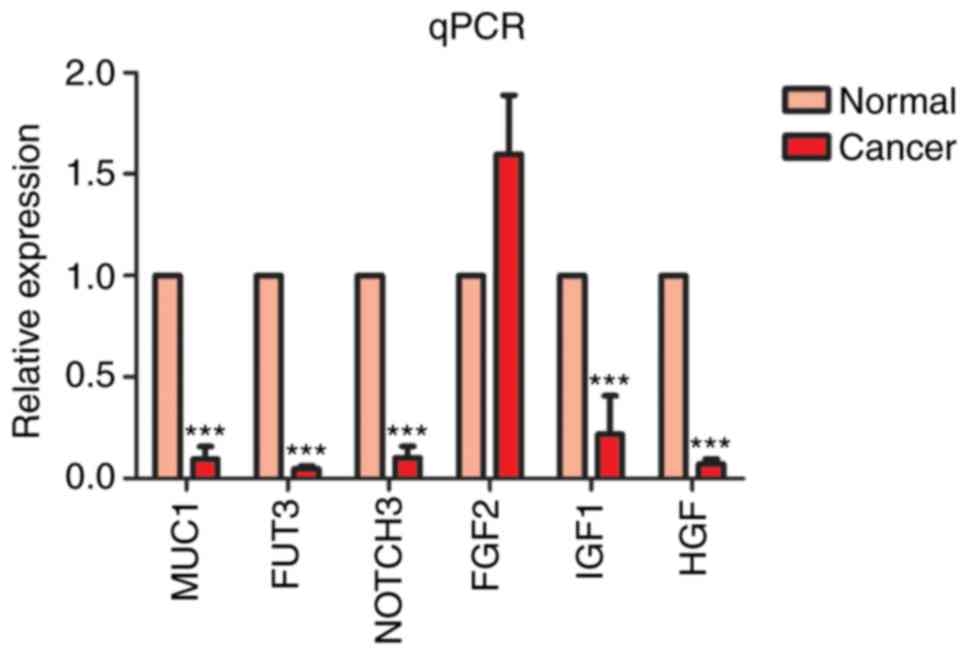 | Figure 10.MUC1, FUT3, NOTCH3, FGF2, IGF1 and
HGF gene validation with qPCR. ***P<0.001 vs. normal. qPCR,
quantitative polymerase chain reaction. MUC1, mucin 1 cell
surface-associated; FUT3, fucosyltransferase 3 (Lewis blood group);
NOTCH3, notch 3; FGF2, fibroblast growth factor 2; IGF1,
insulin-like growth factor 1; HGF, hepatocyte growth factor. |
Discussion
Cervical cancer is one of the most common
malignancies in women worldwide. There are ~490,000 new cases of
cervical cancer in the world each year. In China, 150,000 novel
cases of cervical cancer occur and ~20,000 cervical
cancer-associated mortalities occur each year (17–19).
Previous studies determined that cervical cancer cells were
sensitive to radiotherapy and chemotherapy, and used the two
treatment techniques to improve the survival rate of patients with
cervical cancer (4,20–22).
Simultaneously, radiotherapy was determined to have the better
curative effect on cervical cancer (23–25). A
comprehensive understanding of the molecular mechanism of cervical
cancer is essential to the diagnosis and treatment of the disease.
Since microarrays achieve rapid detection of gene information by
detecting the corresponding position hybridization probes, in the
present study, bioinformatics analysis was used to obtain DEGs from
the GSE26511 dataset of cervical cancer and normal samples. The
function of these differential genes was mainly concentrated in the
‘cell cycle’, ‘biosynthetic process’ and ‘immune response’.
Hispolon induces apoptosis and antitumor effects in cervical cancer
(26), indicating that drugs can
affect the occurrence and development of cervical cancer. In order
to identify the cell tumor pathways associated with early cervical
cancer pelvic lymph node metastasis, GSE26511 was investigated and
it was found that five of the 285 pathways (TGF-β, NFAT, ALK, BAD
and PAR1) were dysregulated in cervical cancer-lymph node-negative
samples, and two pathways (β-catenin and glycosphingolipid
biosynthesis neo lactoseries) were dysregulated in cervical
cancer-lymph node-positive samples (14). A large number of DEGs are enriched on
these pathways. Genes associated with β-catenin, TGF-β, NFAT, ALK
and BAD were found amongst the DEGs.
By observing the results of the bioinformatics
analysis in the present study, a number of genes were found in
cervical cancer that regulated signaling pathways. In the analysis
of the results of the DEGs, transcription factor 4 (TCF4)
expression was observed to confer a significant effect on the
proliferation, apoptosis and radiosensitivity of cervical cancer
cells. It is known that the β-catenin/TCF-4 pathway serves an
important role in promoting the metastasis of gastric cancer
(27). In early-stage cervical
cancer, β-catenin pathways are important in pelvic lymph node
metastasis. RASSF1 is also a member of the DEGs.RASSF1, which gene
encodes a protein similar to the RAS effector proteins.
Hypermethylation of RASSF1A gene is associated with colon cancer
(28).
FGF2 and IGF1 are also members of the upregulated
DEGs. High FGF2 expression is associated with colon cancer
metastasis (29). FGF2 protects the
tumor cells from the antiproliferative effect of gefitinib and
largely prevents reprogramming of the proteome and phosphoproteom.
The process of promoting angiogenesis between IGF1 and cocultured
cells is through the phosphoinositide3 kinase/protein kinase B
(Akt) signaling pathway (30). MUC1
and FUT3 are also members of the downregulated DEGs. MUC1 induces
tamoxifen resistance in estrogen receptor-positive breast cancer
(31). MUC1 expression is associated
with metastatic recurrence in postoperative patients with
esophageal squamous cell cancer (32). Experimental results show that microRNA
expression vector targeting of the FUT3 gene can effectively
inhibit cell proliferation, migration and invasion ability
differences.
By observing the results of the present
bioinformatics analysis, it was found that there were numerous
genes in cervical cancer regulating the cell cycle. In the analysis
of the KEGG results, PAK6 and serine/threonine-protein kinase PAK6
expression clearly demonstrated a significant effect on the
proliferation, apoptosis and radiosensitivity of cervical cancer
cells. PAK6 is specifically required for carcinoma cell-cell
dissociation downstream of HGF for DU145 prostate cancer and HT29
colon cancer cells. In previous studies, PAK6 inhibition resulted
in a significant reduction in the survival of prostate cancer cells
(33,34). However, the high expression of PAK6
has tumor suppressor abilities and is a potential mechanism in the
pathogenesis of hepatocellular carcinoma (35,36). By
contrast, the DEGs CLCX12 and BCL2 were downregulated in Fig. 6 in the present study. The BCL2 marker
is overexpressed in uterine smooth muscle tumors, and CXCL12
(37–39) overexpression improves neurobehavioral
recovery following ischemic stroke through a variety of mechanisms,
including the promotion of endothelial progenitor cell function in
animal models (40).
By observing the results of the present
bioinformatics analysis, immune response-related genes and pathways
from WGCNA were found. The DEGs that were in the brown module,
including AKT, cluster of differentiation (CD)46, MYC
proto-oncogene bHLH transcription factor (MYC) and LIF receptor α
(LIFR), were significantly enriched in ‘immune response’. These
genes are necessary for the immune response to maintain cell
stability. The high expression of LIFR (41) is an independent prognostic marker for
primary melanoma (41,42). MYC regulation in the cervical cancer
immune response has an irreplaceable role (43–47). The
protein encoded by the CD46 gene is a type I membrane protein that
is part of the complement system. The encoded protein has cofactor
activity to protect the host cell from complement damage. CD46 is a
membrane cofactor expressed on all nuclear human cells and serves
an important role in inhibiting autoimmune responses and protecting
host cells from complement-mediated attacks (48).
In conclusion, in the present study, DEGs of the
GSE26511 dataset were assessed by systematic bioinformatics
analysis followed by RT-qPCR validation of a number of the key
genes in clinical samples. The study was successfully able to
reveal biomarkers, indicating that DEGs have important effects for
the development and progress of cervical cancer. However, further
experiments are required to confirm the function of the identified
genes in cervical cancer.
Acknowledgements
Not applicable.
Funding
The present study was supported by the National
Natural Sciences Foundation of China (grant nos. 81672685,
81402270, 81272975 and 81672993) and the Beijing Xisike Clinical
Oncology Research Foundation (grant no. Y-HR2016096).
Availability of data and materials
All data generated or analyzed during this study are
included in this published article.
Authors' contributions
YG and YZ designed the study. CZ, SX, LL, SL, YX, KC
and HC conducted the experiments. YX, KC and HC analyzed the data.
The manuscript was drafted by YG and YZ. All authors read and
approved the final manuscript.
Ethics approval and consent to
participate
Written informed consent was obtained from each
patient, and experimental protocols were approved by the
Institutional Review Board of the Cancer Hospital of Hunan
Province.
Consent for publication
All participants provided written informed consent
for publication.
Competing interests
The authors declare that they have no competing
interests.
Glossary
Abbreviations
Abbreviations:
|
DEGs
|
differentially expressed genes
|
|
GEO
|
gene expression omnibus
|
|
GO
|
gene ontology
|
|
KEGG
|
Kyoto encyclopedia of genes and
genomes
|
|
PPI
|
protein-protein interaction
|
|
WGCNA
|
weighted gene co-expression network
analysis
|
References
|
1
|
Yang M, Wang M, Li X, Xie Y, Xia X, Tian
J, Zhang K and Tang A: Wnt signaling in cervical cancer? J Cancer.
9:1277–1286. 2018. View Article : Google Scholar : PubMed/NCBI
|
|
2
|
Torre LA, Bray F, Siegel RL, Ferlay J,
Lortet-Tieulent J and Jemal A: Global cancer statistics, 2012. CA
Cancer J Clin. 65:87–108. 2015. View Article : Google Scholar : PubMed/NCBI
|
|
3
|
Crosbie EJ, Einstein MH, Franceschi S and
Kitchener HC: Human papillomavirus and cervical cancer. Lancet.
382:889–899. 2013. View Article : Google Scholar : PubMed/NCBI
|
|
4
|
Shelley C, Gore M, Tan S, Thomas K and
Eeles R: Cervical spine fractures in patients undergoing palliative
radiotherapy to the cervical spine: Iimplications for Practice.
Clin Oncol (R Coll Radiol). Apr 10–2018.(Epub ahead of print).
View Article : Google Scholar : PubMed/NCBI
|
|
5
|
Isono-Nakata R, Tsubamoto H, Ueda T, Inoue
K and Shibahara H: Bevacizumab with metronomic chemotherapy of
low-dose oral cyclophosphamide in recurrent cervical cancer: Four
cases. Gynecol Oncol Rep. 24:57–60. 2018. View Article : Google Scholar : PubMed/NCBI
|
|
6
|
Sevin BU, Nadji M, Lampe B, Lu Y,
Hilsenbeck S, Koechli OR and Averette HE: Prognostic factors of
early stage cervical cancer treated by radical hysterectomy.
Cancer. 76 10 Suppl:S1978–S1986. 1995. View Article : Google Scholar
|
|
7
|
Einstein MH, Park KJ, Sonoda Y, Carter J,
Chi DS, Barakat RR and Abu-Rustum NR: Radical vaginal versus
abdominal trachelectomy for stage IB1 cervical cancer: A comparison
of surgical and pathologic outcomes. Gynecol Oncol. 112:73–77.
2009. View Article : Google Scholar : PubMed/NCBI
|
|
8
|
Plante M, Gregoire J, Renaud MC and Roy M:
The vaginal radical trachelectomy: An update of a series of 125
cases and 106 pregnancies. Gynecol Oncol. 121:290–297. 2011.
View Article : Google Scholar : PubMed/NCBI
|
|
9
|
Kim CH, Abu-Rustum NR, Chi DS, Gardner GJ,
Leitao MJ Jr, Carter J, Barakat RR and Sonoda Y: Reproductive
outcomes of patients undergoing radical trachelectomy for
early-stage cervical cancer. Gynecol Oncol. 125:585–588. 2012.
View Article : Google Scholar : PubMed/NCBI
|
|
10
|
Bentivegna E, Gouy S, Maulard A, Chargari
C, Leary A and Morice P: Oncological outcomes after
fertility-sparing surgery for cervical cancer: A systematic review.
Lancet Oncol. 17:e240–e253. 2016. View Article : Google Scholar : PubMed/NCBI
|
|
11
|
Baiocchi G, de Brot L, Faloppa CC, Mantoan
H, Duque MR, Badiglian-Filho L, Da CA and Kumagai LY: Is
parametrectomy always necessary in early-stage cervical cancer?
Gynecol Oncol. 146:16–19. 2017. View Article : Google Scholar : PubMed/NCBI
|
|
12
|
Zhang J, Yao T, Lin Z and Gao Y: Aberrant
methylation of MEG3 functions as a potential Plasma-based biomarker
for cervical cancer. Sci Rep. 7:62712017. View Article : Google Scholar : PubMed/NCBI
|
|
13
|
Chen AH, Qin YE, Tang WF, Tao J, Song HM
and Zuo M: MiR-34a and miR-206 act as novel prognostic and therapy
biomarkers in cervical cancer. Cancer Cell Int. 17:632017.
View Article : Google Scholar : PubMed/NCBI
|
|
14
|
Noordhuis MG, Fehrmann RS, Wisman GB,
Nijhuis ER, van Zanden JJ, Moerland PD, Ver Loren van Themaat E,
Volders HH, Kok M, Ten Hoor KA, et al: Involvement of the TGF-beta
and beta-catenin pathways in pelvic lymph node metastasis in
early-stage cervical cancer. Clin Cancer Res. 17:1317–1330. 2011.
View Article : Google Scholar : PubMed/NCBI
|
|
15
|
Langfelder P and Horvath S: WGCNA: An R
package for weighted correlation network analysis. Bmc
Bioinformatics. 9:5592008. View Article : Google Scholar : PubMed/NCBI
|
|
16
|
Livak KJ and Schmittgen TD: Analysis of
relative gene expression data using real-time quantitative PCR and
the 2(-Delta Delta C(T)) method. Methods. 25:402–408. 2001.
View Article : Google Scholar : PubMed/NCBI
|
|
17
|
Zeng H, Chen W, Zheng R, Zhang S, Ji JS,
Zou X, Xia C, Sun K, Yang Z, Li H, et al: Changing cancer survival
in China during 2003–15: A pooled analysis of 17 population-based
cancer registries. Lancet Glob Health. 6:e555–e567. 2018.
View Article : Google Scholar : PubMed/NCBI
|
|
18
|
Chen JG, Chen HZ, Zhu J, Yang YL, Zhang
YH, Huang PX, Chen YS, Zhu CY, Yang LP, Shen K, et al: Cancer
survival in patients from a hospital-based cancer registry, China.
J Cancer. 9:851–860. 2018. View Article : Google Scholar : PubMed/NCBI
|
|
19
|
Shu T, Zhao D, Li B, Wang Y, Liu S, Li P,
Zuo J, Bai P, Zhang R and Wu L: Prognostic evaluation of
postoperative adjuvant therapy for operable cervical cancer: 10
years' experience of national cancer center in China. Chin J Cancer
Res. 29:510–520. 2017. View Article : Google Scholar : PubMed/NCBI
|
|
20
|
Cardoso MFS, Castelletti CHM, Lima-Filho
JL, Martins DBG and Teixeira JAC: Putative biomarkers for cervical
cancer: SNVs, methylation and expression profiles. Mutat Res.
773:161–173. 2017. View Article : Google Scholar : PubMed/NCBI
|
|
21
|
Wei LC, Li X, Zhang Y, Dang YZ, Li WW, Li
JP, Zhao LN, Liu SJ, Li X and Shi M: Individualized pelvic
lymphadenectomy should follow neoadjuvant concurrent
chemoradiotherapy for locally advanced cervical cancer. Medicine
(Baltimore). 97:e03312018. View Article : Google Scholar : PubMed/NCBI
|
|
22
|
Li F, Guo H, Qiu H, Liu S, Wang K, Yang C,
Tang C, Zheng Q and Hou Y: Urological complications after radical
hysterectomy with postoperative radiotherapy and radiotherapy alone
for cervical cancer. Medicine (Baltimore). 97:e01732018. View Article : Google Scholar : PubMed/NCBI
|
|
23
|
Wang Y, Kong W, Lv N, Li F, Chen J, Jiao
S, Ding D, Zhao H and Song D: Incidence of radiation enteritis in
cervical cancer patients treated with definitive radiotherapy
versus adjuvant radiotherapy. J Cancer Res Ther. 14
Suppl:S120–S124. 2018. View Article : Google Scholar : PubMed/NCBI
|
|
24
|
Yang S, Xing L, Gu L, Cheng H, Feng Y and
Zhang Y: Combination of RIZ1 overexpression and radiotherapy
contributes to apoptosis and DNA damage of HeLa and SiHa cervical
cancer cells. Basic Clin Pharmacol Toxicol. Mar 25–2018.(Epub ahead
of print). View Article : Google Scholar
|
|
25
|
Moelle U, Mathewos A, Aynalem A,
Wondemagegnehu T, Yonas B, Begoihn M, Addissie A, Unverzagt S,
Jemal A, Thomssen C, et al: Cervical cancer in Ethiopia: The effect
of adherence to radiotherapy on survival. Oncologist. Mar
22–2018.(Epub ahead of print). View Article : Google Scholar : PubMed/NCBI
|
|
26
|
Hsin MC, Hsieh YH, Wang PH, Ko JL, Hsin IL
and Yang SF: Hispolon suppresses metastasis via autophagic
degradation of cathepsin S in cervical cancer cells. Cell Death
Dis. 8:e30892017. View Article : Google Scholar : PubMed/NCBI
|
|
27
|
Zhuang K, Yan Y, Zhang X, Zhang J, Zhang L
and Han K: Gastrin promotes the metastasis of gastric carcinoma
through the β-catenin/TCF-4 pathway. Oncol Rep. 36:1369–1376. 2016.
View Article : Google Scholar : PubMed/NCBI
|
|
28
|
Chen HC, Huang HY, Chen YL, Lee KD, Chu
YR, Lin PY, Hsu CC, Chu PY, Huang TH, Hsiao SH and Leu YW:
Methylation of the tumor suppressor genes HIC1 and RassF1A clusters
independently from the methylation of polycomb target genes in
colon cancer. Ann Surg Oncol. 24:578–585. 2017. View Article : Google Scholar : PubMed/NCBI
|
|
29
|
Sako A, Kitayama J, Yamaguchi H, Kaisaki
S, Suzuki H, Fukatsu K, Fujii S and Nagawa H: Vascular endothelial
growth factor synthesis by human omental mesothelial cells is
augmented by fibroblast growth factor-2: Possible role of
mesothelial cell on the development of peritoneal metastasis. J
Surg Res. 115:113–120. 2003. View Article : Google Scholar : PubMed/NCBI
|
|
30
|
Wang Q, Zhang F and Hong Y: Blocking of
autocrine IGF-1 reduces viability of human umbilical cord
mesenchymal stem cells via inhibition of the Akt/Gsk-3β signaling
pathway. Mol Med Rep. 17:4681–4687. 2018.PubMed/NCBI
|
|
31
|
Merikhian P, Ghadirian R, Farahmand L,
Mansouri S and Majidzadeh AK: MUC1 induces tamoxifen resistance in
estrogen receptor-positive breast cancer. Expert Rev Anticancer
Ther. 17:607–613. 2017. View Article : Google Scholar : PubMed/NCBI
|
|
32
|
Ye Q, Yan Z, Liao X, Li Y, Yang J, Sun J,
Kawano T, Wang X, Cao Z, Wang Z and Huang L: MUC1 induces
metastasis in esophageal squamous cell carcinoma by upregulating
matrix metalloproteinase 13. Lab Invest. 91:778–787. 2011.
View Article : Google Scholar : PubMed/NCBI
|
|
33
|
Morse EM, Sun X, Olberding JR, Ha BH,
Boggon TJ and Calderwood DA: PAK6 targets to cell-cell adhesions
through its N-terminus in a Cdc42-dependent manner to drive
epithelial colony escape. J Cell Sci. 129:380–393. 2016. View Article : Google Scholar : PubMed/NCBI
|
|
34
|
Zhang M, Siedow M, Saia G and Chakravarti
A: Inhibition of p21-activated kinase 6 (PAK6) increases
radiosensitivity of prostate cancer cells. Prostate. 70:807–816.
2010.PubMed/NCBI
|
|
35
|
Fang ZP, Jiang BG, Gu XF, Zhao B, Ge RL
and Zhang FB: P21-activated kinase 5 plays essential roles in the
proliferation and tumorigenicity of human hepatocellular carcinoma.
Acta Pharmacol Sin. 35:82–88. 2014. View Article : Google Scholar : PubMed/NCBI
|
|
36
|
Liu W, Liu Y, Liu H, Zhang W, Fu Q, Xu J
and Gu J: Tumor suppressive function of p21-activated kinase 6 in
hepatocellular carcinoma. J Biol Chem. 290:28489–28501. 2015.
View Article : Google Scholar : PubMed/NCBI
|
|
37
|
Meng W, Xue S and Chen Y: The role of
CXCL12 in tumor microenvironment. Gene. 641:105–110. 2018.
View Article : Google Scholar : PubMed/NCBI
|
|
38
|
Goffart N, Lombard A, Lallemand F, Kroonen
J, Nassen J, Di Valentin E, Berendsen S, Dedobbeleer M, Willems E,
Robe P, et al: CXCL12 mediates glioblastoma resistance to
radiotherapy in the subventricular zone. Neuro Oncol. 19:66–77.
2017. View Article : Google Scholar : PubMed/NCBI
|
|
39
|
Collins PJ, McCully ML, Martinez-Muñoz L,
Santiago C, Wheeldon J, Caucheteux S, Thelen S, Cecchinato V,
Laufer JM, Purvanov V, et al: Epithelial chemokine CXCL14
synergizes with CXCL12 via allosteric modulation of CXCR4. FASEB J.
31:3084–3097. 2017. View Article : Google Scholar : PubMed/NCBI
|
|
40
|
Chang S, Li Y, Yuan F, Qu M, Song Y, Zhang
Z, Yang GY and Wang Y: Monomeric CXCL12 outperforms its dimeric and
wild type variants in the promotion of human endothelial progenitor
cells' function. Biochem Biophys Res Commun. 488:303–310. 2017.
View Article : Google Scholar : PubMed/NCBI
|
|
41
|
LIFR functions as a metastasis suppressor
in hepatocellular carcinoma by negatively regulating
phosphoinositide 3-kinase/AKT pathway. Carcinogenesis.
36:1201–1212. 2015. View Article : Google Scholar : PubMed/NCBI
|
|
42
|
Ma D, Jing X, Shen B, Liu X, Cheng X, Wang
B, Fu Z, Peng C and Qiu W: Leukemia inhibitory factor receptor
negatively regulates the metastasis of pancreatic cancer cells
in vitro and in vivo. Oncol Rep. 36:827–836. 2016.
View Article : Google Scholar : PubMed/NCBI
|
|
43
|
Xu Y, Jin Y, Liu L, Zhang X, Chen Y and
Wei J: Study of circulating IgG antibodies to peptide antigens
derived from BIRC5 and MYC in cervical cancer. FEBS Open Bio.
5:198–201. 2015. View Article : Google Scholar : PubMed/NCBI
|
|
44
|
Pan X, Karner CM and Carroll TJ: Myc
cooperates with beta-catenin to drive gene expression in nephron
progenitor cells. Development. 144:4173–4182. 2017. View Article : Google Scholar : PubMed/NCBI
|
|
45
|
Qiu S, Liu PY and Liu T: Up-regulation of
LYAR blocks Myc-induced cell death. Cell Cycle. 16:1857–1858. 2017.
View Article : Google Scholar : PubMed/NCBI
|
|
46
|
Lee JE, Rayyan M, Liao A, Edery I and
Pletcher SD: Acute dietary restriction acts via TOR, PP2A, and Myc
signaling to boost innate immunity in drosophila. Cell Rep.
20:479–490. 2017. View Article : Google Scholar : PubMed/NCBI
|
|
47
|
Kim EY, Kim A, Kim SK and Chang YS: MYC
expression correlates with PD-L1 expression in non-small cell lung
cancer. Lung Cancer. 110:63–67. 2017. View Article : Google Scholar : PubMed/NCBI
|
|
48
|
Wang X, Zhang D, Sjölinder M, Wan Y and
Sjölinder H: CD46 accelerates macrophage-mediated host
susceptibility to meningococcal sepsis in a murine model. Eur J
Immunol. 47:119–130. 2017. View Article : Google Scholar : PubMed/NCBI
|