Introduction
Nasopharyngeal carcinoma (NPC) arises from the
epithelial cells of the nasopharynx and it is highly locally
transferable and aggressive (1,2). At
present, the treatment is radiotherapy at early-stage of NPC and
chemo-radiotherapy at advanced stage of the disease (3,4).
Pathophysiological studies on the development of NPC have indicated
that Epstein-Barr virus infection, genetic susceptibility and
environmental factors are the main etiological factors (5–7). However,
the exact mechanisms involved in the progression of NPC have
remained to be fully illustrated. Therefore, it is essential to
determine the underlying molecular mechanisms of NPC and identify
new biomarker for developing novel therapeutic strategies against
NPC.
Long noncoding RNAs (lncRNAs) are >200
nucleotides in length and cannot be translated into proteins
(8–10). Studies have demonstrated that lncRNAs
are involved in various cellular biological processes, including
cell proliferation, cell migration, cell cycle and cell apoptosis
(11–13). Notably, some lncRNAs are dysregulated
and contribute to the development of human cancer. For example,
Gupta et al (14) reported
that HOX transcript antisense RNA was upregulated and promoted cell
metastasis by interacting with the polycomb repressive complex 2 in
breast cancer. Shi et al (15)
indicated that the downregulation of growth arrest specific 5 was
involved in the tumorigenesis and progression of lung cancer. Zhang
et al (16) suggested that
antisense RNA in the INK4 locus 2 was upregulated in human gastric
cancer and was associated with tumor progression.
In the present study, the authors intended to
investigate the expression and role of lncRNA colon cancer
associated transcript 1 (CCAT1) in NPC. First, the expression
levels of CCAT1 in NPC tissues and cell lines were assessed.
Furthermore, the cell proliferation, migratory, invasive and
apoptotic abilities following the knockdown of CCAT1 in NPC cells
were examined. The present study indicates a considerable step
forward in understanding the importance of lncRNAs in NPC and
provides a novel insight concerning the role of CCAT1 in the
progression and development of NPC.
Materials and methods
NPC samples
A total of 35 nasopharyngeal carcinoma tissues from
patients (53.4±6.3 years old; 57.8% male) with NPC and 20 normal
nasopharyngeal epithelial tissues from healthy individuals
(50.9±5.7 years old; 51.3% men) were obtained from the Nuclear
Industry 215 Hospital of Shaanxi Province (Xianyang, China) between
January 2010 and June 2015. None of the patients acquired
radiotherapy or chemotherapy prior to surgery. The tissue samples
were confirmed by histopathological examination in the Nuclear
Industry 215 Hospital of Shaanxi Province. All specimens were
frozen in liquid nitrogen and subsequently stored at −80°C for
further study. Informed consent was obtained from each participant.
The present study was also approved by the hospital Research Ethics
Board of the Nuclear Industry 215 Hospital of Shaanxi Province.
Cell culture and transfection
The human NPC cell lines (SUNE-1 and C666-1) and
human nasopharyngeal epithelial cell line (NP69) were obtained from
Southern Medical University (Guangzhou, China). SUNE-1, C666-1 and
NP69 cells were cultured in DMEM, RPMI-1640 and keratinocyte
serum-free medium (Gibco; Thermo Fisher Scientific, Inc., Waltham,
MA, USA), respectively, and supplemented with 10% fetal bovine
serum (FBS; Gibco; Thermo Fisher Scientific, Inc., Waltham, MA,
USA), 100 µ/ml penicillin and 100 mg/ml streptomycin a humidified
incubator with 5% CO2 atmosphere at 37°C.
The small interfering RNA (siRNA) targeting CCAT1
(si-CCAT1; 5′-AGGGAAACAGGAGCAAUCAUCATTA-3′) and corresponding siRNA
negative control (si-NC; 5′-UUCUCCGAACGUGUCACGUTT-3′) were designed
and purchased from Shanghai GenePharma Co., Ltd., (Shanghai,
China). For CCAT1 knockdown, the cells were seeded at 37°C
overnight and transfected with either 50 nM si-CCAT1 or si-NC using
Lipofectamine® 2000 (Invitrogen; Thermo Fisher
Scientific, Inc.) following the manufacturer's protocols. The cells
were harvested and total RNA was isolated for quantitative
polymerase chain reaction (qPCR) analysis 24 h after
transfection.
RNA extraction and reverse
transcription (RT)-qPCR
The TRIzol solution (Invitrogen; Thermo Fisher
Scientific, Inc.) was used to isolate total RNA from NPC tissues
and cells. Total RNA samples with 260/280 ratios at 1.8–2.1 were
selected for further experiment. Then, the RNA was converted into
cDNA by using the Super-Script III RT (Invitrogen; Thermo Fisher
Scientific, Inc.). The expression levels of CCAT1 was detected by
using a SYBR® Premix Ex TaqTM II kit (Takara Bio, Inc.,
Otsu, Japan) on the LightCycler 480 Real-Time PCR system (Roche
Molecular Diagnostics, Pleasanton, CA, USA). The conditions of qPCR
were as follows: 94°C for 15 min, followed by 45 cycles of 94°C for
10 sec, 60°C for 30 sec, and 72°C for 30 sec. The sequence of the
CCAT1 forward primer was 5′-TTTATGCTTGAGCCTTGA-3′ and reverse
primer was 5′-CTTGCCTGAAATACTTGC-3′. The sequence of the Actb
forward primer was 5′-GGCACCACACCTTCTACAATGA-3′ and reverse primer
was 5′-GGATAGCACAGCCTGGATAGC-3′. The 2−ΔΔCq method was
used to quantify the relative CCAT1 expression (17).
Cell proliferation assay
Human SUNE-1 and C666-1 cells were seeded in 96-well
plates (~5×10³ cells/well). Following transfection for
four time points (0, 24, 48 and 72 h), 20 µl MTT (5 mg/ml;
Sigma-Aldrich; Merck KGaA, Darmstadt, Germany) solution was added
into cells and incubated at 37°C in a humidified chamber with 5%
CO2 for 4 h. Subsequently, 150 µl DMSO (DMSO;
Sigma-Aldrich; Merck KGaA) was added to each well and incubated at
room temperature for 10–15 min. The absorbance was detected by
using an ELISA microplate reader (Model 680; Bio-Rad Laboratories,
Inc., Hercules, CA, USA) at a wavelength of 480 nm with 630 nm as
the reference wavelength. The experiments were performed in
triplicate.
Cell scratch assay
Cell scratch assay was used to determine the
migratory ability of NPC cells. A total of ~5×10⁵ SUNE-1
and C666-1 cells were seeded on 6-well plates for overnight at 37°C
in a humidified chamber with 5% CO2. Then, the SUNE-1
and C666-1 cells were transfected with either si-CCAT1 or si-NC
when the cells were grown to 70–75% confluence. A sterile 100 µl
pipette tip was used to scrape three clear lines through the cell
layer 6 h after transfection. A light microscope (DM4000B; Leica
Microsystems GmbH, Wetzlar, Germany) was used to observe the
migration distance at 0 and 24 h (magnification, ×200). Assays were
repeated at least three times.
Transwell invasion assay
For the invasion assay, 2×10⁵ SUNE-1 and
C666-1 cells were plated in the top chamber onto the Matrigel
coated membrane (BD Biosciences, Franklin Lakes, NJ, USA), and
cultured with DMEM or RPMI-1640 medium. DMEM or RPMI-1640 medium
containing 10% FBS was added to the lower chamber and served as the
chemoattractant. Following incubation at 37°C in a humidified
chamber with 5% CO2 for 48 h, the cells that did not
invade were removed from the upper chamber, and the cells that have
invaded on the lower membrane surface were fixed in methanol,
stained with 0.1% crystal violet (Sigma-Aldrich; Merck KGaA) at
37°C and counted using a DM4000B microscope (magnification ×200).
Each experiment was performed at least three times.
Cell apoptosis assay
For cell apoptosis analysis, ~5×10⁵
SUNE-1 and C666-1 cells were seeded on 6-well plates for overnight
in a humidified chamber with 5% CO2 at 37°C. After 48 h
transfection, the cells were collected and washed twice with cold
PBS. Then, the cells were re-suspended with 1X binding solution
(Invitrogen; Thermo Fisher Scientific, Inc.) and stained with
annexin V-fluorescein isothiocyanate (annexin V; 5 µl; Invitrogen;
Thermo Fisher Scientific, Inc.) and propidium iodide (PI, 3 µl;
Invitrogen; Thermo Fisher Scientific, Inc.). Finally, a flow
cytometer (EPICS, Xl-4; Beckman Coulter, Inc., Brea, CA, USA) was
used to detect the percentage of apoptotic cells after mixing for
20 min at 37°C. The experiments were performed in triplicate.
Caspase 3 ELISA assay
Human SUNE-1 and C666-1 cells were transfected with
si-CCAT1 or si-NC 48 h after transfection. Apoptosis that was
induced by CCAT1 silencing was determined by calculating the
activity of caspase-3 using a caspase-3 ELISA assay kit
(Invitrogen; Thermo Fisher Scientific, Inc.) according to the
manufacturer's protocols. Optical density (OD) values were measured
by using an ELISA microplate reader (Model 680; Bio-Rad
Laboratories, Inc.).
Statistical analysis
Statistical analysis was analyzed by using SPSS
statistical software (version 18.0; SPSS, Inc., Chicago, IL, USA).
All data were presented as the mean ± standard deviation from at
least three independent experiments. The differences between groups
were analyzed by using Student's t-test or one-way analysis of
variance. P<0.05 was considered statistically significant.
Results
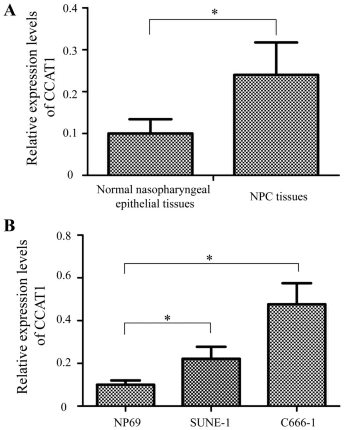
CCAT1 expression is upregulated in NPC
tissues and cell lines
The expression levels of CCAT1 were examined in 35
NPC tissues and 20 non-tumor tissues by RT-qPCR assay. As indicated
in Fig. 1A, the expression of CCAT1
was markedly upregulated in NPC tissues compared with normal
nasopharyngeal epithelial tissues (P<0.05).
The expression levels of CCAT1 were also examined in
two NPC cells (SUNE-1 and C666-1) and the normal nasopharyngeal
epithelial cell line (NP69). As indicated in Fig. 1B, CCAT1 expression was higher in
SUNE-1 and C666-1 cells compared with NP69 cells (P<0.05).
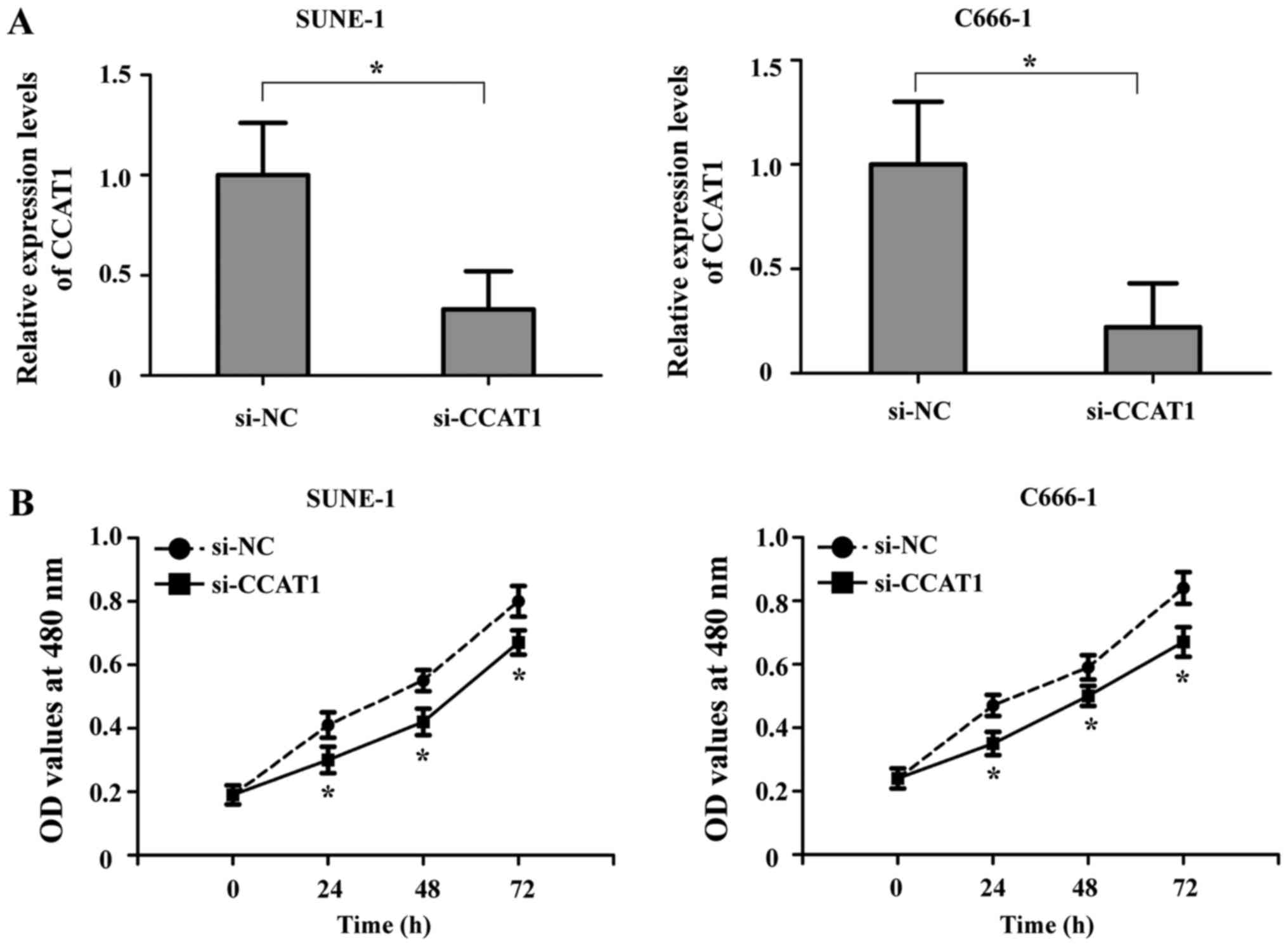
CCAT1 suppression inhibits the
proliferation of NPC cells in vitro
To examine the role of CCAT1 in the development of
NPC, SUNE-1 and C666-1 cells were transfected with si-CCAT1 or
si-NC by using Lipofectamine® 2000. As indicated in
Fig. 2A, CCAT1 expression in SUNE-1
and C666-1 cells was significantly downregulated following
transfection with si-CCAT1 compared with the si-NC group
(P<0.05). Furthermore, the effect of CCAT1 on the growth ability
of NPC cells was checked by MTT assay. The results showed that
proliferation ability of NPC cells was significantly reduced after
treatment with si-CCAT1 compared with the si-NC group (P<0.05;
Fig. 2B).
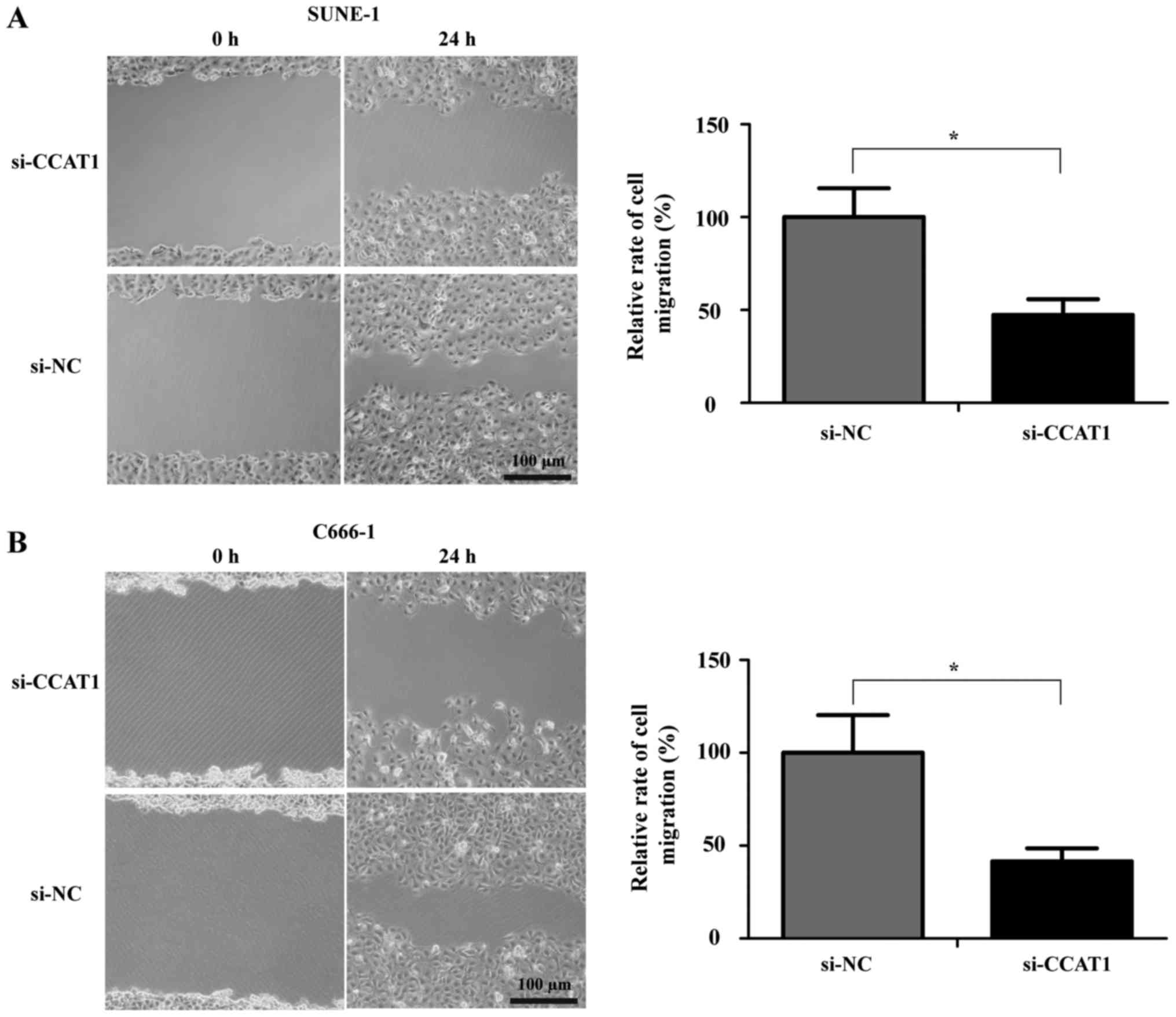
CCAT1 knockdown inhibits the migration
and invasion of NPC cells in vitro
Cell migration was examined by cell scratch assay.
The results revealed that the migratory ability of NPC cells that
were transfected with si-CCAT1 was decreased significantly compared
with the cells that were treated with si-NC. The relative rate of
migration was 47.3±8.4% for si-CCAT1-transfected SUNE-1 cells
(P<0.05; Fig. 3A), and 41.5±7.1%
for si-CCAT1-transfected C666-1 cells (P<0.05; Fig. 3B).
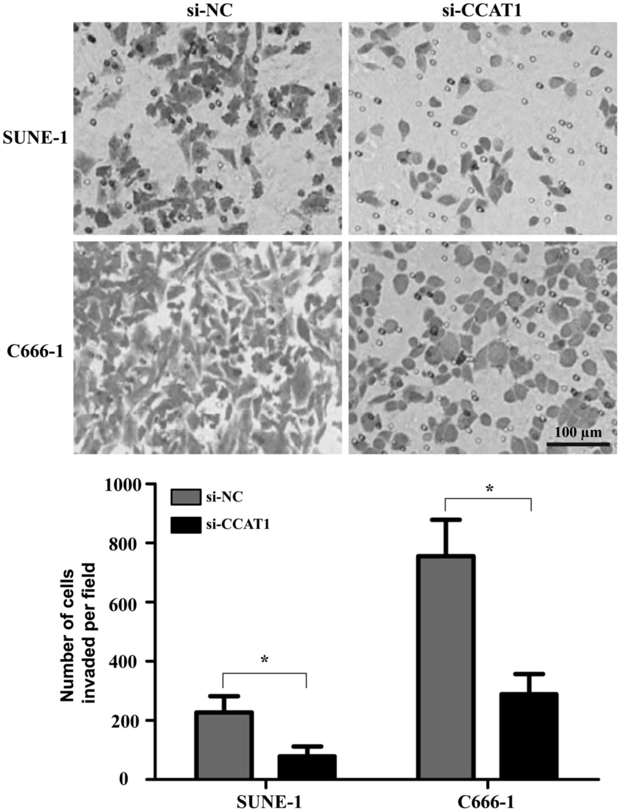
Transwell assay was also used to detect the invasion
of NPC cells following transfection with si-CCAT1 or si-NC. The
results indicated that the knockdown of CCAT1 inhibited the
invasion of SUNE-1 and C666-1 cells (P<0.05; Fig. 4).
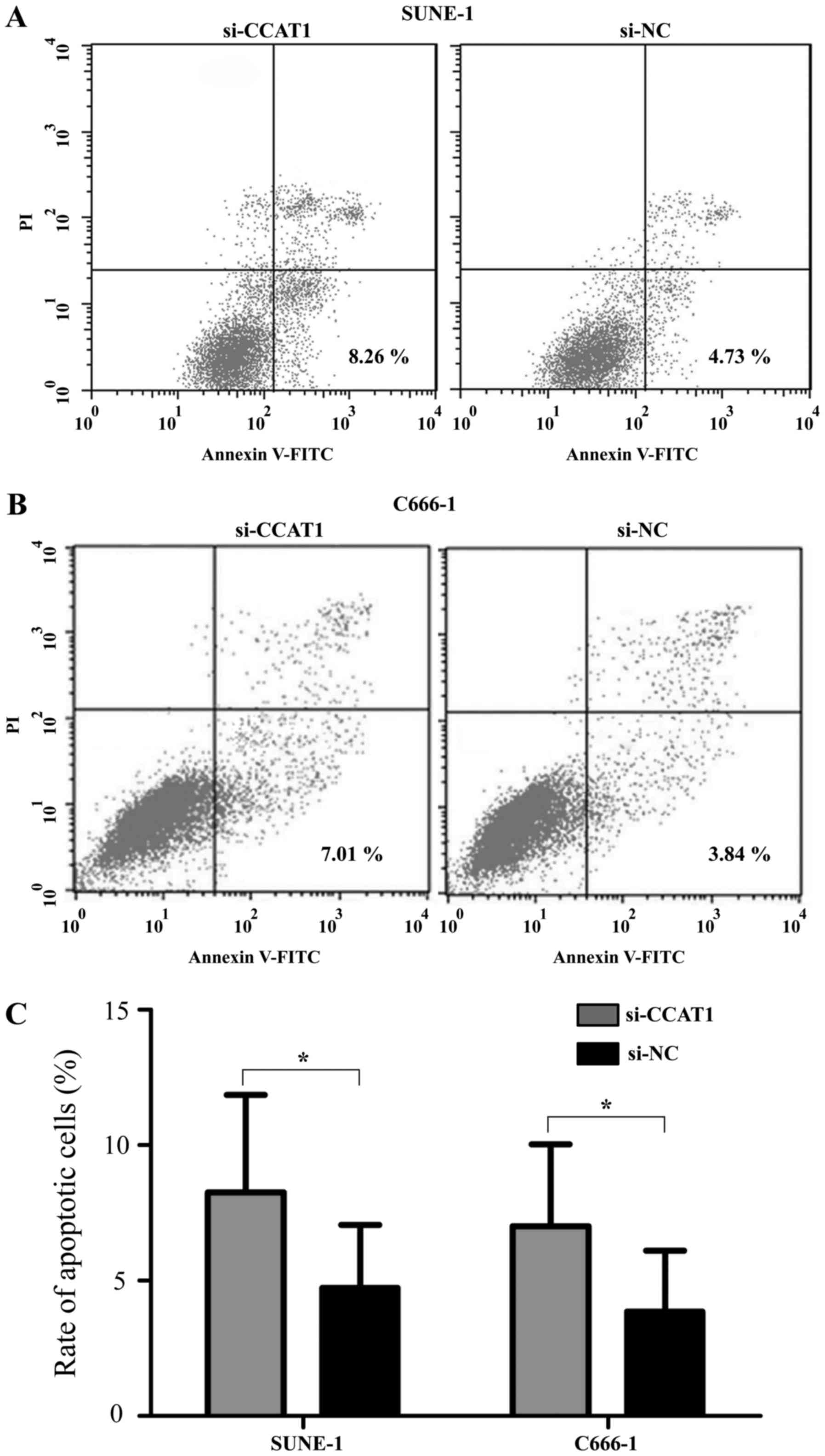
Targeting CCAT1 induces the apoptosis
of NPC cells in vitro
To investigate the effect of CCAT1 on the apoptosis
of NPC cells, flow cytometric assay was performed (Fig. 5). SUNE-1 and C666-1 cells were
transfected with si-CCAT1 or si-NC. As shown in Fig. 5A and C, the apoptotic rate was
increased significantly in the SUNE-1 cells that were transfected
with si-CCAT1 compared with the si-NC group (P<0.05).
Additionally, the apoptotic rate of C666-1 cells that were treated
with si-CCAT1 was markedly higher compared with cells that were
transfected with si-NC (P<0.05; Fig.
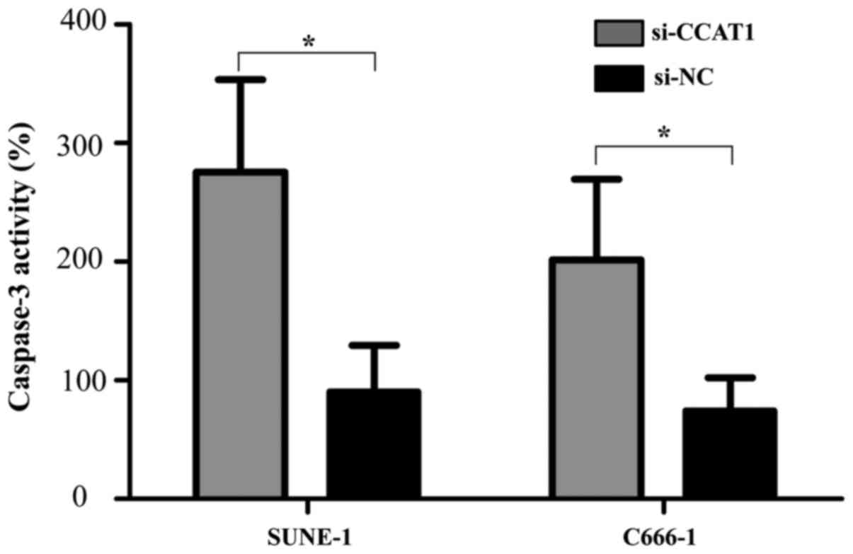
5B and C). The relative activity of caspase-3 was determined by
caspase-3 ELISA assay 48 h after transfection. Consistent with the
data from flow cytometry assay, the relative activity of caspase-3
was increased in SUNE-1 and C666-1 cells that were treated with
si-CCAT1 compared with the si-NC group (P<0.05; Fig. 6).
Discussion
Recently, novel treatment strategies have been
developed for NPC, including molecular targeted therapy,
immunotherapy and gene therapy (18).
However, the long-term prognosis of NPC is still poor due to local
recurrence and early metastasis, and NPC remains a major public
health concern (18,19). A growing number of studies have been
focused on the function of dysregulated lncRNAs in various human
cancers (20,21). An increasing number of studies have
showed that the aberrant expression of lncRNAs are involved in cell
proliferation, metastasis and apoptosis in NPC. For instance, Li
et al (22) reported that long
non-coding RNA H19 regulates enhancer of zeste 2 polycomb
repressive complex 2 subunit expression by interacting with miRNA
(miR)-630 and promoting cell invasion in NPC. Gong et al
(23) revealed that LOC401317
inhibited cell growth and activated apoptosis in NPC cells. Lu
et al (24) illustrated that
nuclear paraspeckle assembly transcript 1 regulated epithelial to
mesenchymal transition and radioresistance in via the miR-204/ZEB1
axis in NPC. Therefore, the identification of novel lncRNAs
expression signatures in the progression and development of NPC
will be helpful to identify novel diagnostic and therapeutic
biomarkers for NPC in the future.
CCAT1 is a recently identified lncRNA with 2628
nucleotide in length, which is located in the vicinity of a
well-known transcription regulator, c-Myc (25). Previous studies have demonstrated that
CCAT1 was significantly upregulated in colon cancer tissues when
compared with matched normal tissues (26). The expression of CCAT1 was also
indicated to be upregulated in patients with breast cancer tissues
compared with adjacent normal tissues (27). In the present study, the expression of
CCAT1 in NPC tissues and normal nasopharyngeal epithelial tissues
were compared. Consistent with the upregulation of CCAT1 in breast
cancer and colon carcinoma from previous reports (26,27), the
data from the present study demonstrated that CCAT1 was also
upregulated in NPC tissues in comparison with normal nasopharyngeal
epithelial tissues.
Previous studies have also reported that CCAT1
promoted the cell proliferation and invasion of hepatocellular
carcinoma cells (28). lnRNA CCAT1
also regulated the development of gallbladder cancer via negative
regulation of microRNA-218-5p (29).
C-Myc-activated lnRNA CCAT1 induced the proliferation and invasion
of colon cancer cells (30). In the
present study, the role of CCAT1 in NPC cells by applying was
examined using loss-of-function experiments. Accordingly, the
present study revealed that the inhibition of CCAT1 suppressed the
proliferation, migration and invasion of NPC cells, and induced the
apoptosis of NPC cells. Therefore, the present data indicated that
CCAT1 acts as an oncogene in NPC.
In conclusion, the present study is the first to
confirm that CCAT1 expression was upregulated in NPC tissues and
cell lines. It was also indicated that the knockdown of CCAT1
significantly suppressed the growth, migratory and invasive
abilities of NPC cells, and promoted cell apoptosis, suggesting
CCAT1 might be a promising biomarker for molecularly targeted
therapy of NPC.
Acknowledgements
Not applicable.
Funding
No funding was received.
Availability of data and materials
The datasets used and/or analyzed during the current
study are available from the corresponding author on reasonable
request.
Authors' contributions
YD and GJ designed and carried out the experiments.
YD and HY analyzed the data. YD and GJ wrote and revised the
manuscript.
Ethics approval and consent to
participate
The present study was approved by the ethics
committee of the Nuclear Industry 215 Hospital of Shaanxi Province.
Written informed consent was obtained from all patients prior to
the start of the study.
Consent for publication
Written informed consent was obtained from all
patients prior to the start of the study.
Competing interests
The authors declare that they have no competing
interest.
References
|
1
|
Wei WI and Sham JS: Nasopharyngeal
carcinoma. Lancet. 365:2041–2054. 2005. View Article : Google Scholar : PubMed/NCBI
|
|
2
|
Torre LA, Bray F, Siegel RL, Ferlay J,
Lortet-Tieulent J and Jemal A: Global cancer statistics, 2012. CA
Cancer J Clin. 65:87–108. 2015. View Article : Google Scholar : PubMed/NCBI
|
|
3
|
Ma BB, Hui EP and Chan AT: Systemic
approach to improving treatment outcome in nasopharyngeal
carcinoma: Current and future directions. Cancer Sci. 99:1311–1318.
2008. View Article : Google Scholar : PubMed/NCBI
|
|
4
|
Perri F, Dell'Oca I, Muto P, Schiavone C,
Aversa C, Fulciniti F, Solla R, Scarpati GD, Buonerba C, Di Lorenzo
G and Caponigro F: Optimal management of a patient with recurrent
nasopharyngeal carcinoma. World J Clin Cases. 2:297–300. 2014.
View Article : Google Scholar : PubMed/NCBI
|
|
5
|
Zeng Z, Huang H, Zhang W, Xiang B, Zhou M,
Zhou Y, Ma J, Yi M, Li X, Li X, et al: Nasopharyngeal carcinoma:
Advances in genomics and molecular genetics. Sci China Life Sci.
54:966–975. 2011. View Article : Google Scholar : PubMed/NCBI
|
|
6
|
Burgos JS: Involvement of the Epstein-Barr
virus in the nasopharyngeal carcinoma pathogenesis. Med Oncol.
22:113–121. 2005. View Article : Google Scholar : PubMed/NCBI
|
|
7
|
Lo KW, To KF and Huang DP: Focus on
nasopharyngeal carcinoma. Cancer Cell. 5:423–428. 2004. View Article : Google Scholar : PubMed/NCBI
|
|
8
|
Johnsson P and Morris KV: Expanding the
functional role of long noncoding RNAs. Cell Res. 24:1284–1285.
2014. View Article : Google Scholar : PubMed/NCBI
|
|
9
|
Lau E: Non-coding RNA: Zooming in on
lncRNA functions. Nat Rev Genet. 15:574–575. 2014. View Article : Google Scholar : PubMed/NCBI
|
|
10
|
Ponting CP, Oliver PL and Reik W:
Evolution and functions of long noncoding RNAs. Cell. 136:629–641.
2009. View Article : Google Scholar : PubMed/NCBI
|
|
11
|
Gibb EA, Brown CJ and Lam WL: The
functional role of long non-coding RNA in human carcinomas. Mol
Cancer. 10:382011. View Article : Google Scholar : PubMed/NCBI
|
|
12
|
Wilusz JE, Sunwoo H and Spector DL: Long
noncoding RNAs: Functional surprises from the RNA world. Genes Dev.
23:1494–1504. 2009. View Article : Google Scholar : PubMed/NCBI
|
|
13
|
Ørom UA, Derrien T, Beringer M, Gumireddy
K, Gardini A, Bussotti G, Lai F, Zytnicki M, Notredame C, Huang Q,
et al: Long noncoding RNAs with enhancer-like function in human
cells. Cell. 143:46–58. 2010. View Article : Google Scholar : PubMed/NCBI
|
|
14
|
Gupta RA, Shah N, Wang KC, Kim J, Horlings
HM, Wong DJ, Tsai MC, Hung T, Argani P, Rinn JL, et al: Long
non-coding RNA HOTAIR reprograms chromatin state to promote cancer
metastasis. Nature. 464:1071–1076. 2010. View Article : Google Scholar : PubMed/NCBI
|
|
15
|
Shi X, Sun M, Liu H, Yao Y, Kong R, Chen F
and Song Y: A critical role for the long non-coding RNA GAS5 in
proliferation and apoptosis in non-small-cell lung cancer. Mol
Carcinog. 54 Suppl 1:E1–E12. 2015. View
Article : Google Scholar : PubMed/NCBI
|
|
16
|
Zhang EB, Kong R, Yin DD, You LH, Sun M,
Han L, Xu TP, Xia R, Yang JS, De W and Chen JF: Long noncoding RNA
ANRIL indicates a poor prognosis of gastric cancer and promotes
tumor growth by epigenetically silencing of miR-99a/miR-449a.
Oncotarget. 5:2276–2292. 2014. View Article : Google Scholar : PubMed/NCBI
|
|
17
|
Livak KJ and Schmittgen TD: Analysis of
relative gene expression data using real-time quantitative PCR and
the 2(-Delta Delta C(T)) method. Methods. 25:402–408. 2001.
View Article : Google Scholar : PubMed/NCBI
|
|
18
|
Spratt DE and Lee N: Current and emerging
treatment options for nasopharyngeal carcinoma. Onco Targets Ther.
5:297–308. 2012.PubMed/NCBI
|
|
19
|
Wei WI and Kwong DL: Current management
strategy of nasopharyngeal carcinoma. Clin Exp Otorhinolaryngol.
3:1–12. 2010. View Article : Google Scholar : PubMed/NCBI
|
|
20
|
Yang G, Lu X and Yuan L: LncRNA: A link
between RNA and cancer. Biochim Biophys Acta. 1839:1097–1109. 2014.
View Article : Google Scholar : PubMed/NCBI
|
|
21
|
Gong Z, Zhang S, Zhang W, Huang H, Li Q,
Deng H, Ma J, Zhou M, Xiang J, Wu M, et al: Long non-coding RNAs in
cancer. Sci China Life Sci. 55:1120–1124. 2012. View Article : Google Scholar : PubMed/NCBI
|
|
22
|
Li X, Lin Y, Yang X, Wu X and He X: Long
noncoding RNA H19 regulates EZH2 expression by interacting with
miR-630 and promotes cell invasion in nasopharyngeal carcinoma.
Biochem Biophys Res Commun. 473:913–919. 2016. View Article : Google Scholar : PubMed/NCBI
|
|
23
|
Gong Z, Zhang S, Zeng Z, Wu H, Yang Q,
Xiong F, Shi L, Yang J, Zhang W, Zhou Y, et al: LOC401317, a
p53-regulated long non-coding RNA, inhibits cell proliferation and
induces apoptosis in the nasopharyngeal carcinoma cell line SUNE-1.
PLoS One. 9:e1106742014. View Article : Google Scholar : PubMed/NCBI
|
|
24
|
Lu Y, Li T, Wei G, Liu L, Chen Q, Xu L,
Zhang K, Zeng D and Liao R: The long non-coding RNA NEAT1 regulates
epithelial to mesenchymal transition and radioresistance in through
miR-204/ZEB1 axis in nasopharyngeal carcinoma. Tumour Biol.
37:11733–11741. 2016. View Article : Google Scholar : PubMed/NCBI
|
|
25
|
Alaiyan B, Ilyayev N, Stojadinovic A,
Izadjoo M, Roistacher M, Pavlov V, Tzivin V, Halle D, Pan H, Trink
B, et al: Differential expression of colon cancer associated
transcript1 (CCAT1) along the colonic adenoma-carcinoma sequence.
BMC Cancer. 13:1962013. View Article : Google Scholar : PubMed/NCBI
|
|
26
|
Mizrahi I, Mazeh H, Grinbaum R, Beglaibter
N, Wilschanski M, Pavlov V, Adileh M, Stojadinovic A, Avital I,
Gure AO, et al: Colon cancer associated transcript-1 (CCAT1)
expression in adenocarcinoma of the Stomach. J Cancer. 6:105–110.
2015. View Article : Google Scholar : PubMed/NCBI
|
|
27
|
Zhang XF, Liu T, Li Y and Li S:
Overexpression of long non-coding RNA CCAT1 is a novel biomarker of
poor prognosis in patients with breast cancer. Int J Clin Exp
Pathol. 8:9440–9445. 2015.PubMed/NCBI
|
|
28
|
Zhu H, Zhou X, Chang H, Li H, Liu F, Ma C
and Lu J: CCAT1 promotes hepatocellular carcinoma cell
proliferation and invasion. Int J Clin Exp Pathol. 8:5427–5434.
2015.PubMed/NCBI
|
|
29
|
Ma MZ, Chu BF, Zhang Y, Weng MZ, Qin YY,
Gong W and Quan ZW: Long non-coding RNA CCAT1 promotes gallbladder
cancer development via negative modulation of miRNA-218-5p. Cell
Death Dis. 6:e15832015. View Article : Google Scholar : PubMed/NCBI
|
|
30
|
He X, Tan X, Wang X, Jin H, Liu L, Ma L,
Yu H and Fan Z: C-Myc-activated long noncoding RNA CCAT1 promotes
colon cancer cell proliferation and invasion. Tumour Biol.
35:12181–12188. 2014. View Article : Google Scholar : PubMed/NCBI
|