Introduction
Liver cancer is one of the most common cancers and
is a leading cause of cancer-associated mortality in several
countries, including China and the United States (1,2). In
2018, liver cancer accounted for 8.2% of all cancer-associated
mortalities and 4.7% of all cancer cases (3). Hepatocellular carcinoma (HCC) is the
most common subtype of liver cancer, accounting for ~90% of all
cases (4). HCC mainly affects
patients with chronic liver diseases, such as hepatitis B vs. (HBV)
or hepatitis C vs. (HCV)-induced cirrhosis (5). With the popularization of viral
screening programs, the incidence of HCC is expected to decrease in
the future (6). However, other
factors, such as alcohol and tobacco consumption are also closely
associated with HCC (7). Thus, other
prevention and treatment approaches are still required.
In addition to viral infections, molecular factors
also play critical roles in the initiation and development of HCC
(8,9). Previous studies have demonstrated that
certain molecules in HCC can be used to develop novel therapeutic
targets, which aim to suppress cancer development by regulating
gene expression (10,11). It is well-known that long non-coding
RNAs (lncRNAs) exert their functions in cancer biology by
regulating gene expression (12),
suggesting that they may act as promising targets for targeted
therapy (13). Thus, functional
characterization of lncRNAs in cancers is required. lncRNA
TONSL-AS1 acts as a tumor suppressor in gastric cancer; however,
its role in HCC remains unknown (14). It was hypothesized that microRNA
(miRNA/miR)-135a, a critical player in cancer biology (15), targets TONSL-AS1 in HCC. Thus, the
present study aimed to investigate the role of TONSL-AS1 in HCC and
determine the potential interactions between TONSL-AS1 and miR-135a
in HCC.
Materials and methods
Patients and tissue samples
A total of 64 patients with HCC (38 men and 26
women; mean age, 52.1±6.9 years; age range, 40–64 years) at the
First Affiliated Hospital of Shenzhen University were enrolled in
the present study between January 2013 and January 2015. The
inclusion criteria were as follows: i) Patients with pathologically
confirmed HCC; ii) patients who underwent curative surgical
resection and iii) patients >18 years. The exclusion criteria
were as follows: i) Patients receiving preoperative chemotherapy or
radiotherapy and ii) patients who have two or more primary tumors
simultaneously or asynchronously. Prior to therapy, HCC tissues and
paired adjacent normal tissues (>5 cm away from cancer tissues)
were collected from all patients via biopsy. All the histological
diagnoses for HCC and normal tissues were confirmed by two
pathologists from the First Affiliated Hospital of Shenzhen
University and immediately subjected to total RNA extraction
following collection. Based on the medical records of all patients,
HBV infection was observed in 26 cases, HCV infection was observed
in 16 cases, both HBV and HCV infections were observed in 16 cases,
and no infections were observed in 6 cases. The present study was
approved by the Ethics Committee of the First Affiliated Hospital
of Shenzhen University (Shenzhen, China; approval no. 2013006) and
written informed consent was provided by all patients prior to the
study start.
Treatment and follow-up
Based on the tumor-node-metastasis (TNM) staging
system (16), of the 64 patients
included, 28 cases were in stages I and II, while 36 cases were in
stages III and IV. Treatment approaches were determined based on
the TNM stage and health conditions of the patients. From June 30,
2015, all patients were followed-up for 5 years in a monthly manner
through telephone to record their survival. The survival status of
patients was recorded during follow-up until June 30, 2020.
Patients who died of causes unrelated to HCC were excluded.
Survival analysis
Survival analysis was performed using the
Kaplan-Meier method and log-rank test. Patients were divided into
high and low TONSL-AS1 expression groups (n=32/group and median
TONSL-AS1 expression in HCC tissues was set as the cut-off
value).
IntaRNA2.0
The interaction between TONSL-AS1 and miR-135a was
predicted using IntaRNA2.0 (http://rna.informatik.uni-freiburg.de/IntaRNA).
Cell lines and cell transfection
The HCC cell lines, SNU-398 and SNU-475, were
purchased from the American Type Culture Collection. Cells were
maintained in RPMI-1640 medium (Sigma, India; 90%) supplemented
with 10% fetal bovine serum (MP Biomedicals), at 37°C with 5%
CO2 and 95% humidity. Cells were collected once they
reached ~85% confluence and used in subsequent experiments.
The TONSL-AS1 expressing vector was constructed with
pcDNA3.1 vector as the backbone (Invitrogen; Thermo Fisher
Scientific, Inc.). miR-135a mimic and negative control (NC) miRNA
were synthesized by Invitrogen; Thermo Fisher Scientific, Inc.
Vector (1 µg) or miRNA (50 nM) were transfected into SNU-398 and
SNU-475 cells using Lipofectamine® 2000 reagent
(Invitrogen; Thermo Fisher Scientific, Inc.), according to the
manufacturer's instructions. Transfection with empty vector or NC
miRNA served as the control groups, and control cells were
untransfected cells. Cells were cultured for 48 h at 37°C with 5%
CO2 and harvested for subsequent experimentation.
Sequences of transfection vectors were as follows: miR-135a mimics
forward, 5′-UAUGGCUUUUUAUUCCUAUGUGA-3′ and reverse,
5′-GUGCCGAGGAUUAGGGAUAUACU-3′; and NC mimics forward,
5′-UUCUCCGAACGUGUCACGUTT−3′ and reverse,
5′-ACGUGACACGUUCGGAGAATT−3′.
RNA preparation and reverse
transcription-quantitative (RT-q)PCR
Total RNA was extracted from tissues and cells using
RiboZol™ reagent (Amresco, LLC). RNA samples were digested to
remove genomic DNA using DNase I (Invitrogen; Thermo Fisher
Scientific, Inc.), at 37°C for 100 min. RNA integrity was assessed
using 5% urine-PAGE gel.
Total RNA was reverse transcribed into cDNA using
the PrimeScript RT reagent kit (Takara Bio, Inc.) at 37°C for 15
min. qPCR was subsequently performed using LightCycler®
480 SYBR Green I Master (Roche Diagnostics GmbH). The following
thermocycling conditions were used for qPCR: Initial denaturation
at 95°C for 30 sec, followed by 40 cycles at 95°C for 5 sec and at
60°C for 45 sec (17). The following
primer sequences were used for qPCR: TONSL-AS1 forward,
5′-AGCCCTCTCTCCCTGCACCAC−3′ and reverse,
5′-AGCTGAGCACCTTCCTGAAACCC-3′; miR-135a forward,
5′-AGCATAATACAGCAGGCACAGAC-3′ and reverse,
5′-AAAGGTTGTTCTCCACTCTCTCAC−3′; and U6 forward,
5′-GCUUCGGCAGCACAUAUACUAAAAU-3′ and reverse,
5′-CGCUUCACGAAUUUGCGUGUCAU-3′. TONSL-AS1 expression was measured
with 18S rRNA as the internal control.
miR-135a expression levels were measured as follows:
i) Addition of poly (A); ii) RT of miRNA and iii) miRNA qPCR. U6
was used as the internal control. Relative expression levels were
calculated using the 2−ΔΔcq method (18). All experiments were performed in
triplicate.
Cell proliferation assay
The Cell Counting Kit-8 (CCK-8) assay (cat. no.
ab228554; Abcam) was performed to assess the proliferative ability
of SNU-398 and SNU-475 cells. Cells were seeded into 96-well plates
at a density of 3,000 cells/well (in 0.1 ml medium) and cultured at
37°C, and optical density (OD) values were measured at a wavelength
of 450 nm every 24 h for a total of 4 days. CCK-8 solution was
added to each well for 2 h to reach 10% concentration, prior to
analyzing cell proliferation.
Statistical analysis
All experiments were performed in triplicate and
data are presented as the mean ± standard deviation. Differences
between two groups were compared using an unpaired two-tailed
Student's t-test, while one-way ANOVA and Tukey's post hoc test
were used to compare differences between multiple groups. Unpaired
Student's t-test was performed to assess the significance of cell
proliferation assay. Pearson's correlation coefficient was used to
determine the correlation between TONSL-AS1 and miR-135a expression
levels. Survival analysis was performed using the Kaplan-Meier
method and log-rank test. P<0.05 was considered to indicate a
statistically significant difference.
Results
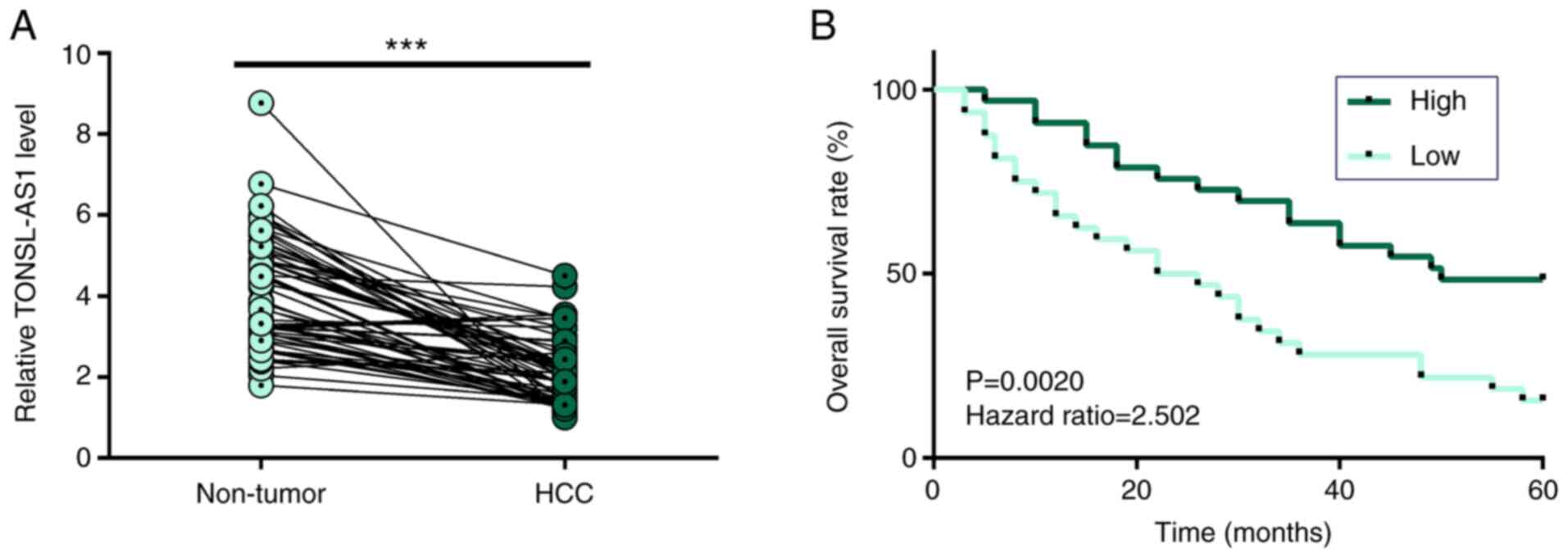
Downregulation of TONSL-AS1 expression
predicts poor survival of patients with HCC
RT-qPCR analysis was performed to detect TONSL-AS1
expression in HCC tissues and paired adjacent normal tissues from
64 patients with HCC. The results demonstrated that TONSL-AS1
expression was significantly downregulated in HCC tissues compared
with adjacent normal tissues (P<0.001; Fig. 1A). In addition, survival analysis
demonstrated that patients with low TONSL-AS1 expression had
significantly lower overall survival rates than those with high
TONSL-AS1 expression (Fig. 1B).
Notably, no significant differences in TONSL-AS1 expression were
observed between patients in different clinical stages or patients
with (mean value, 3.23±0.97) or without HBV/HCV (mean value,
3.04±0.72) infections (P>0.05) (data not shown). Taken together,
these results suggest that TONSL-AS1 may be an independent
prognostic factor for HCC.
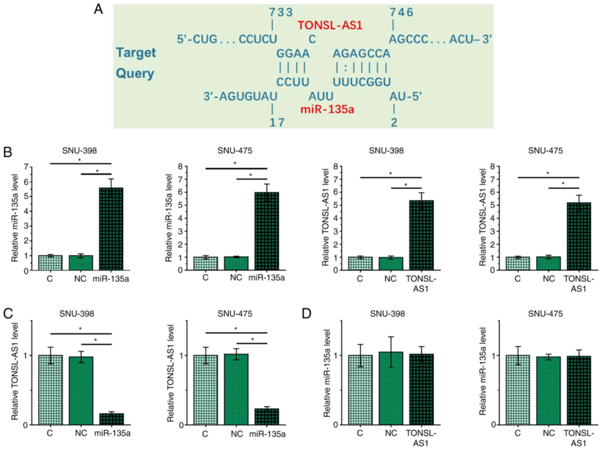
miR-135a targets TONSL-AS1 in SNU-398
and SNU-475 cells
The interaction between miR-135a and TONSL-AS1 was
predicted using IntaRNA2.0. The results indicated that miR-135a
targets TONSL-AS1 (Fig. 2A). To
further investigation their interaction, SNU-398 and SNU-475 cells
were transfected with miR-135a mimic or TONSL-AS1 expression
vector, and transfection efficiency was assessed via RT-qPCR
analysis 48 h post-transfection (P<0.05; Fig. 2B). The results demonstrated that
overexpression of miR-135a downregulated TONSL-AS1 expression
(Fig. 2C), while overexpression of
TONSL-AS1 had no effect on miR-135a expression (Fig. 2D).
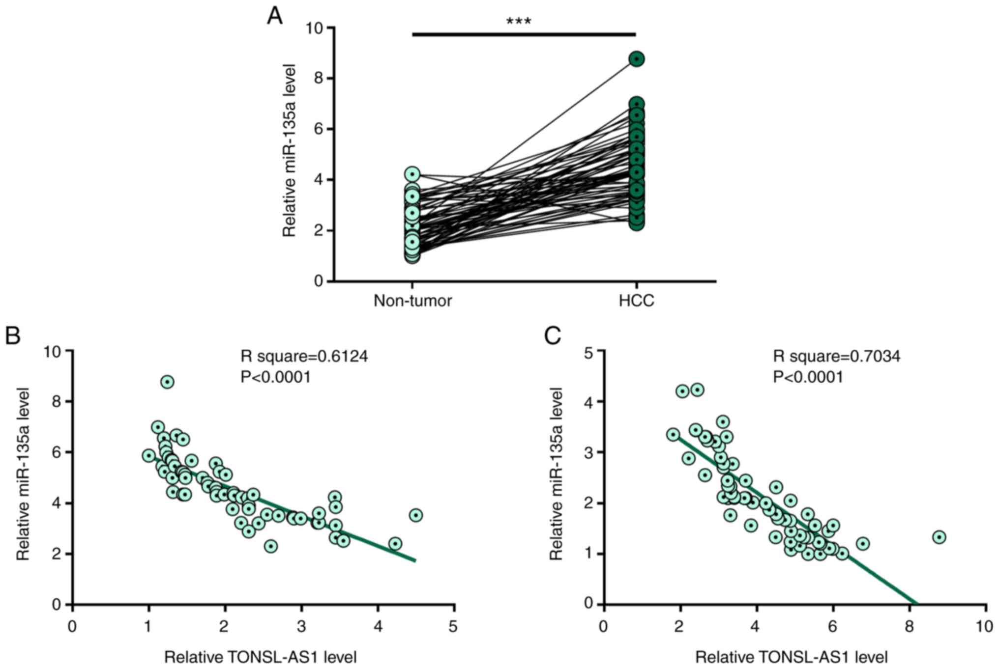
TONSL-AS1 and miR-135a are inversely
correlated with each other
RT-qPCR analysis was performed to detect miR-135a
expression in HCC tissues and paired adjacent normal tissues from
64 patients with HCC. The results demonstrated that miR-135a
expression was significantly upregulated in HCC tissues compared
with adjacent normal tissues (P<0.001; Fig. 3A). In addition, Pearson's correlation
coefficient analysis demonstrated that TONSL-AS1 and miR-135a
expression levels were inversely correlated with each other in both
HCC tissues (Fig. 3B) and adjacent
normal tissues (Fig. 3C).
Collectively, these results suggest that TONSL-AS1 and miR-135a
interact with each other.
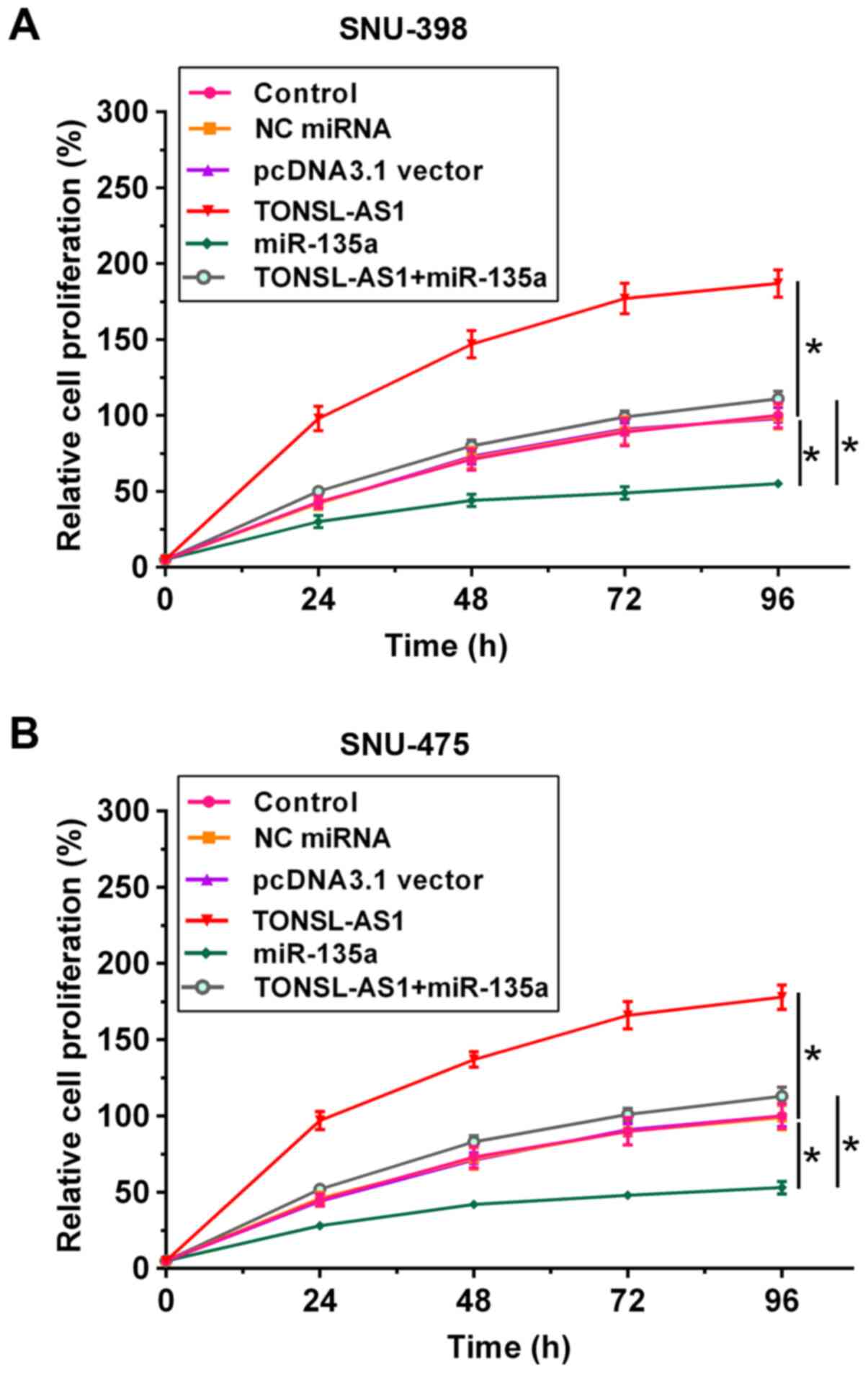
miR-135a targets TONSL-AS1 to promote
HCC cell proliferation
The CCK-8 assay was performed to assess the effects
of miR-135a and TONSL-AS1 on the proliferative ability of HCC
cells. The results demonstrated that overexpression of miR-135a
increased the proliferation of SNU-398 and SNU-475 cells that
compared with the control group. Conversely, overexpression of
TONSL-AS1 decreased cell proliferation. In addition, overexpression
of miR-135a attenuated the effect of overexpression of TONSL-AS1 on
cell proliferation (P<0.05; Fig. 4A
and B).
Discussion
The present study aimed to investigate the
interaction between miR-135a and TONSL-AS1 in HCC. The results
demonstrated that miR-135a and TONSL-AS1 expression levels were
altered in HCC. In addition, miR-135a may target TONSL-AS1 to
promote the proliferation of HCC cells.
A recent study demonstrated the involvement of
lncRNA TONSL-AS1 in HCC (14). It
has been demonstrated that TONSL-AS1 expression is downregulated in
HCC, and overexpression of TONSL-AS1 inhibits cancer cell invasion,
migration and proliferation (15).
To the best of our knowledge, the present study was the first to
investigate the role of TONSL-AS1 in HCC. The results of the
present study demonstrated that overexpression of TONSL-AS1
decreased the proliferation of HCC cells, suggesting that TONSL-AS1
may act as a tumor suppressor in HCC.
It is estimated that <15% of patients with HCC
survive >5 years following the initial diagnosis, even with
effective treatment (19). Due to
the lack of effective therapeutic approaches, the low survival rate
is unlikely to be significantly improved in the future. The results
of the present study demonstrated that high TONSL-AS1 expression
was closely associated with favorable survival outcomes in patients
with HCC. Thus, measuring TONSL-AS1 expression prior to therapy may
assist the prognosis of patients with HCC, thereby guiding the
determination of treatment approaches. However, further clinical
studies are required to confirm the results presented here.
miR-135a plays different roles in different types of
cancer (15,20). For example, miR-135a targets HOXA10
to promote breast cancer cell invasion and migration (15). In addition, miR-135a targets PHLPP2
and FOXO1 to promote the proliferation of bladder cancer cells
(20). The results of the present
study demonstrated that miR-135a expression was upregulated in HCC,
which in turn promoted cancer cell proliferation. Thus, miR-135a is
likely to act as an oncogene in HCC. These results are consistent
with previous findings that miR-135a promotes the progression of
HCC (21,22). However, these previous studies have
only reported the role of miR-135a in enhancing the metastasis of
HCC, whereas the present study suggests that miR-135a may also
accelerate the growth of HCC.
Previous studies have demonstrated that miR-135a
targets protein-coding genes to participate in cancer biology
(23–25). The results of the present study
demonstrated that miR-135a targeted TONSL-AS1 to participate in
HCC. Taken together, these results provide insight on the
functionality of miR-135a in cancer biology.
The present study is not without limitations. The
effects of TONSL-AS1/miR-135a on several signaling pathways remain
to be elucidated. In addition, the targets of miR-135a associated
with HCC need to be investigated. Thus, a comprehensive analysis is
required in the future. In conclusion, the results of the present
study suggest that TONSL-AS1 expression is downregulated and
miR-135a expression is upregulated in HCC. In addition, miR-135a
can target TONSL-AS1 to promote HCC cell proliferation.
Acknowledgements
Not applicable.
Funding
The present study was supported by the Science and
Technology Planning Project of Guangdong Province (grant no.
2014A020212701), the Medical Scientific Research Foundation of
Guangdong Province (grant no. B2014326), the Medical Scientific
Research Foundation of Guangdong Province (grant no. A2017338), the
Chinese Foundation for Hepatitis Prevention and Control (grant no.
TQGB20180220) and Shenzhen Municipal Science and Technology
Foundation (grant no. JCYJ20190806162818268).
Availability of data and materials
The datasets used and/or analyzed during the present
study are available from the corresponding author upon reasonable
request.
Authors' contributions
XD and JC acquired funding, performed the
experiments, analyzed the data and drafted the initial manuscript.
NZ, JC and YZ performed the experiments, and performed the
literature review and clinical research. CL and YN managed the
project, and performed the literature review and clinical research.
XD and YN confirmed the authenticity of all the raw data. All
authors have read and approved the final manuscript.
Ethics approval and consent to
participate
The present study was approved by the Ethics
Committee of the First Affiliated Hospital of Shenzhen University
(Shenzhen, China; approval no. 2013006) and written informed
consent was provided by all patients prior to the study start.
Patient consent for publication
Not applicable.
Competing interests
The authors declare that they have no competing
interests.
References
|
1
|
Zuo TT, Zheng RS, Zhang SW, Zeng HM and
Chen WQ: Incidence and mortality of liver cancer in China in 2011.
Chin J Cancer. 34:508–513. 2015. View Article : Google Scholar : PubMed/NCBI
|
|
2
|
Altekruse SF, Henley SJ, Cucinelli JE and
McGlynn KA: Changing hepatocellular carcinoma incidence and liver
cancer mortality rates in the United States. Am J Gastroenterol.
109:542–553. 2014. View Article : Google Scholar : PubMed/NCBI
|
|
3
|
Bray F, Ferlay J, Soerjomataram I, Siegel
RL, Torre LA and Jemal A: Global cancer statistics 2018: GLOBOCAN
estimates of incidence and mortality worldwide for 36 cancers in
185 countries. CA Cancer J Clin. 68:394–424. 2018. View Article : Google Scholar : PubMed/NCBI
|
|
4
|
Kulik L and El-Serag HB: Epidemiology and
management of hepatocellular carcinoma. Gastroenterology.
156:477–491.e1. 2019. View Article : Google Scholar : PubMed/NCBI
|
|
5
|
Hiotis SP, Rahbari NN, Villanueva GA,
Klegar E, Luan W, Wang Q and Yee HT: Hepatitis B vs hepatitis C
infection on viral hepatitis-associated hepatocellular carcinoma.
BMC Gastroenterol. 12:642012. View Article : Google Scholar : PubMed/NCBI
|
|
6
|
Lu T, Seto WK, Zhu RX, Lai CL and Yuen MF:
Prevention of hepatocellular carcinoma in chronic viral hepatitis B
and C infection. World J Gastroenterol. 19:8887–8894. 2013.
View Article : Google Scholar : PubMed/NCBI
|
|
7
|
Saran U, Humar B, Kolly P and Dufour JF:
Hepatocellular carcinoma and lifestyles. J Hepatol. 64:203–214.
2016. View Article : Google Scholar : PubMed/NCBI
|
|
8
|
Tarocchi M, Polvani S, Marroncini G and
Galli A: Molecular mechanism of hepatitis B virus-induced
hepatocarcinogenesis. World J Gastroenterol. 20:11630–11640. 2014.
View Article : Google Scholar : PubMed/NCBI
|
|
9
|
Marra M, Sordelli IM, Lombardi A, Lamberti
M, Tarantino L, Giudice A, Stiuso P, Abbruzzese A, Sperlongano R,
Accardo M, et al: Molecular targets and oxidative stress biomarkers
in hepatocellular carcinoma: An overview. J Transl Med. 9:1712011.
View Article : Google Scholar : PubMed/NCBI
|
|
10
|
Sawey ET, Chanrion M, Cai C, Wu G, Zhang
J, Zender L, Zhao A, Busuttil RW, Yee H, Stein L, et al:
Identification of a therapeutic strategy targeting amplified FGF19
in liver cancer by oncogenomic screening. Cancer Cell. 19:347–358.
2011. View Article : Google Scholar : PubMed/NCBI
|
|
11
|
Pan W, Li W, Zhao J, Huang Z, Zhao J, Chen
S, Wang C, Xue Y, Huang F, Fang Q, et al: lncRNA-PDPK2P promotes
hepatocellular carcinoma progression through the PDK1/AKT/Caspase 3
pathway. Mol Oncol. 13:2246–2258. 2019. View Article : Google Scholar : PubMed/NCBI
|
|
12
|
Esteller M: Non-coding RNAs in human
disease. Nat Rev Genet. 12:861–874. 2011. View Article : Google Scholar : PubMed/NCBI
|
|
13
|
Li CH and Chen Y: Targeting long
non-coding RNAs in cancers: Progress and prospects. Int J Biochem
Cell Biol. 45:1895–1910. 2013. View Article : Google Scholar : PubMed/NCBI
|
|
14
|
Wang P, Yang X, Zhao L, Liu D, Liu J and
Ding Y: A novel long non-coding RNA TONSL-AS1 regulates progression
of gastric cancer via activating TONSL. Exp Cell Res.
382:1114532019. View Article : Google Scholar : PubMed/NCBI
|
|
15
|
Chen Y, Zhang J, Wang H, Zhao J, Xu C, Du
Y, Luo X, Zheng F, Liu R, Zhang H and Ma D: miRNA-135a promotes
breast cancer cell migration and invasion by targeting HOXA10. BMC
Cancer. 12:1112012. View Article : Google Scholar : PubMed/NCBI
|
|
16
|
Park S, Choi S, Cho YA, Sinn DH, Kim JM,
Park CK and Ha SY: Evaluation of the American joint committee on
cancer (AJCC) 8th edition staging system for hepatocellular
carcinoma in 1,008 patients with curative resection. Cancer Res
Treat. 52:1145–1152. 2020.PubMed/NCBI
|
|
17
|
Lang N, Wang C, Zhao J, Shi F, Wu T and
Cao H: Long non-coding RNA BCYRN1 promotes glycolysis and tumor
progression by regulating the miR-149/PKM2 axis in non-small-cell
lung cancer. Mol Med Rep. 21:1509–1516. 2020.PubMed/NCBI
|
|
18
|
Livak KJ and Schmittgen TD: Analysis of
relative gene expression data using real-time quantitative PCR and
the 2(-Delta Delta C(T)) method. Methods. 25:402–408. 2001.
View Article : Google Scholar : PubMed/NCBI
|
|
19
|
Bertuccio P, Turati F, Carioli G,
Rodriguez T, La Vecchia C, Malvezzi M and Negri E: Global trends
and predictions in hepatocellular carcinoma mortality. J Hepatol.
67:302–309. 2017. View Article : Google Scholar : PubMed/NCBI
|
|
20
|
Mao XP, Zhang LS, Huang B, Zhou SY, Liao
J, Chen LW, Qiu SP and Chen JX: Mir-135a enhances cellular
proliferation through post-transcriptionally regulating PHLPP2 and
FOXO1 in human bladder cancer. J Transl Med. 13:862015. View Article : Google Scholar : PubMed/NCBI
|
|
21
|
Liu S, Guo W, Shi J, Li N, Yu X, Xue J, Fu
X, Chu K, Lu C, Zhao J, et al: MicroRNA-135a contributes to the
development of portal vein tumor thrombus by promoting metastasis
in hepatocellular carcinoma. J Hepatol. 56:389–396. 2012.
View Article : Google Scholar : PubMed/NCBI
|
|
22
|
Zeng YB, Liang XH, Zhang GX, Jiang N,
Zhang T, Huang JY, Zhang L and Zeng XC: miRNA-135a promotes
hepatocellular carcinoma cell migration and invasion by targeting
forkhead box O1. Cancer Cell Int. 16:632016. View Article : Google Scholar : PubMed/NCBI
|
|
23
|
Xie Y, Li F, Li Z and Shi Z: miR-135a
suppresses migration of gastric cancer cells by targeting
TRAF5-mediated NF-κB activation. Onco Targets Ther. 12:975–984.
2019. View Article : Google Scholar : PubMed/NCBI
|
|
24
|
Yang C, Zheng X, Ye K, Sun Y, Lu Y, Fan Q
and Ge H: miR-135a inhibits the invasion and migration of
esophageal cancer stem cells through the hedgehog signaling pathway
by targeting Smo. Mol Ther Nucleic Acids. 19:841–852. 2020.
View Article : Google Scholar : PubMed/NCBI
|
|
25
|
Wang J, Yang J, Zhang H, Liao Y, Xu D and
Ma S: Effects of miR-135a-5p and miR-141 on proliferation, invasion
and apoptosis of colorectal cancer SW620 cells. Oncol Lett.
20:914–920. 2020. View Article : Google Scholar : PubMed/NCBI
|