Introduction
There are trace amounts of heavy metals in
cosmetics. Heavy metals such as mercury (Hg), which is added to
skin-whitening cosmetics, may cause acute or chronic damage to
human cells. Hg, a divalent metal with no known biological
function, may cause several deleterious effects in adults (1,2), as
well as in developing organisms (3,4), which
primarily involve the central nervous system (5–7) and
the kidneys (1,8,9). Young
animals seem to be more sensitive to Hg toxicity than adults,
particularly during the first days following birth. Hg is also a
widespread environmental and industrial pollutant that induces
severe adverse effects in humans as well as the environment
(10). Its carcinogenic activity
has been well-documented. Hg is also known to alter the
intracellular redox homeostasis (11,12),
which is recognized as a factor that determines cell fate (13). The outcome of cells exposed to
Hg-containing compounds depends on the chemical characteristics of
the compound, as well as on its dosage, accounting for the various
results reported in the literature, ranging from improved cell
survival to apoptosis and necrosis.
Keratinocytes have long been considered the
structural backbone of the epidermis; however, there is increasing
evidence that they play an active role in the pathogenesis of skin
damage by heavy metals (14).
Available histopathological (15)
and cytotoxicological (16–18) studies describing keratinocyte damage
by mercury chloride (HgCl2) are currently limited. This
underlines the importance of investigating the direct cytotoxic
effects of the metals on keratinocytes, as well as intracellular
damage, for which available data are limited.
Metallothioneins (MTs) are ubiquitous, low-molecular
weight proteins, rich in cysteine residues. Their high content of
sulfhydrilic amino acids (∼30%) gives these proteins unique
metal-binding properties (19,20).
Factors such as exposure to toxic or essential metals (3,21–23),
stress (24,25), radiation (26) and other agents (27,28),
promote the synthesis of these molecules (29). With respect to their biological
functions and due to the metal affinity of their sulfhydryl groups,
it is believed that MTs possess antioxidant properties (26,30),
are involved in the homeostasis of essential metals such as zinc
(Zn) and copper (Cu) (20,29) and act as detoxifying agents from
metal ions (20,31,32).
In this study, we investigated the cytotoxicity of
HgCl2 to human keratinocytes, using human
keratinocyte-derived HaCaT cells as an experimental model. In
addition, we focused on HgCl2-induced HaCaT cell damage
and examined the expression of MTs.
Materials and methods
Materials
Human keratinocyte-derived (HaCaT) cells were
obtained from the Food Industry Research and Development Institute
(Hsinchu, Taiwan). Dulbecco’s modified Eagle’s medium (DMEM),
heat-inactivated fetal calf serum (FCS), penicillin-streptomycin
solution and trypsin-EDTA solution were purchased from Life
Technologies Corporation (Carlsbad, CA, USA). Sterile
dimethylsulfoxide (DMSO)
3-(4,5-dimethylthiazol-2-yl)-2,5-diphenyltetrazolium bromide (MTT)
and HgCl2 were purchased from Sigma-Aldrich (St. Louis,
MO, USA).
Cell culture
HaCaT cells were grown in DMEM supplemented with
heat-inactivated FCS (10%; v/v), streptomycin (100 U/ml) and
penicillin (0.1 mg/ml), in a humidified atmosphere of 5%
CO2 at 37°C. The culture medium was changed three times
a week. The cells were subcultured following trypsinization and
seeded in 6-well plate at a density of 1×10⁵ cells per
cm2.
Cells treated with HgCl2
The keratinocytes were treated with HgCl2
(0.25–1.5 μM) at 37°C for 24 h. When the non-treated control
cells were grown confluently, the cell groups were prepared for
cell viability assay or MT western blot analysis.
MTT assay
The cell viability was monitored following treatment
with various concentrations of HgCl2. MTT was used to
quantify the metabolically active living cells. Mitochondrial
dehydrogenases metabolize MTT to a purple formazan dye, which was
measured photometrically at 570 nm using a spectrophotometer
(33).
Western blot analysis for MT protein
expression
Cell homogenates were prepared by sonication of
cells in 600 μl of ice-cold lysis buffer, containing 50 mM
Tris-HCl (pH 8.0), 150 mM NaCl, 0.02% sodium azide, 100
μg/ml PMSF, 1 μg/ml aprotinin and 1% NP-40.
Homogenates were clarified by centrifugation at 20,000 × g for 45
min at 4°C. Total protein concentration was determined using the
BCA (Bio-Rad, Hercules, CA, USA) assay. Samples (50 μg of
total protein) from HaCaT cells treated for 24 h with various
concentrations of HgCl2 were analyzed for human MT
proteins, using sodium dodecyl sulfate-polyacrylamide gel
electrophoresis (SDS-PAGE; Laemmli, 1970) in 10–20% gradient gels.
Proteins were electrophoretically transferred to nitrocellulose
membranes. The resulting membranes were incubated in 2.5%
glutaraldehyde for 1 h and then washed 3 times for 5 min in
phosphate buffer (8.1 mM Na2HPO4, 1.2 mM
KH2PO4, 2.7 mM KCl, pH 7.4). Monoethanolamine
(50 mM) was added to the third wash solution to quench residual
glutaraldehyde reactivity. MT proteins were detected by Immun-Star
Chemiluminescent Protein Detection Systems (Bio-Rad). A monoclonal
antibody to polymerized equine renal MT (Dako, Carpinteria, CA,
USA) was used for immunodetection.
Statistical analysis
Means ± standard error (SE) were calculated in
triplicate. A statistical significance between the groups was
determined by the Student’s t-test. P<0.05 was considered to
indicate a statistically significant difference between the two
groups.
Results
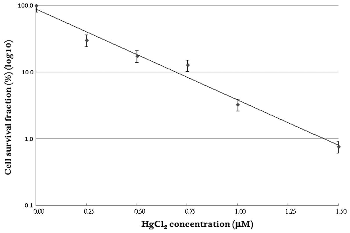
Cell survival fractions of HaCaT cells
treated with HgCl2 at various concentrations
Comparison of cell survival fractions in HaCaT cells
treated with HgCl2 at various concentrations from 0.25
to 1.5 μM is shown in Fig.
1. The cell survival fraction was 38.08% when the keratinocytes
were treated with 0.25 μM of HgCl2. The cell
survival fractions were 17.59, 12.76, 3.29 and 0.77%, when the
keratinocytes were treated with 0.5, 0.75, 1 and 1.5 μM of
HgCl2, respectively. For each concentration
investigated, a linear characteristic concentration-response curve
was observed, with decreased cell survival at increasing
concentrations of HgCl2 on a semi-log scale.
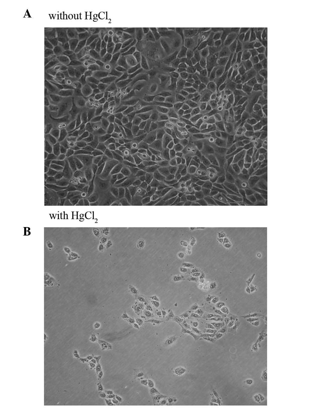
Effect of HgCl2 on HaCaT cell
morphology
Keratinocytes were treated with HgCl2 for
24 h or left untreated. The HaCaT cell morphology is shown in
Fig. 2. The cell membrane of
untreated cells is clear and intact (Fig. 2A), whereas that of
HgCl2-treated cells is unclear and interrupted (Fig. 2B).
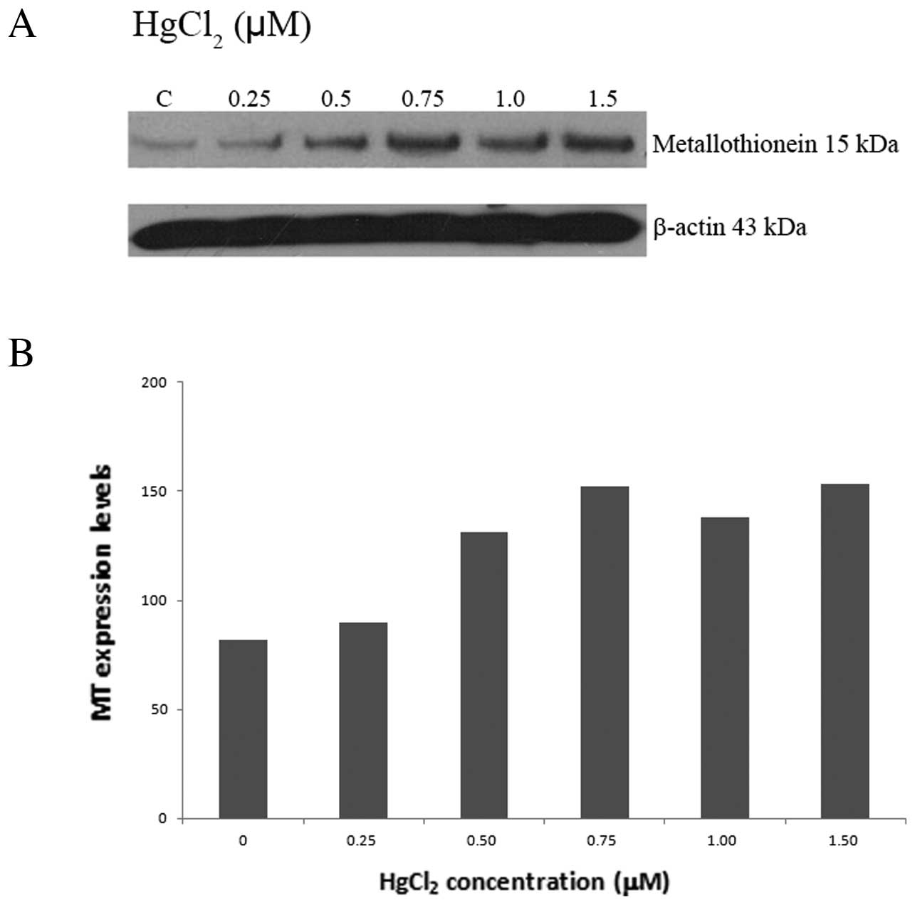
Effect of HgCl2 on MT
expression
MT expression levels in HaCaT cells treated with
various concentrations of HgCl2 are presented in
Fig. 3. MT expression levels
increased significantly with the increase in the concentrations of
HgCl2.
Discussion
The purpose of this study was to assess the
cytotoxicity of HgCl2 to the keratinocytes, as well as
MT expression in HgCl2-treated keratinocytes. The
results demonstrated that exposure of HaCaT cells to
HgCl2 resulted in dose-dependent cell death and distinct
cell membrane damage. Reports of Hg poisoning due to exposure to
skin-whitening creams, ointments and soaps have increased
significantly over the past few years. Furthermore, since people
with lighter skin tone may represent a higher status in certain
cultures, skin-whitening cosmetics are widely used by women to
enhance their appeal (34–36). Otto et al(36) detected high Hg concentrations in the
blood and urine of Balkan refugees of varying ages who had been
exposed to a Hg-based skin-bleaching ointment.
We have demonstrated that exposure of keratinocytes
to HgCl2 resulted in cell membrane damage. Picoli et
al(37) also investigated the
effect of HgCl2 on gap junction intercellular
communication (GJIC) in cultured human keratinocytes. They
demonstrated that subcytotoxic concentrations of HgCl2,
as low as 10 nM, may cause inhibition of the GJIC. In addition,
they demonstrated that HgCl2-treated keratinocytes
exhibited a decrease in free thiols and accumulation of
mitochondria-derived reactive oxygen species, albeit no effect on
the respiratory chain activity was observed.
This study has demonstrated that MT expression may
be induced by HgCl2 in HaCaT cells. Kramer et
al(38) demonstrated that MT
may be induced by Hg+2 in neuronal cells and induced MT
decreases the rate of metal binding to other structures, providing
protection against metal toxicity (39). Apart from Hg, MT also plays a role
in the homeostasis of essential metals such as Zn and Cu, the
detoxication of toxic metals such as Cadmium (Cd) and protection
against oxidative stress (40–42).
Richards et al(43) and
McCormick et al(44)
demonstrated that plasma zinc concentrations were related to MT
expression, further suggesting an association with cellular zinc
homeostasis. Ogra et al(41)
demonstrated that cell viability was significantly decreased in
MT-null cells compared to wild-type cells by Cu(I)-specific
chelator treatment (41). They also
showed that MT expression levels were increased by Cu(I)-specific
chelator treatment in wild-type cells. Thus, MT was induced under
Cu-deficient conditions, in order to maintain the activities of
intracellular cuproenzymes, such as cytochrome c oxidase and
Cu/Zinc superoxidase dismutase. Urani et al(42) showed that MT expression was
upregulated following exposure to CdCl2, with a
saturation curve at 48 as well as 72 h. High levels of MT possibly
confer an acquired tolerance to stress and protection against cell
injury, as demonstrated by the low cytotoxicity values.
In conclusion, our results demonstrated that
exposure of HaCaT cells to HgCl2 resulted in significant
dose-dependent cell death and cell membrane damage. Moreover, MT
expression may be induced by HgCl2 in HaCaT cells. This
suggests that MT protects the keratinocytes against
HgCl2-induced toxicity.
Acknowledgements
This study was supported by grant no.
NSC 98-2314-B-238-001 from the National Science Council and grant
no. VIT-98-CM-01 from Vanung University, Taiwan.
References
|
1
|
Emanuelli T, Rocha JB, Pereira ME,
Porciúncula LO, Morsch VM, Martins AF and Souza DO: Effect of
mercuric chloride intoxicationand dimercaprol treatment on
δ-aminolevulinate dehydratase from brain, liver and kidney of adult
mice. Pharmacol Toxicol. 79:136–143. 1996.
|
|
2
|
Shigematsu J, Yasuda T, Goto Y, Tanaka K,
Tobimatsu S and Kato M: Recovery of brain dysfunction after
methylmercury exposure in rats. J Neurol Sci. 182:61–68. 2000.
View Article : Google Scholar : PubMed/NCBI
|
|
3
|
Peixoto NC, Roza T, Flores EM and Pereira
ME: Effects of zinc and cadmium on HgCl2-δ-ALA-D
inhibition and Hg levels in tissues of suckling rats. Toxicol Lett.
146:17–25. 2003.PubMed/NCBI
|
|
4
|
Roza T, Peixoto NC, Welter A, Flores EM
and Pereira ME: 2,3-Dimercapto-1-propanol does not alter the
porphobilinogen synthase inhibition but decreases the mercury
content in liver and kidney of suckling rats exposed to
HgCl2. Basic Clin Pharmacol Toxicol. 96:302–308. 2005.
View Article : Google Scholar : PubMed/NCBI
|
|
5
|
Pereira ME, Morsch VM, Christofari RS and
Rocha JB: Methyl mercury exposure during post-natal brain growth
alters behavioral response to SCH 23390 in young rats. Bull Environ
Contam Toxicol. 63:256–262. 1999. View Article : Google Scholar : PubMed/NCBI
|
|
6
|
Rocha JB, Rocha LK, Emanuelli T and
Pereira ME: Effect of mercuric chloride and lead acetate treatment
during the second stage of rapid post-natal brain growth on the
behavioral response to chlorpromazine and on δ-ALA-D activity in
weaning rats. Toxicol Lett. 125:143–150. 2001.PubMed/NCBI
|
|
7
|
Peixoto NC, Roza T, Morsch VM and Pereira
ME: Behavioral alterations induced by HgCl2 depend on
the postnatal period of exposure. Int J Devl Neurosci. 25:39–46.
2007. View Article : Google Scholar : PubMed/NCBI
|
|
8
|
Magos L, Webb M and Butler WH: The effect
of cadmium pretreatment on the nephrotoxic action and kidney uptake
of mercury in male and female rats. Br J Exp Pathol. 55:589–594.
1974.PubMed/NCBI
|
|
9
|
Peixoto NC and Pereira ME: Effectiveness
of ZnCl2in protecting against nephrotoxicity induced by
HgCl2in newborn rats. Ecotoxicol Environ Saf.
66:441–446. 2007.
|
|
10
|
Clarkson TW and Magos L: The toxicology of
mercury and its chemical compounds. Crit Rev Toxicol. 36:609–662.
2006. View Article : Google Scholar : PubMed/NCBI
|
|
11
|
Valko M, Morris H and Cronin MT: Metals,
toxicity and oxidative stress. Curr Med Chem. 12:1161–1208. 2005.
View Article : Google Scholar : PubMed/NCBI
|
|
12
|
Valko M, Rhodes CJ, Moncol J, Izakovic M
and Mazur M: Free radicals, metals and antioxidants in oxidative
stress-induced cancer. Chem Biol Interact. 160:1–40. 2006.
View Article : Google Scholar : PubMed/NCBI
|
|
13
|
D’Autréaux B and Toledano MB: ROS as
signalling molecules: mechanisms that generate specificity in ROS
homeostasis. Nat Rev Mol Cell Biol. 8:813–824. 2007.PubMed/NCBI
|
|
14
|
Luger TA, Schwarz T, Krutmann J, Kirnbauer
R, Neuner P, Kock A, Urbanski A, Borth W and Schauer E:
Interleukin-6 is produced by epidermal cells and plays an important
role in the activation of human T-lymphocytes and natural killer
cells. Ann NY Acad Sci. 557:405–414. 1989. View Article : Google Scholar : PubMed/NCBI
|
|
15
|
Willis CM, Stephens CJ and Wilkinson JD:
Epidermal damage induced by irritants in man: a light and electron
microscopic study. J Invest Dermatol. 93:695–699. 1989. View Article : Google Scholar : PubMed/NCBI
|
|
16
|
Picardo M, Zompetta C, De Luca C,
Cristaudo A, Cannistraci C, Faggioni A and Santucci B:
Nickel-keratinocyte interaction: a possible role in sensitization.
Br J Dermatol. 122:729–735. 1990. View Article : Google Scholar : PubMed/NCBI
|
|
17
|
Little MC, Gawkrodger DJ and MacNeil S:
Chromium- and nickel-induced cytotoxicity in normal and transformed
human keratinocytes: an investigation of pharmacological approaches
to the prevention of Cr(VI)-induced cytotoxicity. Br J Dermatol.
134:199–207. 1996. View Article : Google Scholar : PubMed/NCBI
|
|
18
|
Brosin A, Wolf V, Mattheus A and Heise H:
Use of XTT-assay to assess the cytotoxicity of different
surfactants and metal salts in human keratinocytes (HaCaT):
Afeasible method for in vitro testing of skin irritants. Acta Derm
Venereol. 77:26–28. 1997.
|
|
19
|
Chan J, Huang Z, Merrifield ME, Salgado MT
and Stillman MJ: Studies of metal binding reactions in
metallothioneins by spectroscopic, molecular biology, and molecular
modelling techniques. Coord Chem Rev. 233–234:319–339. 2002.
|
|
20
|
Dabrio M, Rodríguez AR, Bordin G, Bebianno
MJ, De Ley M, Sestáková I, Vasák M and Nordberg M: Recent
developments in quantification methods for metallothionein. J Inorg
Biochem. 88:123–134. 2002. View Article : Google Scholar : PubMed/NCBI
|
|
21
|
Goering PL and Fowler BA: Metal
constitution of metallothionein influences inhibition of
δ-aminolaevulinic acid dehydratase (porphobilinogen synthase) by
lead. Biochem J. 245:339–345. 1987.PubMed/NCBI
|
|
22
|
Pedersen SN, Pedersen KL, Hojrup P,
Knudsen J and Depledge MH: Induction and identification of
cadmium-, zinc- and copper-metallothioneins in the shore crab
Carcinus maenas (L.). Comp Biochem Physiol C Pharmacol
Toxicol Endocrinol. 120:251–259. 1998. View Article : Google Scholar : PubMed/NCBI
|
|
23
|
Bebianno MJ and Langston WJ:
Metallothionein induction in mussels exposed to a metal mixture.
Metallothionein IV (Advances in Life Sciences). Klaassen CD:
Birkhäuser Verlag; Basel, Switzerland: pp. 187–194. 2009
|
|
24
|
Kondoh M, Tsukahara R, Kuronaga M,
Higashimoto M, Takiguchi M and Sato M: Enhancement of MT synthesis
by leptin in fasted mice. Life Sci. 71:2425–2433. 2002. View Article : Google Scholar : PubMed/NCBI
|
|
25
|
Kondoh M, Kamada K, Kuronaga M,
Higashimoto M, Takiguchi M, Watanabe Y and Sato M: Antioxidant
property of metallothionein in fasted mice. Toxicol Lett.
143:301–306. 2003. View Article : Google Scholar : PubMed/NCBI
|
|
26
|
Cai L, Satoh M, Tohyama C and Cherian MG:
Metallothionein in radiation exposure: its induction and protective
role. Toxicology. 132:85–98. 1999. View Article : Google Scholar : PubMed/NCBI
|
|
27
|
Rojas P, Cerutis DR, Happe HK, Murrin LC,
Hao R, Pfeiffer RF and Ebadi M: 6-Hydroxydopamine-mediated
induction of rat brain metallothionein I mRNA. Neurotoxicology.
17:323–334. 1996.PubMed/NCBI
|
|
28
|
Theocharis SE, Margeli AP, Skaltsas SD,
Spiliopoulou CA and Koutselinis AS: Induction of metallothionein in
the liver of carbon tetrachloride intoxicated rats: an
immunohistochemical study. Toxicology. 161:129–138. 2001.
View Article : Google Scholar : PubMed/NCBI
|
|
29
|
Dunn MA, Blalock TL and Cousins RJ:
Metallothionein. Proc Soc Exp Biol Med. 185:107–119. 1987.
View Article : Google Scholar
|
|
30
|
Cai L and Cherian MG: Zinc-metallothionein
protects from DNA damage induced by radiation better than
glutathione and copper- or cadmium-metallothioneins. Toxicol Lett.
136:193–198. 2003. View Article : Google Scholar : PubMed/NCBI
|
|
31
|
Stillman MJ: Metallothioneins. Coord Chem
Rev. 144:461–511. 1995. View Article : Google Scholar
|
|
32
|
Yoshida M, Satoh M, Shimada A, Yasutake A,
Sumi Y and Tohyama C: Pulmonary toxicity caused by acute exposure
to mercury vapor is enhanced in metallothionein-null mice. Life
Sci. 64:1861–1867. 1999. View Article : Google Scholar : PubMed/NCBI
|
|
33
|
Green LM, Reade JL and Ware CF: Rapid
colorimetric assay for cell viability: application to the
quantitation of cytotoxic and growth inhibitory lymphokines. J
Immunol Methods. 70:257–268. 1984. View Article : Google Scholar : PubMed/NCBI
|
|
34
|
Jovanovic S, Maisner V, Horras-Hun G,
Gabrio T and Schwenk M: Poisoning of a family by a
mercury-containing ointment. Gesundheitswesen. 59:405–408. 1997.(In
German).
|
|
35
|
Weldon MM, Smolinski MS, Maroufi A, Hasty
BW, Gilliss DL, Boulanger LL, Balluz LS and Dutton RJ: Mercury
poisoning associated with a Mexican beauty cream. West J Med.
173:15–18. 2000. View Article : Google Scholar : PubMed/NCBI
|
|
36
|
Otto M, Ahlemeyer C, Tasche H and von
Mehlendahl KE: Endemic mercury burden caused by a bleaching
ointment in Balkan refugees. Gesundheitswesen. 56:686–689. 1994.(In
German).
|
|
37
|
Piccoli C, D’Aprile A, Scrima R, Ambrosi
L, Zefferino R and Capitanio N: Subcytoxic mercury chloride
inhibits gap junction intercellular communication by a redox- and
phosphorylation-mediated mechanism. Free Radical Bio Med.
52:916–927. 2012. View Article : Google Scholar
|
|
38
|
Kramer KK, Zoelle JT and Klaassen CD:
Induction of metallothionein mRNA and protein in primary murine
neuron cultures. Toxicol Appl Pharmacol. 141:1–7. 1996. View Article : Google Scholar : PubMed/NCBI
|
|
39
|
Cherian MG and Nordberg M: Cellular
adaptation in metal toxicology and metallothionein. Toxicology.
28:1–15. 1983. View Article : Google Scholar : PubMed/NCBI
|
|
40
|
Davis SR and Cousins RJ: Metallothionein
expression in animals: a physiological perspective on function. J
Nutr. 130:1085–1088. 2000.PubMed/NCBI
|
|
41
|
Ogra Y, Aoyama M and Suzuki KT: Protective
role of metallothionein against copper depletion. Arch Biochem
Biophys. 451:112–118. 2006. View Article : Google Scholar : PubMed/NCBI
|
|
42
|
Urani CM, Melchioretto P, Canevali C,
Morazzoni F and Gribaldo L: Metallothionein and hsp70 expression in
HepG2 cells after prolonged cadmium exposure. Toxicol In Vitro.
21:314–319. 2007. View Article : Google Scholar : PubMed/NCBI
|
|
43
|
Richards MP and Cousins RJ: Mammalian zinc
homeostasis: requirement for RNA and metallothionein synthesis.
Biochem Biophys Res Commun. 64:1215–1223. 1975. View Article : Google Scholar : PubMed/NCBI
|
|
44
|
McCormick CC, Menard MP and Cousins RJ:
Induction of hepatic metallothionein by feeding zinc to rats of
depleted zinc status. Am J Physiol. 240:E414–E421. 1981.PubMed/NCBI
|