Introduction
Canine malignant mammary gland tumors (CMMGTs) are
the most common malignancies observed in females, with a low
post-surgery overall survival rate (OSR) (1,2).
Statistical analyses indicate that in canine mammary gland tumors,
the recurrence risks of malignant tumors are 13-fold higher than
benign tumors (3). Currently,
numerous studies are being conducted on CMMGTs and it has been
revealed that the biological behavior of CMMGTs is similar to that
of the human breast cancer (HBC) (4). The development of molecular profiling
in cancer allows the identification of significant molecular
pathways and the description of novel biomarkers (5).
Biomarkers have specific roles in the determination
of OSR and are recognized as a novel targeted therapy. Therefore,
the assessment of biomarkers is considered to be useful in cancer
research. By contrast, novel molecular pathways have aided in
achieving therapeutic goals. Thus far, numerous significant
molecular pathways in mammary gland tumors have been recognized and
are being extensively studied (4).
As aforementioned, there are similarities between HBC and CMMGTs.
Therefore, CMMGTs are considered to be an appropriate model for the
development of recombinant drugs and monoclonal antibodies for the
treatment of HBC (6). Tumoral
xenograft models have been recommended as the preclinical phase of
cancer, however, increasing evidence suggests that spontaneous
CMMGTs have more similarities with HBC (3).
In recent years, the vimentin biomarker has been
extensively investigated in cancers. The overexpression of vimentin
filaments in types of cancer of an epithelial origin is abnormal
and represents phenotypic changes of malignant epithelial cells
into mesenchymal cells, known as epithelial-mesenchymal transition
(EMT) (7). Although EMT has been
identified to occur during embryogenesis and wound healing, its
role in cancers of an epithelial origin, along with some of its
mechanisms, has been recently identified (8). There is numerous evidence indicating
that overexpression of EMT-related genes in the epithelial
malignant cells increases the invasiveness and metastatic potential
(7,8). Furthermore, the findings of HBC
studies have demonstrated that overexpression of the vimentin
filaments in breast malignant cells correlates with a poor
prognosis and that EMT leads to unfavorable clinicopathological
features in patients (5).
Currently, multiple biomarkers are applied for
detection, prognosis and prediction of cancer. Based on the
numerous similarities between CMMGTs and HBC, a previous study has
examined the accuracy of the markers for CMMGTs (9).
The present study aimed to assess the correlation
between vimentin overexpression and the clinicopathological
features of CMMGTs, and to validate the associated CMMGT model.
Materials and methods
CMMGTs and immunohistochemistry (IHC)
analysis
In the retrospective observational study, the
investigators were blinded to all the pathological diagnostic
stages. A total of 42 CMMGTs obtained from bitches that underwent
surgery in several small animal clinics in Tehran (Iran) between
2009 and 2013 were collected and studied. The inclusion criteria
were CMMGTs that adhered to the Goldschmidt et al (10) guideline, with histology test results
of malignant epithelial neoplasms or special types of malignant
epithelial neoplasms; CMMGTs with complete clinical data; and
tumors in the initial stage. Recurrent tumors were excluded from
the study. All the recorded clinical data, including the age of the
canines, tumor size, affected area of the breast, lymph node
involvement or recognizable metastases, and the presence or absence
of invasion, were collected. Paraffin sections or blocks of the
specimens were prepared and the slides were stained with
hematoxylin and eosin (H&E) and observed under a light
microscope to confirm the malignancy of tumors. Blocks, 4-μm thick,
underwent IHC staining performed with vimentin (clone: Vim 3B4;
Dako, Carpinteria, CA, USA), Ki-67 (clone: MIB-1; Dako) and cluster
of differentiation 34 (CD34) (clone: QBEnd 10; Dako) antibodies
according to the manufacturer’s instructions.
Clinical evaluation
The tumor size of the tissues were examined and
classified as T1, T2 and T3 (T1<3, T2=3–5 and T3>5 cm)
according to the criteria recommended by Cassali et al
(9). The regional lymph nodes were
classified as N0, N1 and N2 according to the classification
developed by Cassali et al (9). The clinical cancer stages (stages
I–IV) [tumor-node-metastasis (TNM)] were determined according to
the system described by Cassali et al (9).
Histological analysis
The tumors were graded from I to III using a
guideline based on the study by Goldschmidt et al (10), as well as tubule formation, nuclear
pleomorphism and mitotic index (9).
Vascular invasion was considered positive if the H&E staining
revealed clear malignant cells in the vessels. The margin status of
the paraffin blocks subjected to H&E staining was investigated
and all the specimens were classified as free (0) and involved (+1)
based on the absence or presence of the deep margin,
respectively.
The rate of membrane immunoreactivity of the
vimentin filaments in the malignant epithelial cells was
interpreted according to the intensity and degree of reactivity.
The canine fibrosarcoma tumor was used as the positive control. The
pathologist randomly selected 10 fields and rated the intensity and
degree of reactivity under magnification, ×400, based on the
positive control. A three-tiered scoring system was conducted as 0
(negative), +1 (weak positive) and +2 (strong positive) (11).
Angiogenesis evaluations were performed as
previously described by Dhakal et al (12) and da Silva et al (13) for the microvessel density (MVD)-CD34
by counting the number of endothelial cells and immunoreactive
lumens in four hot-spot regions under magnification, ×100, and
determining the average value. The results were reported as
negative (0; MVD<20), weak positive (+1; 20≤MVD≤ 40) and strong
positive (+2; MVD> 40) (12,13).
For the evaluation of tumor proliferation using the
Ki-67 antibody, 10 fields showing 100 malignant epithelial cells
were randomly selected and the number of immunoreactive nucleoli
was determined under magnification, ×400. The proliferation index
results were reported as percentages [negative (0), <5; weak
positive (+1), 5–15; strong positive (+2), >15%] (9).
Statistical analysis
The results are presented as mean ± standard error
of the mean. The ordinal scale was employed for the measurement of
medians. Linear regression was used to determine the correlation
between the overexpression of the vimentin filaments and tumor
size, tumor stage, tumor grade, vascular invasion, margin status,
tumor angiogenesis and tumor proliferation. All the statistical
analyses were performed using BioStat® 2008 software
(AnalystSoft, Inc., Vancouver, BC, Canada). P<0.05 was
considered to indicate a statistically significant difference.
Results
Tumor stage and IHC analyses
The mean age of the canines included in the present
study was 7.6±0.9 years and there were 35 (83.2%) tumors involved
the abdominal region. In terms of the involvement of the mammary
glands, 29 (69.0%) had only one mammary gland involved. In
addition, mamectomy (40.5%) was the most common surgery technique.
The tumor sizes were T1, T2 and T3 at 47.6, 42.8 and 9.6%,
respectively. According to the TNM system, tumor staging showed
that stage I tumors had a frequency of 33.3% and stage I tumors,
which were the most common clinical stage II noted in the present
study, had a frequency of 52.4% (Table
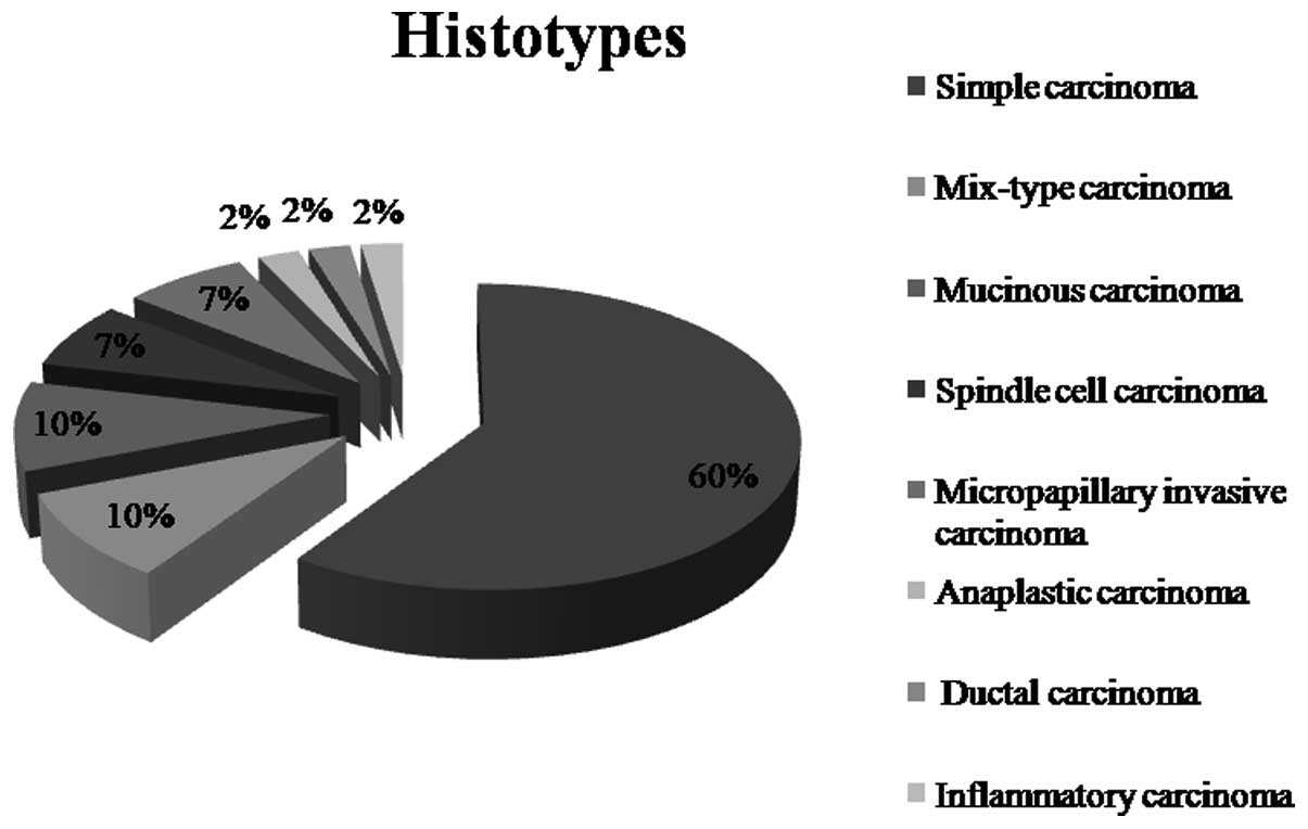
I). Based on the guideline recommended by Goldschmidt et
al (10), the histopathological
findings revealed that 59.5% of the tumors were simple carcinoma.
The other histological features are shown in Fig. 1. The grade II tumors were observed
most frequently with a frequency of 54.8%. Furthermore, 45.8% of
the tumors had margin involvement and a positive vascular invasion
was reported in 52.4% of the tumors (Table I).
 | Table IClinical and pathological results of
canine malignant mammary gland tumors in the present study. |
Table I
Clinical and pathological results of
canine malignant mammary gland tumors in the present study.
| Vimentin | |
|---|
|
| |
|---|
| Variables (n=42)
score | Positivea (n=18) | Negative (n=24) | P-value |
|---|
| Grade |
| I | 2 | 10 | 0.021 |
| II | 10 | 13 | |
| III | 6 | 1 | |
| Stage |
| I | 7 | 9 | NS |
| II | 8 | 13 | |
| III | 3 | 2 | |
| IV | 0 | 0 | |
| Tumor size |
| T1 | 7 | 13 | 0.030 |
| T2 | 8 | 10 | |
| T3 | 3 | 1 | |
| Margin status |
| Free | 6 | 17 | NS |
| Involved | 12 | 7 | |
| Vascular
invasion |
| Present | 14 | 10 | 0.043 |
| Not present | 4 | 14 | |
| Ki-67 |
| Low | 3 | 13 | 0.001 |
| Moderate | 6 | 8 | |
| High | 9 | 3 | |
| MVD-CD34 |
| Low | 1 | 8 | 0.043 |
| Moderate | 5 | 15 | |
| High | 12 | 1 | |
The IHC results showed that 12 (28.6%) and six
(14.3%) of the specimens were strong- and weak-positive for the
vimentin biomarker, respectively. The findings of Ki-67 and
MVD-CD34 analyses are illustrated in Table I. Although the statistical analysis
represented a significant association between vimentin filaments
overexpression and the tumor size (r=0.71, P=0.03), tumor grade
(r=0.80, P=0.021), angiogenesis (r=0.57, P=0.043), proliferation
coefficient (r=0.06, P=0.001) and vascular invasion (r=0.76,
P=0.043), there was no statistical correlation between the vimentin
filaments and the cancer stage and margin status (Tables I and II).
| Table IICorrelation of vimentin filaments
expression with clinocopathological features of the canine
malignant mammary.a |
Table II
Correlation of vimentin filaments
expression with clinocopathological features of the canine
malignant mammary.a
| Vimentin filaments
expression |
|---|
| Tumor staging | − |
| Tumor grading | + |
| Tumor size | + |
| Tumor margin
status | − |
| Vascular
invasion | + |
| Tumor
proliferation | + |
| Tumor
angiogenesis | + |
Discussion
The result of the present study revealed that the
overexpression of the vimentin filaments was associated with
numerous clinicopathological features observed in tumors with
unfavorable characteristics. Some studies have demonstrated that
the presence of vimentin biomarkers is linked to a poor prognosis
in HBC (7,11,14).
The study by Hemalatha et al (11) indicated that overexpression of
vimentin is not associated with tumor grade and Ki-67, whereas Vora
et al (14) concluded that
the invasion potency is increased by the overexpression of vimentin
and the loss of cytokeratin in breast cancer. However, Yamashita
et al (7) evaluated the
expression of vimentin in invasive ductal carcinoma of sporadic
breast cancer and revealed that the overexpression of vimentin
increased in the subtype of triple-negative breast cancer and was
associated with invasiveness potency (8).
EMT is a process in which the epithelial cells lose
their epithelial characteristics and acquire mesenchymal
characteristics. This mechanism occurs in breast cancer and results
in the reduction or loss of cadherin adhesion molecules, loss of
luminal cytokeratin, enhancement of basal cytokeratin,
manifestation of different proteins, including α-smooth muscle
actin, changes in the expression of matrix metalloproteinase genes
and transforming growth factor-β (TGF-β) and increased expression
of vimentin, thus facilitating migration and metastasis of
malignant cells (14–16).
The evaluation of vimentin expression in breast
cancer is considered to be beneficial for determining the
prognosis. Furthermore, analysis of the vimentin expression has
therapeutic value as the tumor progression may be controlled if
vimentin and its upstream line are targeted (17) and such mechanisms would be traceable
in CMMGTs. In the study by Yoshida et al (16) on a canine mammary cancer cell line,
it was concluded that an increase in TGF-β induced vimentin
expression and increased invasion potency. Krol et al
(18) examined five cell lines in
canine mammary cancer and found that the cell culture of these cell
lines underwent genetic alterations favoring EMT. Terra et
al (19) analyzed benign and
malignant canine mammary glands and observed that vimentin
overexpression had a direct association with the malignancy of
tumors.
Numerous studies on vimentin demonstrated that EMT
occurs in humans, as well as canines, and that vimentin expression
is associated with tumor malignancy (14,15,17).
However, unlike the previous studies, in the present study, no
association was observed between vimentin expression and cancer
stage, similar to the study by Terra et al (19). In addition, there was no correlation
between vimentin expression and margin status, thus warranting
further studies in this area.
Similar to the findings of the present study,
certain recent studies have indicated that vimentin filament
expression in HBC and CMMGTs is associated with the severity of
cancer. Thus far, numerous studies have demonstrated the similarity
between HBC and CMMGTs and a majority of them have concluded that
the biological behavior of CMMGTs is similar to that of HBC
(3,4,6). Pinho
et al (6) indicated that
CMMGTs can be regarded as an HBC animal model and that HBC and
CMMGTs have similar biomarkers, thus making the canine mammary
tumor an appropriate model for epidemiological, genetic and
therapeutic studies on HBC (4).
Although there are breast xenograft models, there
appears to be a gap between the in vivo and in vitro
models, and canine mammary gland tumor models could be the
appropriate choice. Several studies have supported the association
between the biomarkers and clinicopathological characteristics of
HBC and CMMGTs (3,4). The study by Muhammadnejad et al
(3) indicated that HER-2/neu
changes in CMMGTs are similar to those in HBC and Queiroga et
al (4) have also investigated
this association.
Thus, spontaneous canine mammary tumor models appear
to be an appropriate animal model for breast cancer research and
the results of the present study could aid to reinforce the model.
To make advancement in the treatment of canine cancers, vimentin
could be targeted in the CMMGT models in the future. In addition,
the results obtained could be helpful in the treatment of HBC.
However, ethical issues should be considered in modeling studies
and the 3Rs (reduction, refinement and replacement) should be
considered in canine oncology studies. In addition, the oncology
studies designed should also be in aid of the treatment for cancers
in canines.
Acknowledgements
The authors express their gratitude to Tehran
Veterinary Hospital and the Small Animal Hospital of Faculty of
Veterinary Medicine of Tehran University. Also, thanks to Dr
E’temad Moghaddam, Pathology Lab, and Miss Morsali for their great
technical support for the IHC staining.
References
|
1
|
Santos AA, Lopes CC, Ribeiro JR, Martins
LR, Santos JC, Amorim IF, et al: Identification of prognostic
factors in canine mammary malignant tumours: a multivariable
survival study. BMC Vet Res. 9:12013. View Article : Google Scholar : PubMed/NCBI
|
|
2
|
Karayannopoulou M, Kaldrymidou E,
Constantinidis TC and Dessiris A: Adjuvant post-operative
chemotherapy in bitches with mammary cancer. J Vet Med A Physiol
Pathol Clin Med. 48:85–96. 2001. View Article : Google Scholar : PubMed/NCBI
|
|
3
|
Muhammadnejad A, Keyhani E, Mortazavi P,
Behjati F and Haghdoost IS: Overexpression of HER-2/neu in
malignant mammary tumors; translation of clinicopathological
features from dog to human. Asian Pac J Cancer Prev. 13:6415–6421.
2012. View Article : Google Scholar : PubMed/NCBI
|
|
4
|
Queiroga FL, Raposo T, Caravalho MI, Prada
J and Pires I: Canine mammary tumours as a model to study human
breast cancer: most recent findings. In Vivo. 25:455–465.
2011.PubMed/NCBI
|
|
5
|
Karihtala P, Auvinen P, Kauppila S,
Haapasaari KM, Jukkola-Vuorinen A and Soini Y: Vimentin, zeb1 and
Sip1 are up-regulated in triple-negative and basal-like breast
cancers: association with an aggressive tumour phenotype. Breast
Cancer Res Treat. 138:81–90. 2013. View Article : Google Scholar : PubMed/NCBI
|
|
6
|
Pinho SS, Carvalho S, Cabral J, Reis CA
and Gärtner F: Canine tumors: a spontaneous animal model of human
carcinogenesis. Transl Res. 159:165–172. 2012. View Article : Google Scholar : PubMed/NCBI
|
|
7
|
Yamashita N, Tokunaga E, Kitao H,
Hisamatsu Y, Taketani K, Akiyoshi S, et al: Vimentin as a poor
prognostic factor for triple-negative breast cancer. J Cancer Res
Clin Oncol. 139:739–746. 2013. View Article : Google Scholar : PubMed/NCBI
|
|
8
|
Kokkinos MI, Wafai R, Wong MK, Newgreen
DF, Thompson EW and Waltham M: Vimentin and epithelial-mesenchymal
transition in human breast cancer - observations in vitro and in
vivo. Cells Tissues Organs. 185:191–203. 2007. View Article : Google Scholar : PubMed/NCBI
|
|
9
|
Cassali GD, Lavalle GE, De Nardi AB,
Ferreira E, Bertagnolli AC, Estrela-Lima A, et al: Consensus for
the diagnosis, prognosis and treatment of canine mammary tumors.
Braz J Vet Pathol. 4:153–180. 2011.
|
|
10
|
Goldschmidt M, Pena L, Rasotto R and
Zappulli V: Classification and grading of canine mammary tumors.
Vet Pathol. 48:117–131. 2011. View Article : Google Scholar : PubMed/NCBI
|
|
11
|
Hemalatha A, Suresh TN and Kumar ML:
Expression of vimentin in breast carcinoma, its correlation with
Ki67 and other histopathological parameters. Indian J Cancer.
50:189–194. 2013. View Article : Google Scholar : PubMed/NCBI
|
|
12
|
Dhakal HP, Bassarova A, Naume B,
Synnestvedt M, Borgen E, Kaaresen R, et al: Breast carcinoma
vascularity: a comparison of manual microvessel count and Chalkley
count. Histol Histopathol. 24:1049–1059. 2009.PubMed/NCBI
|
|
13
|
da Silva BB, Lopes-Costa PV, dos Santos
AR, de Sousa-Júnior EC, Alencar AP, Pires CG, et al: Comparison of
three vascular endothelial markers in the evaluation of microvessel
density in breast cancer. Eur J Gynaecol Oncol. 30:285–288.
2009.PubMed/NCBI
|
|
14
|
Vora HH, Patel NA, Rajvik KN, Mehta SV,
Brahmbhatt BV, Shah MJ, et al: Cytokeratin and vimentin expression
in breast cancer. Int J Biol Markers. 24:38–46. 2009.PubMed/NCBI
|
|
15
|
Moustakas A and Heldin P: TGFβ and
matrix-regulated epithelial to mesenchymal transition. Biochim
Biophys Acta. Feb 18–2014.(Epub ahead of print).
|
|
16
|
Yoshida K, Saito T, Kamida A, Matsumoto K,
Saeki K, Mochizuki M, et al: Transforming growth factor-β
transiently induces vimentin expression and invasive capacity in a
canine mammary gland tumor cell line. Res Vet Sci. 94:539–541.
2013.
|
|
17
|
Satelli A and Li S: Vimentin in cancer and
its potential as a molecular target for cancer therapy. Cell Mol
Life Sci. 68:3033–3046. 2011. View Article : Google Scholar : PubMed/NCBI
|
|
18
|
Król M, Pawłowski KM, Szyszko K,
Maciejewski H, Dolka I, Manuali E, Jank M and Motyl T: The gene
expression profiles of canine mammary cancer cells grown with
carcinoma-associated fibroblasts (CAFs) as a co-culture in vitro.
BMC Vet Res. 8:352012.PubMed/NCBI
|
|
19
|
Terra EM, Magalhães GM, Rodrigues MP,
Amorim RL, Rocha NS and Costa MT: Immunohistochemical expression of
TGFβ, E-cadherin and vimentin in benign and malignant neoplasias of
canine mammary gland. BMC Proceedings. 7(Suppl 2): P202013.
|