Introduction
Ischemic heart diseases are leading causes of
fatality worldwide, and irreversible and widespread loss of
myocardial cells and subsequent ventricular remodeling induced by
acute myocardial infarction are the main elements resulting in
chronic heart failure and permanent loss of labor force (1). There are 17.3 million people who
succumbed of ischemic heart diseases in 2008 and the number is
estimated to be 23.6 million by 2030 (2). Revascularization could improve the
prognosis of the patients with acute myocardial infarction
(1), however, reperfusion could induce
additional injury, which is sometimes extremely severe or fatal and
diminishes the benefits of revascularization. Basic studies
(3–6) and
small-sample clinical data (7,8) have verified that ischemic
postconditioning (IPOC) could attenuate injury induced by
ischemia/reperfusion (IR) and certain studies (8–10) have
indicated that the mitochondrial KATP
(mitoKATP) channel plays an important role in
cardioprotection effects of IPOC, but its molecular mechanisms are
not completely clear thus far. Connexin 43 (Cx43; a gap junction
protein located mainly in the ventricular myocardium in mammals)
plays an important role in intercellular electrical conduction and
cell survival, but whether the changes of membrane Cx43 are
involved in IPOC-induced cardioprotective effects and the
corresponding details is unclear. Therefore, exploring the effects
and mechanisms could aid the understanding of the deeply molecular
mechanisms of IPOC, thus providing information for constructing an
effective strategy to enhance cardioprotection.
Therefore, the present study was performed to
investigate the changes of membrane Cx43, cellular ultrastructure,
infarct size, reperfusion arrhythmia in different states of IR and
IPOC to determine potential mechanisms and associated details of
IPOC.
Materials and methods
Study approval
The study was reviewed and approved by the
Institutional Ethics Committee of the Anzhen Hospital (Beijing,
China) and Beijing Institute of Heart, Lung and Blood Vessel
Diseases on Animal Resource (Beijing, China), and conformed to the
guiding principles of ‘Guide for the Care and Use of Laboratory
Animals’ (National Institutes of Health Publication no. 83–23,
revised 1996) during, maintaining and using the animals.
Animal model and experiment
protocol
All the rats were randomly divided into 5 groups to
receive different treatments (n=16 in each group): i) Sham; ii) IR
group, 30-min ischemia followed by reperfusion for 180 min; iii) IR
+ IPOC group, after the 30-min ischemia, IPOC was performed through
application of 5 intermittent cycles of 10-sec reperfusion and
10-sec ischemia immediately followed by reperfusion for 178 min 20
sec; iv) IR + diazoxide group, diazoxide (30 µmol/l) was
administrated intravenously in the first 10 min of reperfusion; and
v) IR + IPOC + 5-hydroxydecanoate acid (5-HD) group, 5-HD (300
µmol/l) was administrated intravenously in the first 10 min of
reperfusion.
The IR model was prepared with 8-week-old male
Sprague-Dawley rats weighing 260–310 g (Laboratory Animal Division,
Medicine Department, Beijing University, Beijing, China), as
described previously (11). The
sternum was incised and the heart was exposed subsequent to
anesthetizing with 1% pentobarbital (40 mg/kg body weight) through
peritoneal injection and support from a breathing machine. The left
coronary artery anterior descending branch was found and marked
with silk and ligated with ligator to establish the rat IR model
(baseline electrocardiography was recorded at 10 min after marking
with silk). The coronary arteries of the rats in the sham group
were only marked and not ligated.
Occurrence of arrhythmia in the
different groups
Electrocardiograms of the rats in the different
groups at baseline and during the experiment were monitored
continuously (electrocardiograph 2E31A; SAN-EI TECH Ltd., Chiba,
Japan) and arrhythmia events were recorded.
Measurement of myocardial creatases,
nitric oxide (NO) and malondialdehyde (MDA) in the different
groups
The blood was obtained from abdominal aorta
immediately after 3-h reperfusion, and the serum levels of NO, MDA,
creatine kinase (CK), CK-MB and lactate dehydrogenase (LDH) in the
different groups were measured with assay kits (Jiancheng
Technology Co., Ltd., Nanjing, China) according to the
manufacturer's instructions.
Evaluation of infarct size in the
different groups
Myocardial infarct size was measured as described
previously (12). After having
finished reperfusion, the left anterior descending artery was
reoccluded and Evans blue dye was administered intravenously to
stain the normal region of the left ventricle (LV) and the heart
was rapidly excised. LV tissue was isolated and cut into 10
cross-sectional pieces of equal thickness. The non-stained LV area
at risk (AAR) was separated from the surrounding blue-stained LV
normal zone and the two regions were separately incubated at 37°C
for 15 min in 1% triphenyltetrazolium chloride (TTC) in 0.1 M
phosphate buffer adjusted to pH 7.4. The tissues were fixed
overnight in 10% formaldehyde. AAR and blue-stained LV normal zone
regions were weighed for determination of AAR/LV. TTC stains living
tissue a deep red color, but necrotic tissue is TTC-negative and
appears white within the AAR slices. Each slice was scanned with a
commercial scanner (Canoscan LiDE 60; Canon Inc., Tokyo, Japan) and
infarct and non-infarct areas were measured using an image analysis
program (Image-Pro Plus Version 6.0; Media Cybernetics, Georgia,
MD, USA). Myocardial infarct size was expressed as a percentage of
the AAR.
Western blot analysis of membrane Cx43
expression in the different groups
The rats were sacrificed respectively at 1- and 3-h
after reperfusion by intraperitoneal injection of 3% pentobarbital
sodium with the dose of 100 mg/kg, their hearts were taken
immediately and LV tissues below the ligation point were obtained
and prepared. The samples were homogenized in Protein Extraction
reagents (Pierce 78510; Pierce Biotechnology, Inc., Waltham, MA,
USA) and harvested in 50 µl of sample buffer, boiled and sonicated.
Protein lysates were separated on 10% sodium dodecyl
sulphate-polyacrylamide gel electrophoresis. Following transfer to
the polyvinylidene fluoride membranes and blocking with skimmed
milk (5% w/v), the blots were incubated with primary antibodies of
Cx43 and GAPDH (C8093; Sigma-Aldrich, St. Louis, MO, USA; and
G5262; Sigma, Santa Clara, CA, USA, respectively) overnight at 4°C.
GAPDH served as the internal reference. Primary antibody binding
was detected with an enhanced chemiluminescence western blotting
kit (Amersham Biosciences, Piscataway, NJ, USA) and quantified by
laser densitometry using the Typhoon 9400 fluorescent scanner
together with ImageQuant 5.0 software (Amersham Biosciences) as
previously described (13).
Immunohistochemistry analysis of
membrane Cx43 in the different groups
Left ventricular tissue below the ligation point was
fixed with 10% formaldehyde solution at 1- and 3-h after
reperfusion, respectively, embedded in epoxy resin and sectioned,
and subsequently processed with the immunohistochemistry kit
(HaoYang Bioscience Co., Tianjing, China) according to the
manufacturer's instructions (anti-Cx43 antibody was purchased from
Santa Cruz Biotechnology, Inc., Dallas, TX, USA; sc-271837),
stained with diaminobenzidine (DAB) and counterstained with
hematoxylin. Subsequently, the samples were observed with a
microscope (BX51; Olympus, Tokyo, Japan).
Ultrastructure of the myocardial
cell
The ultrastructure of the myocardial cell was
observed with a transmission electron microscope as described
previously (14). For electron
microscopic examination, following fixation in glutaraldehyde, the
sample was refixed in 1% osmium tetroxide, embedded in epoxy resin,
sectioned at 0.1-µm and double-stained with uranium acetate and
lead citrate. The sections were observed with a transmission
electron microscope (Hitachi-600; Hitachi High-Technologies Corp.,
Tokyo, Japan).
Apoptosis analysis in the myocardium
in the different groups
The tissue sample in each group was embedded in
paraffin, sectioned at 4-µm, blocked with 3% hydrogen peroxide and
incubated for 60 min with terminal deoxynucleotidyl transferase
dUTP nick end labeling reaction mixture (45 µl Enzyme solution +
405 µl Label solution) at 37°C in the dark. Subsequently, the
sample was stained with converter-peroxidase solution and DAB
substrate, and counterstained with hematoxylin. Apoptosis in each
group was analyzed by observing with a light microscope (Motic
BA400; Motic, Xiamen, China) and the apoptosis index was
calculated.
Statistical analysis
All the values are expressed as mean ± standard
error of the mean. Differences in continuous variables between 2
groups were analyzed via the Student's t-test and differences
between ≥3 groups were evaluated via one-way analysis of variance
with Bonferroni correction; differences in categorical data were
assessed by the χ2 test, and in the case of low cell
counts (<5) the Fisher's exact test was used instead of
χ2 test. P<0.05 was considered to indicate a
statistically significant difference.
Results
Various arrhythmias in different
groups
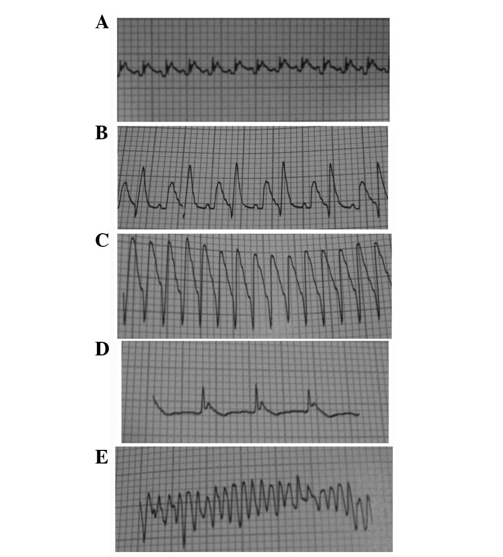
No arrhythmia event occurred in the sham group and
the occurrence rate of the arrhythmia events was 62.5, 25.0, 37.5
and 50.0% in the IR, IR + IPOC, IR + diazoxide and IR + IPOC + 5-HD
groups, respectively. There was a significant difference between
the groups (Table I). The arrhythmias
that happened during IR included ventricular premature beat
(coupled rhythm), ventricular tachycardia, atrioventricular block
and ventricular fibrillation (Fig.
1).
 | Table I.Occurrence of arrhythmia in the
different groups. |
Table I.
Occurrence of arrhythmia in the
different groups.
|
|
|
| During reperfusion,
times |
|
|---|
|
|
|
|
|
|
|---|
| Group | No. | During ischemia,
times | 1 h | 2 h | 3h | Occurence rate of
arrhythmia during reperfusion, % |
|---|
| A | 16 | 0 | 0 | 0 | 0 | 0.0 |
| B | 16 | 18 | 12 | 2 | 0 |
62.5a,b |
| C | 16 | 17 | 6 | 1 | 0 |
25.0a,c |
| D | 16 | 19 | 8 | 1 | 0 |
37.5a–c |
| E | 16 | 19 | 9 | 1 | 0 |
50.0a–d |
Levels of creatases, NO and MDA in the
different groups
After 3-h reperfusion, the serum levels of CK,
CK-MB, LDH, NO and MDA increased significantly in the IR group.
IPOC and diazoxide impaired the increases, and 5-HD attenuated, but
did not completely abolish, the effects of IPOC. There was no clear
difference in the levels of NO and MDA between the IR + IPOC and IR
+ diazoxide groups (51.7 vs. 45.5 µmol/l and 6.93 vs. 6.55 nM,
respectively; P>0.05), but the levels of CK and CK-MB in the IR
+ diazoxide group was higher than that in the IR + IPOC group
(3,286.9 vs. 3,775.9 U/l and 670.2 vs. 747.3 U/l, respectively;
P<0.05) (Table II).
| Table II.Serum levels of NO, MDA, CK, CK-MB and
LDH in the different groups. |
Table II.
Serum levels of NO, MDA, CK, CK-MB and
LDH in the different groups.
| Group | No. | NO, µmol/1 | MDA, nM | CK, U/1 | CK-MB, U/1 | LDH, U/1 |
|---|
| A | 14 |
26.0±9.7 |
3.88±0.88 |
896.3±125.5 |
214.6±87.7 |
260.4±98.6 |
| B | 9 |
86.1±29.0a |
11.08±3.22a |
5,283.9±1,348.2a |
985.1±223.0a |
1,135.2±327.6a |
| C | 13 |
51.7±12.6b,c |
6.93±1.77b,c |
3,286.9±949.8b–d |
670.2±237.6b–d |
780.7±249.1b,c |
| D | 12 |
45.5±12.9b,c |
6.55±2.46b,c |
3,775.9±1,034.6b,c,e |
727.3±304.4b,c,e |
813.4±278.3b,c |
| E | 10 |
70.8±14.5b,d,e |
8.69±1.47b,d,e |
4,633.3±1,177.3b,d,e |
886.8±275.4b,d,e |
1,002.8±376.7d,e |
Infarct size in the different
groups
After 3-h reperfusion, the infarct size was
43.3±9.7% in the IR group, and IPOC and diazoxide decreased the
size (10.9 and 15.5 vs. 43.3%, respectively; P<0.05), and 5-HD
attenuated, but not completely abolished, the effects of IPOC (37.7
vs. 43.3%, P<0.05). The infarct size in the IR + IPOC group was
smaller than that in the IR + diazoxide group, but this was not
significant (Table III).
| Table III.Infarct size in the different
groups. |
Table III.
Infarct size in the different
groups.
| Group | No. | Infarct size,
% |
|---|
| A | 4 |
0.0±0.0 |
| B | 4 |
43.3±9.7a |
| C | 4 |
10.9±6.3a–c |
| D | 4 |
15.5±9.5a–c |
| E | 4 |
37.7±6.7a,b,d,e |
Membrane Cx43 expression in the
different groups after 1- and 3-h reperfusion
Total Cx43 (t-Cx43) and phosphorylated Cx43 (p-Cx43)
in the IR group decreased, and in particular, p-Cx43 decreased
significantly (42.7 vs. 16.8% and 38.1 vs. 6.4%, after 1- and 3-h
reperfusion respectively, P<0.01). IPOC and diazoxide partly
restored the levels of t-Cx43 and p-Cx43, and 5-HD alleviated, but
not completely abolished, the effects of IPOC (16.8 vs. 21.7% and
6.4 vs. 14.2%, after 1- and 3-h reperfusion, respectively,
P<0.05). The p-Cx43 level in the IR + IPOC group was lower than
that in the IR + diazoxide group after 1-h reperfusion (26.1 vs.
29.4%, P>0.05), however, it was reversed after 3-h reperfusion
and the p-Cx43 level in the IR + IPOC group was significantly
higher than that in the IR + diazoxide group (32.8 vs. 18.7%,
P<0.01), which suggested that mitoKATP plays an
important role in the expression of Cx43 during the early phase of
IR. IPOC still significantly restored the expression of Cx43
through other pathways besides opening mitoKATP during
the late phase of IR and the benefit of single opening
mitoKATP was limited in the late phase of IR (Figs. 2 and 3).
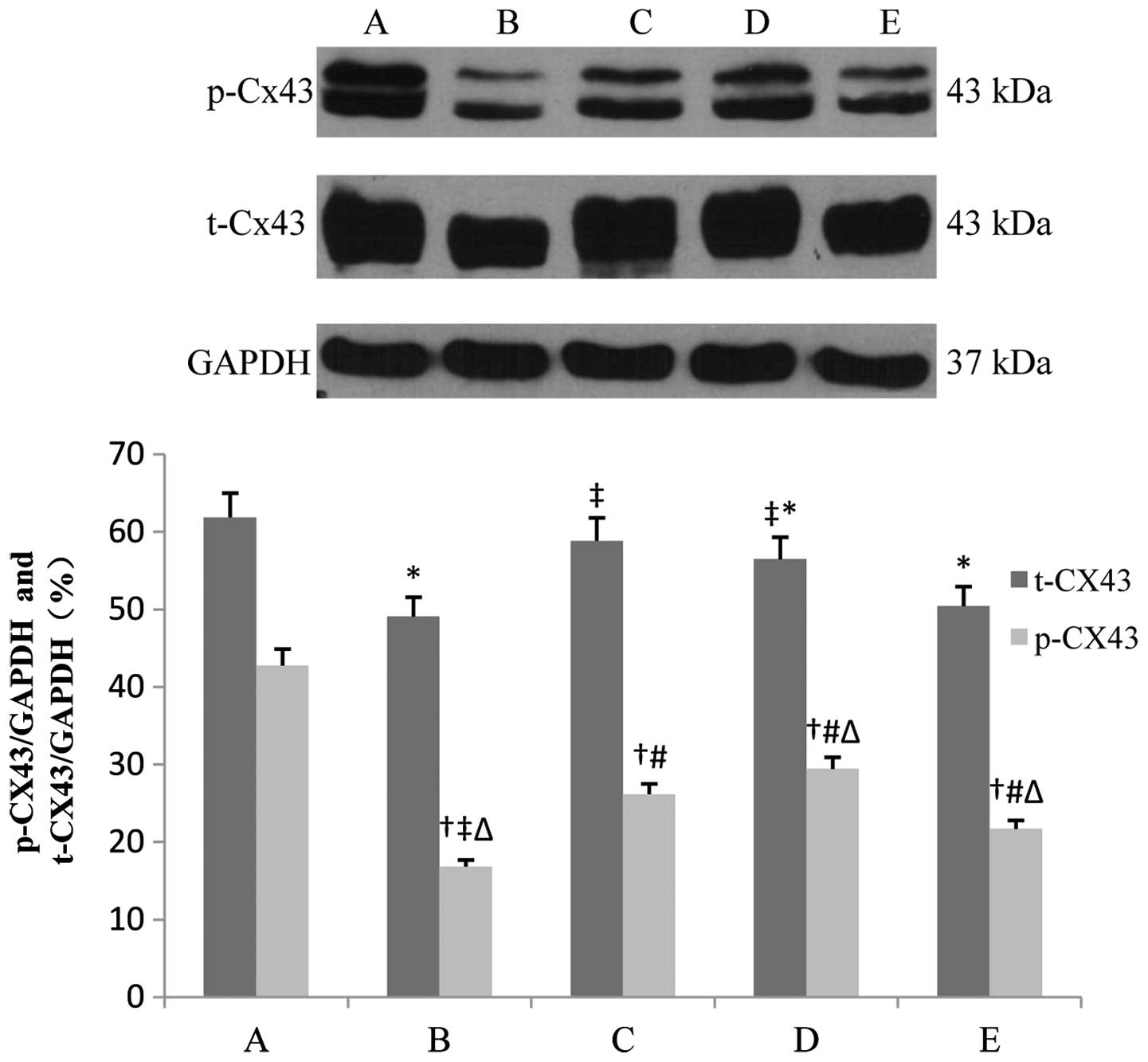 | Figure 2.Expression of t-Cx43 and p-Cx43 in
each group after 1-h reperfusion. Bar graphs showing the relative
expression of t-Cx43 and p-Cx43 over GAPDH following densitometric
scanning for western blot analysis. Data are represented as mean ±
standard deviation for 3 different experiments. Lanes A-E are the
sham, IR, IR + IPOC, IR + diazoxide and IR + IPOC + 5-HD groups,
respectively. Cx43, connexin 43; t-Cx43, total Cx43; p-Cx43,
phosphorylated Cx43; IR, ischemia/reperfusion; IPOC, ischemic
postconditioning; 5-HD, 5-hydroxydecanoate acid. *P<0.05 vs.
t-Cx43 in group A; †P<0.05 vs. p-Cx43 in group A;
‡P<0.05 vs. t-Cx43 in group B; #P<0.05
vs. p-Cx43 in group B; △P<0.05 vs. p-Cx43 in group
C. |
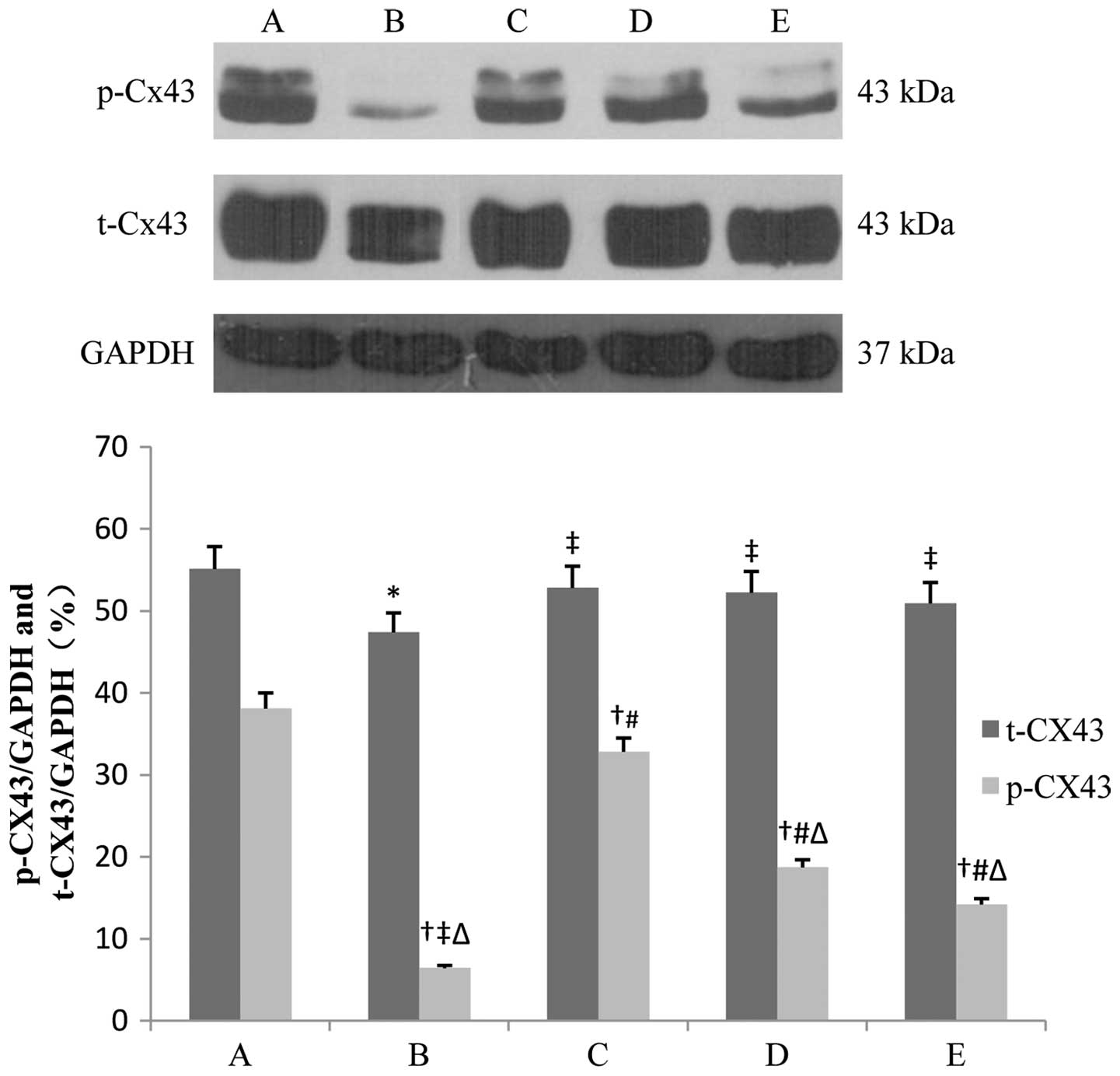 | Figure 3.Expression of t-Cx43 and p-Cx43 in
each group after 3-h reperfusion. Bar graphs showing the relative
expression of t-Cx43 and p-Cx43 over GAPDH following densitometric
scanning for western blot analysis. Data are represented as mean ±
standard deviation for 3 different experiments. Lanes A-E are the
sham, IR, IR + IPOC, IR + diazoxide and IR + IPOC + 5-HD groups,
respectively. Cx43, connexin 43; t-Cx43, total Cx43; p-Cx43,
phosphorylated Cx43; IR, ischemia/reperfusion; IPOC, ischemic
postconditioning; 5-HD, 5-hydroxydecanoate acid. *P<0.05 vs.
t-Cx43 in group A; †P< 0.05 vs. p-Cx43 in group A;
‡P< 0.05 vs. t-Cx43 in group B; #P<
0.05 vs. p-Cx43 in group B; △P< 0.05 vs. p-Cx43 in
group C. |
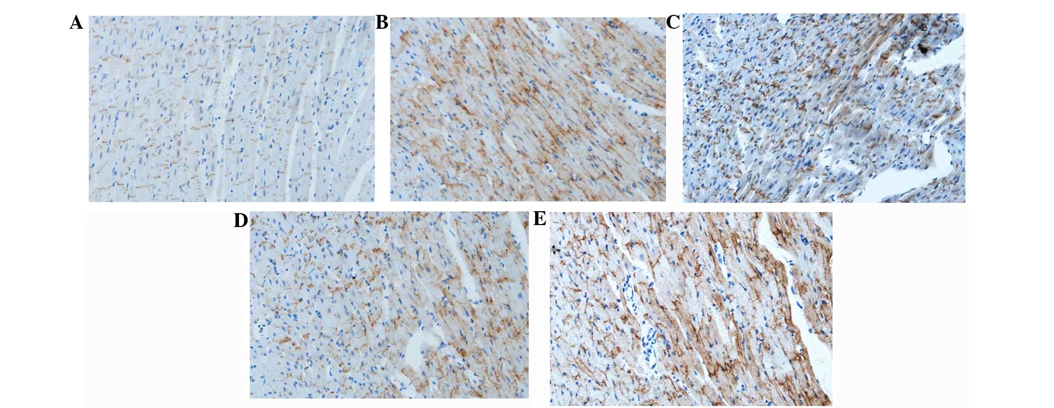
Distribution of membrane Cx43 in the
different groups after 3-h reperfusion
In the sham group, membrane Cx43 was located mainly
in the intercalated disc between proximate myocardial cells and
there was hardly any Cx43 at the cell lateral membrane. After
ischemia and 3-h reperfusion, Cx43 was distributed on the whole
surface of the cells, and was not limited in the intercalated disc.
The structure of the intercalated disc was unclear. Cx43 was also
redistributed, but the majority of them were still located in the
intercalated disc in the IR + IPOC group. There was also Cx43 at
the cell lateral membrane, but Cx43 was mainly in the intercalated
disc. The intercalated disc was sparse in the IR + diazoxide group.
There was significant redistribution of Cx43 and less Cx43 in the
disc in the IR + IPOC + 5-HD group (Fig.
4).
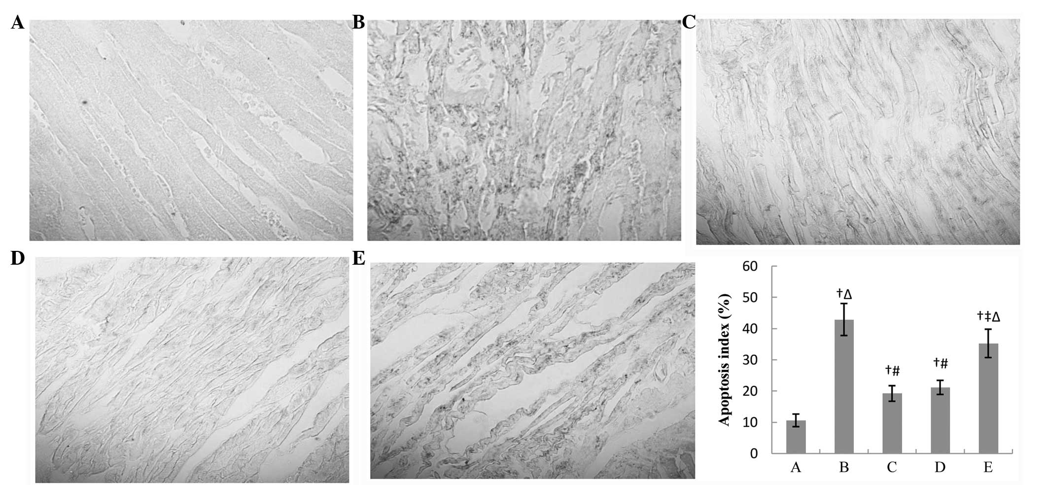
Changes of the ultrastructures of
myocardial cells after 3-h reperfusion
In the sham group, the structure of the intercalated
disc was clear and there was a regular array of myofilaments,
echelonment of Z line and normal mitochondrion. After ischemia and
3-h reperfusion, there was a disrupted and textureless intercalated
disc, dissolved myocardial cells and condensed nucleus and
megamitochondrion or the mitochondrion with a vacuole. IPOC
attenuated the pathological changes significantly and the majority
of the disc had an intact structure. Diazoxide also alleviated the
changes induced by IR, but there were only fewer discs whose
structures were clear. 5-HD largely impaired the IPOC-induced
improvement in ultrastructure and there were disrupted
myofilaments, flocculent mitochondrion and the structure of the
disc was not intact (Fig. 5).
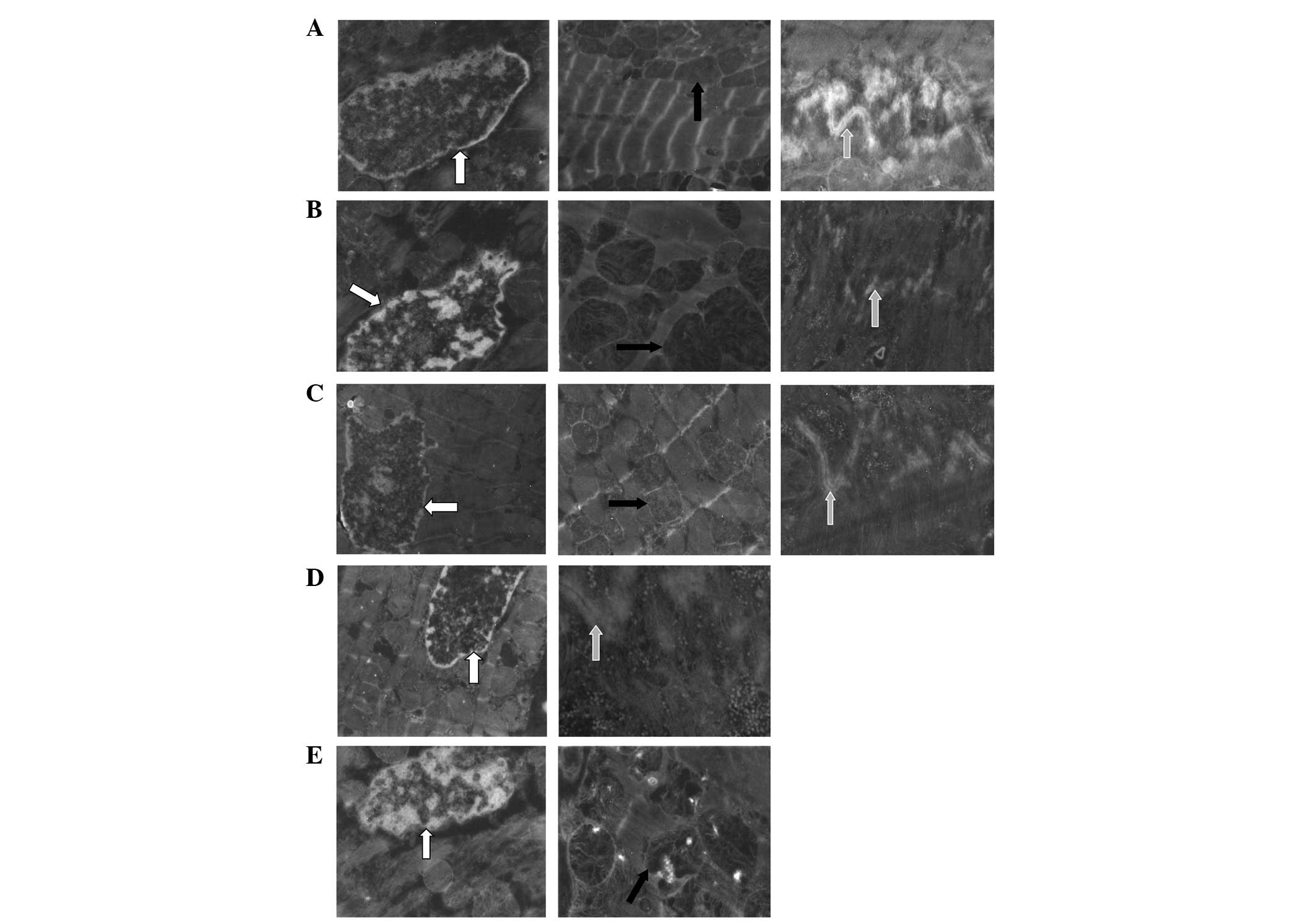 | Figure 5.Ultrastructure of the myocardial cell
in each group. Cell nucleus is indicated by the white arrow,
mitochondrion by the black arrow and intercalated disc by the gray
arrow. These pictures indicate that the structure of the
intercalated disc was clear and there was a regular array of
myofilaments, echelonment of Z line and normal mitochondrion in the
sham group. There was a disrupted and textureless intercalated
disc, dissolved myocardial cells and condensed nucleus and
megamitochondrion or the mitochondrion with a vacuole in the IR
group. IPOC significantly attenuated the pathological changes
induced by IR and the majority of the disc had an intact structure.
Diazoxide also alleviated the changes, but there were only fewer
discs in which their structures were clear. 5-HD largely impaired
the IPOC-induced improvement in ultrastructure and there were
disrupted myofilaments, flocculent mitochondrion and the structure
of the disc was not intact. (A) Sham, (B) IR, (C) IR + IPOC, (D) IR
+ diazoxide and (E) IR + IPOC + 5-HD groups. IR,
ischemia/reperfusion; IPOC, ischemic postconditioning; 5-HD,
5-hydroxydecanoate acid. |
Apoptosis in the different groups
after 3-h reperfusion
There were numerous black apoptotic cells and
evidently disrupted myocardial fibers in the IR group. IPOC
attenuated the apoptosis significantly (19.2 vs. 42.9%, P<0.01)
and there was a relatively regular array of myocardial cells.
Following treatment with diazoxide, the apoptosis was similar to
the IR + IPOC group. 5-HD attenuated the effects of IPOC and there
was more significant apoptosis and disorderly myocardial array in
the IR + IPOC + 5-HD group than that in the IR + IPOC group. The
effects of IPOC were not abolished completely, and the apoptosis
index in the IR + IPOC + 5-HD group was still lower than that in
the IR group (33.2 vs. 42.9%, P<0.05) (Fig. 6).
Discussion
The present study investigated the expression and
distribution of Cx43, ultrastructure and apoptosis in the
myocardium. The levels of myocardial creatases, NO and MDA, infarct
size and arrhythmia events for the different treatments of IR, IR +
IPOC, IR + diazoxide or IR + IPOC + 5-HD were assessed. The results
indicated that IPOC decreased infarct size and the levels of
myocardial creatases, NO and MDA, and improved the expression and
distribution of Cx43, ultrastructure and apoptosis. Diazoxide could
partly simulate the effects, and 5-HD attenuated, but not
completely abolished, the effects of IPOC, which suggested that
cell membrane Cx43 is also involved in the process of IPOC-induced
cardioprotection. The improvement of membrane Cx43 is much more
dependent on mitoKATP in the earlier phase of IPOC than
that in the late phase of IPOC, and to the best of our knowledge,
this is the first report of this.
Previous data have indicated that IPOC could induce
cardioprotection by inhibiting the expression of matrix
metalloproteinase-2 (15), decreasing
no-reflow (8), inhibiting apoptosis
(16) and attenuating inflammatory
response (5), and certain studies
indicated that mitoKATP plays a crucial role in
IPOC-induced cardioprotection (9,10,17). The present study indicated that IPOC
decreased infarct size and the levels of myocardial creatases,
improved the expression and distribution of Cx43 and apoptosis.
Opening mitoKATP with diazoxide partly simulated the
effects of IPOC, and an inhibitor of mitoKATP, 5-HD,
attenuated, but not abolished completely, the effects of IPOC,
which suggested that mitoKATP plays an important role.
However, there are other independent key pathways in IPOC-induced
cardioprotection.
Cx43, a gap junction protein located mainly in
ventricular myocardium in mammals, plays an important role in
intercellular electric conduction and cell survival. Previous
studies have indicated that mitochondrial Cx43 may be involved in
mitoKATP-mediated IPOC (18,19), but
whether the changes of membrane Cx43 are involved in
cardioprotection of mitoKATP-mediated IPOC and the
corresponding details are not clear. The present study indicated
that IR decreased expression of t-Cx43 and p-Cx43 (particularly the
latter) and induced the redistribution of Cx43 on the cell surface,
and IPOC attenuated the decreases. Diazoxide partly restored the
levels of Cx43, and 5-HD alleviated the effects of IPOC. Therefore,
mitoKATP plays a more important role in expression of
Cx43 during the earlier phase of IR, whereas the benefit of single
opening mitoKATP was limited in the late phase of
IR.
Reperfusion injury is a problem in treatment for the
patients with acute myocardial infarction and IPOC could attenuate
the injury, but its mechanism is not clear. The present study
indicated that cell membrane Cx43 is also involved in the process
of IPOC-induced cardioprotection and the improvement of membrane
Cx43 is much more dependent on mitoKATP in the earlier
phase of IPOC. Therefore, it is important that appropriate drugs
should be selected in different phases of IR to protect membrane
Cx43 against injury.
In conclusion, cell membrane Cx43 is also involved
in the process of IPOC-induced cardioprotection and the improvement
of membrane Cx43 is much more dependent on mitoKATP in
the earlier phase of IPOC than that in the late phase of IPOC.
The present study verified that cell membrane Cx43
is also involved in the process of IPOC-induced cardioprotection,
and the detailed mechanisms of the changes of membrane Cx43 in IPOC
would be a future task, thus find new potential targets for
attenuating IR.
Acknowledgements
The present study was supported by the Development
Funds of Capital Medicine (grant no. 2009-3022), Chinese Natural
Science Foundation Grants (no. 81100142), Open Topic Funds Grants
of Essential Laboratory in Cardiovasular Remodeling and
Transforming Medicine, National Ministry of Education (no.
2010XGCS02), Cultivating Project Grants of Beijing for Highly
Talented Men of Medicine (no. 2014-3-042), and the Superintendent
Cultivating Funds Grants of Beijing Anzhen Hospital (no. 2010F03).
The study was also supported by the Beijing Anzhen Hospital,
Capital University of Medical Sciences and Beijing Institute of
Heart Lung and Blood Vessel Diseases. The authors thank Dr TieMin
Ma for his valuable suggestions regarding the rat IR model and Dr
ZhuQing Jia for her assistance of the ultrastructure analysis with
electron microscope.
References
|
1
|
Roger VL, Go AS, Lloyd-Jones DM, Benjamin
EJ, Berry JD, Borden WB, Bravata DM, Dai S, Ford ES, Fox CS, et al
American Heart Association Statistics Committee and Stroke
Statistics Subcommittee: Heart disease and stroke statistics - 2012
update: A report from the American Heart Association. Circulation.
125:e2–e220. 2012. View Article : Google Scholar : PubMed/NCBI
|
|
2
|
Mendis S, Puska P and Norrving BO: Global
Atlas on Cardiovascular Disease Prevention and ControlWorld Health
Organization; Geneva: 2011
|
|
3
|
Jiang ZH, Zhang TT and Zhang JF:
Protective effects of fasudil hydrochloride post-conditioning on
acute myocardial ischemia/reperfusion injury in rats. Cardiol J.
20:197–202. 2013. View Article : Google Scholar : PubMed/NCBI
|
|
4
|
Wei C, Li H, Han L, Zhang L and Yang X:
Activation of autophagy in ischemic postconditioning contributes to
cardioprotective effects against ischemia/reperfusion injury in rat
hearts. J Cardiovasc Pharmacol. 61:416–422. 2013. View Article : Google Scholar : PubMed/NCBI
|
|
5
|
Wang NP, Pang XF, Zhang LH, Tootle S,
Harmouche S and Zhao ZQ: Attenuation of inflammatory response and
reduction in infarct size by postconditioning are associated with
downregulation of early growth response 1 during reperfusion in rat
heart. Shock. 41:346–354. 2014. View Article : Google Scholar : PubMed/NCBI
|
|
6
|
Tu Y, Wan L, Fan Y, Wang K, Bu L, Huang T,
Cheng Z and Shen B: Ischemic postconditioning-mediated miRNA-21
protects against cardiac ischemia/reperfusion injury via PTEN/Akt
pathway. PLoS One. 8:e758722013. View Article : Google Scholar : PubMed/NCBI
|
|
7
|
Crimi G, Pica S, Raineri C, Bramucci E, De
Ferrari GM, Klersy C, Ferlini M, Marinoni B, Repetto A, Romeo M, et
al: Remote ischemic post-conditioning of the lower limb during
primary percutaneous coronary intervention safely reduces enzymatic
infarct size in anterior myocardial infarction: A randomized
controlled trial. JACC Cardiovasc Interv. 6:1055–1063. 2013.
View Article : Google Scholar : PubMed/NCBI
|
|
8
|
Mewton N, Thibault H, Roubille F, et al:
Postconditioning attenuates no-reflow in STEMI patients. Basic Res
Cardiol. 108:3832013. View Article : Google Scholar : PubMed/NCBI
|
|
9
|
Shimizu S, Oikawa R, Tsounapi P, Inoue K,
Shimizu T, Tanaka K, Martin DT, Honda M, Sejima T, Tomita S, et al:
Blocking of the ATP sensitive potassium channel ameliorates the
ischaemia-reperfusion injury in the rat testis. Andrology.
2:458–465. 2014. View Article : Google Scholar : PubMed/NCBI
|
|
10
|
Mykytenko J, Reeves JG, Kin H, Wang NP,
Zatta AJ, Jiang R, Guyton RA, Vinten-Johansen J and Zhao ZQ:
Persistent beneficial effect of postconditioning against infarct
size: Role of mitochondrial K(ATP) channels during reperfusion.
Basic Res Cardiol. 103:472–484. 2008. View Article : Google Scholar : PubMed/NCBI
|
|
11
|
Mukhopadhyay P, Mukherjee S, Ahsan K,
Bagchi A, Pacher P and Das DK: Restoration of altered microRNA
expression in the ischemic heart with resveratrol. PLoS One.
5:e157052010. View Article : Google Scholar : PubMed/NCBI
|
|
12
|
Ichinomiya T, Cho S, Higashijima U,
Matsumoto S, Maekawa T and Sumikawa K: High-dose fasudil preserves
postconditioning against myocardial infarction under hyperglycemia
in rats: Role of mitochondrial KATP channels. Cardiovasc Diabetol.
11:282012. View Article : Google Scholar : PubMed/NCBI
|
|
13
|
Pogwizd SM, Qi M, Yuan W, Samarel AM and
Bers DM: Upregulation of Na(+)/Ca(2+) exchanger expression and
function in an arrhythmogenic rabbit model of heart failure. Circ
Res. 85:1009–1019. 1999. View Article : Google Scholar : PubMed/NCBI
|
|
14
|
Sui H, Wang W, Wang PH and Liu LS:
Protective effect of antioxidant ebselen (PZ51) on the cerebral
cortex of stroke-prone spontaneously hypertensive rats. Hypertens
Res. 28:249–254. 2005. View Article : Google Scholar : PubMed/NCBI
|
|
15
|
Liu ZZ, Kong JB, Li FZ, Ma LL, Liu SQ and
Wang LX: Ischemic postconditioning decreases matrix
metalloproteinase-2 expression during ischemia-reperfusion of
myocardium in a rabbit model: A preliminary report. Exp Clin
Cardiol. 18:e99–e101. 2013.PubMed/NCBI
|
|
16
|
Sun H, Guo T, Liu L, Yu Z, Xu W, Chen W,
Shen L, Wang J and Dou X: Ischemic postconditioning inhibits
apoptosis after acute myocardial infarction in pigs. Heart Surg
Forum. 13:E305–E310. 2010. View Article : Google Scholar : PubMed/NCBI
|
|
17
|
Penna C, Mancardi D, Rastaldo R, Losano G
and Pagliaro P: Intermittent activation of bradykinin B2 receptors
and mitochondrial KATP channels trigger cardiac postconditioning
through redox signaling. Cardiovasc Res. 75:168–177. 2007.
View Article : Google Scholar : PubMed/NCBI
|
|
18
|
He Y, Zeng ZY, Zhong GQ, Li JY, Li WK and
Li W: Mitochondrial connexin43 and postconditioning protection in
rabbits underwent myocardial ischemia/reperfusion injury. Zhonghua
Xin Xue Guan Bing Za Zhi. 38:357–362. 2010.(In Chinese). PubMed/NCBI
|
|
19
|
Rottlaender D, Boengler K, Wolny M, et al:
Connexin 43 acts as a cytoprotective mediator of signal
transduction by stimulating mitochondrial KATP channels in mouse
cardiomyocytes. J Clin Invest. 120:1441–1453. 2010. View Article : Google Scholar : PubMed/NCBI
|