Introduction
Varicella-zoster virus (VZV) belongs to the
α-herpesvirus family (1,2). VZV can infect humans and vertebrates
(including chimpanzees and gorillas). The primary VZV infection in
humans results in chickenpox. Following the recovery from
chickenpox, the VZV becomes dormant in the sensory ganglia-like
dorsal root ganglia (3,4). In certain conditions, VZV can reactivate
and cause herpes zoster. There is a difference in onset time of
primary VZV infection in different geographic regions (4). Generally, the infection time in people
who have lived in temperate regions is earlier than those who have
lived in tropical regions. In temperate regions, the first
infection of VZV is mostly found in children, and in the tropical
regions, the first infection is delayed until adulthood.
The genome of VZV is a linear duplex DNA molecule
(5). The genome of VZV contained
~25,000 base pairs, including ≥70 open reading frames (ORFs).
Genetic variations have been identified in the genome of VZV
strains (6–13). Based on the specific single-nucleotide
polymorphisms (SNPs) in ORFs 1, 21, 50 and 54, VZV strains have
been divided into the following genotypes: A (Africa/Asia), B and C
(Europe and North America), and J (Japanese) (6). By analyzing a short region of nucleotides
[447 base pairs (bp)] in ORF 22, VZV strains could be divided into
genotype E (Europe), genotype isolates J (Japanese) and genotype M
(Mosaic) (13). The genotype M strains
can be further divided into genotypes of M1, M2, M3 and M4
(13–15). The R5 variable regions (R5A and R5B)
between ORF60 and ORF61 are also used to distinguish the genotypes
of VZV strains (10,11). By phylogenetic analysis of the complete
VZV genomes, the VZV strains could be broadly grouped into 5 clades
(clades 1 and 3: European/North American strains; clade 2: Asian
strains, particularly for the strains from Japan; clade 4: Certain
strains from Europe; and clade 5: Indian strains) (16).
Until now, the VZV strains in China were not
extensively studied. In the study by Loparev et al (13), 6 VZV samples in China were analyzed.
The 3 samples in South China were all type M2. The other 3 samples
in North China were all type E. Liu et al (17) collected 19 VZV samples from the
patients with zoster or varicella in Hefei city of Anhui province,
which is located in the middle Eastern part of China (18). The collected VZV isolates were grouped
into genotype J or J1. The genotype of R5A and R5B were found in
these isolates by analyzing the R5 variable region. In the present
study, 42 VZV isolates were collected from Yunnan province of
Southwestern China. Using the described genotyping method of Liu
et al (17), a genetic
characterization of the VZV strains was made in Southwestern
China.
Materials and methods
Patients and clinical samples
The VZV isolates were collected from the 42
ambulatory and hospitalized patients with herpes zoster in The
First People's Hospital of Yunnan Province (The Affiliated Hospital
of Kunming University of Science and Technology, Kunming, Yunnan,
China) between August 2013 and December 2014. Vesicle fluid was
collected from skin lesions, and the collected samples were stored
at −20°C. The patients were all Han Chinese in the Yunnan province
of Southwestern China. The study was approved by the Institutional
Ethics Committee of the First People's Hospital of Yunnan Province.
The written informed consent was signed and obtained from all the
patients who participated. The study was performed according to the
principles of the Declaration of Helsinki. Genomic DNA of VZV was
extracted from vesicle fluid from skin lesions using a viral DNA
extraction kit (Da An Gene Co., Ltd., of Sun Yat-Sen University,
Guangzhou, China). The collected isolates were first detected by
nested polymerase chain reaction (PCR) and quantitative PCR, as
described by Weidmann et al (19). The results of the nested PCR and
quantitative PCR showed that all the collected isolates exhibited
the VZV gene.
PCR and sequencing
PCR was performed in a 25-µl reaction mixture
containing 10X LA PCR Buffer II (Mg2+ Plus), 2.5 units
of TaKaRa LA Taq (Takara Bio, Inc., Dalian, China), 50 µM of each
deoxyribonucleotide, 0.2 µM of each primer and 50 ng DNA. The
primers for amplifying and sequencing the fragments of the VZV
genes referred to the previous studies by Liu et al
(17), Barrett-Muir et al
(6), Hawrami and Breuer (10) and Loparev et al (13), and are shown in Table I. The conditions for PCR amplification
was as follows: A denaturation cycle at 94°C for 5 min, followed by
35 cycles at 94°C for 30 sec, annealing at 55°C for 30 sec and
extension at 72°C for 40 sec, and ending with a final extension at
72°C for 7 min.
 | Table I.Primers used for amplifying the
fragments in the VZV genes. |
Table I.
Primers used for amplifying the
fragments in the VZV genes.
| Gene | Primer sequences
(5′→3′) | Size, bp | Note | (Refs.) |
|---|
| ORF1 | F:
TCAGCTGGCTTTTCTAAGAATTCG | 506 | PCR; sequencing | (9) |
|
| R:
TATTTTTGGGATCCGCAATGTC |
| PCR; Sequencing |
|
| ORF21 | F:
TGGCGCGGTTTAAATGAATTGA | 503 | PCR; sequencing | (9) |
|
| R:
CACGTGTAGCTCCAAAAACCTAGG |
| PCR; sequencing |
|
| ORF22 | F:
GGGTTTTGTATGAGCGTTGG | 447 | PCR; sequencing | (9) |
|
| R:
CCCCCGAGGTTCGTAATATC |
| PCR; sequencing |
|
| ORF38 | F:
AAGTTTCAGCCAACGTGCCAATAAA | 647 | PCR; RFLP analysis
(PstI) | (9) |
|
| R:
AGACGCGCTTAACGGAAGTAACG |
| PCR; RFLP analysis
(PstI) |
|
| ORF54 | F:
CGTAATGCATAACAGGCCAACAC | 497 | PCR; RFLP analysis
(BglI); sequencing |
(9) |
|
| R:
AAACCTGGCGTCAAACATTACA |
| PCR; RFLP analysis
(BglI); sequencing |
|
| R5 | F:
GGCAAATACTTAGACCGTTTT | 359 (R5A) | PCR | (10) |
|
| R:
TAATGGACTTTTAATGGATTG | 471 (R5B) | PCR |
|
| ORF62 | F:
TTCCCACCGCGGCACAAACA | 268 | PCR; RFLP analysis
(SmaI) | (12) |
|
| R:
GGTTGCTGGTGTTGGACGCG |
| PCR; RFLP analysis
(SmaI) |
|
The PCR products of ORFs 1, 21, 22 and 54 in the VZV
genes were purified using Genomic DNA Purification kits (Tiangen
Biotech, Beijing, China) and were sequenced using the forward and
reverse primers and the Big Dye Terminator version 3.1 Cycle
Sequencing kit (Applied Biosystems, Carlsbad, CA, USA) on an ABI
PRISM 3730 DNA sequencer (Applied Biosystems).
Restriction enzyme reactions
Restriction endonuclease digestion was performed in
a 20 µl mixture containing 7 µl PCR products, 1 µl endonuclease
(BglI, PstI or SmaI; Takara Bio Inc.), 2 µl of the accompanying 10X
endonuclease buffer and 13 µl sterile water. The reaction mixture
for BglI and PstI was incubated at 37°C for 3 h. The reaction
mixture for SmaI was incubated at 30°C for 3 h. The digested
products of BglI, PstI and SmaI were resolved by 6% polyacrylamide
gels and were silver stained prior to visualization. The genotyping
data were further validated by sequencing 2 randomly selected VZV
samples.
Results
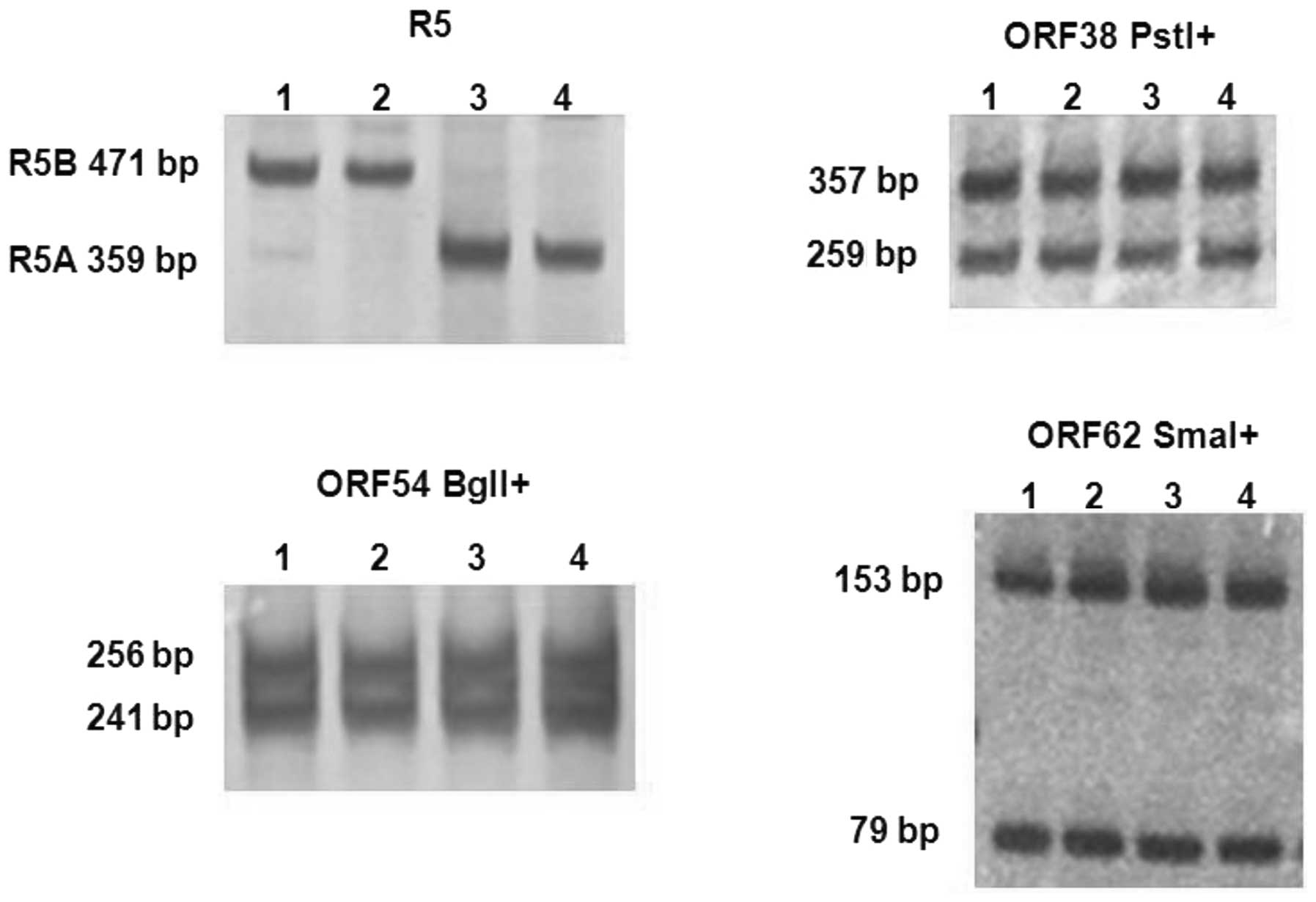
Restriction fragment length
polymorphism (RFLP) analysis of ORF 38, 54 and 62
The specific SNPs in the VZV strains that are
located in ORFs 38, 54 and 62 have been used as genetic markers in
the study of VZV epidemiology. The PCR products of ORFs 38, 54 and
62 in the VZV genes were amplified and they were 647, 497 and 268
bp, respectively. RFLP analysis was used to determine the genotype
of ORF38 (PstI), ORF54 (BglI) and ORF62 (SmaI) in the collected VZV
isolates, as shown in Fig. 1. The PCR
products of ORF38 were digested by PstI, and produced 2 fragments
of 357 and 290 bp. The digestion of PCR products of ORF54 by BglI
yielded 2 fragments of 256 and 241 bp. Similarly, the PCR products
of ORF62 were digested by SmaI, and produced 3 fragments of 153, 79
and 36 bp. These data indicated that the VZV isolates collected in
the study contained the cleaving site of PstI, BglI and SmaI, and
that the collected isolates were all BglI positive
(BglI+), PstI+ and SmaI+ (Table II).
| Table II.Genotype of the VZV strains in the
present study and other studies. |
Table II.
Genotype of the VZV strains in the
present study and other studies.
| VZV strain | ORF38
(PstI) | ORF54
(BglI) | ORF62
(SmaI) | R5 type (%) | SNP in ORF22 | SNP in ORFs 1, 21
and 54 |
|---|
| MLS (17) | + | + | − | R5A | M1 | A1 |
| v-Oka (17) | − | + | + | R5B | J | J2 |
| p-Oka (17) | − | + | − | R5B | J | J1 |
| VZV isolates
from | + | + | − | R5A (47.4) | J | J1 |
| Anhui city of China
(18) |
|
|
| R5B (52.6) |
|
|
| VZV isolates
from | + | + | + | R5A (46.4) |
J
(41/42) | J1 (41/42) |
| Yunnan province
(present study) |
|
|
| R5B (53.6) | M2 (1/42) | A1 (1/42) |
Analysis of the R5 variable
region
The R5 variable region has been shown to be vary
among different VZV strains. The R5 variable region was amplified
by PCR and analyzed by electrophoresis to determine its
distribution in the collected VZV isolates. The representative
results are shown in Fig. 1. The PCR
products of R5A were 359 bp, while the PCR products of R5B were 471
bp. In the collected VZV isolates, R5A and R5B were observed. The
percentages of R5A and R5B were 46.4 and 53.6%, respectively, in
the collected isolates.
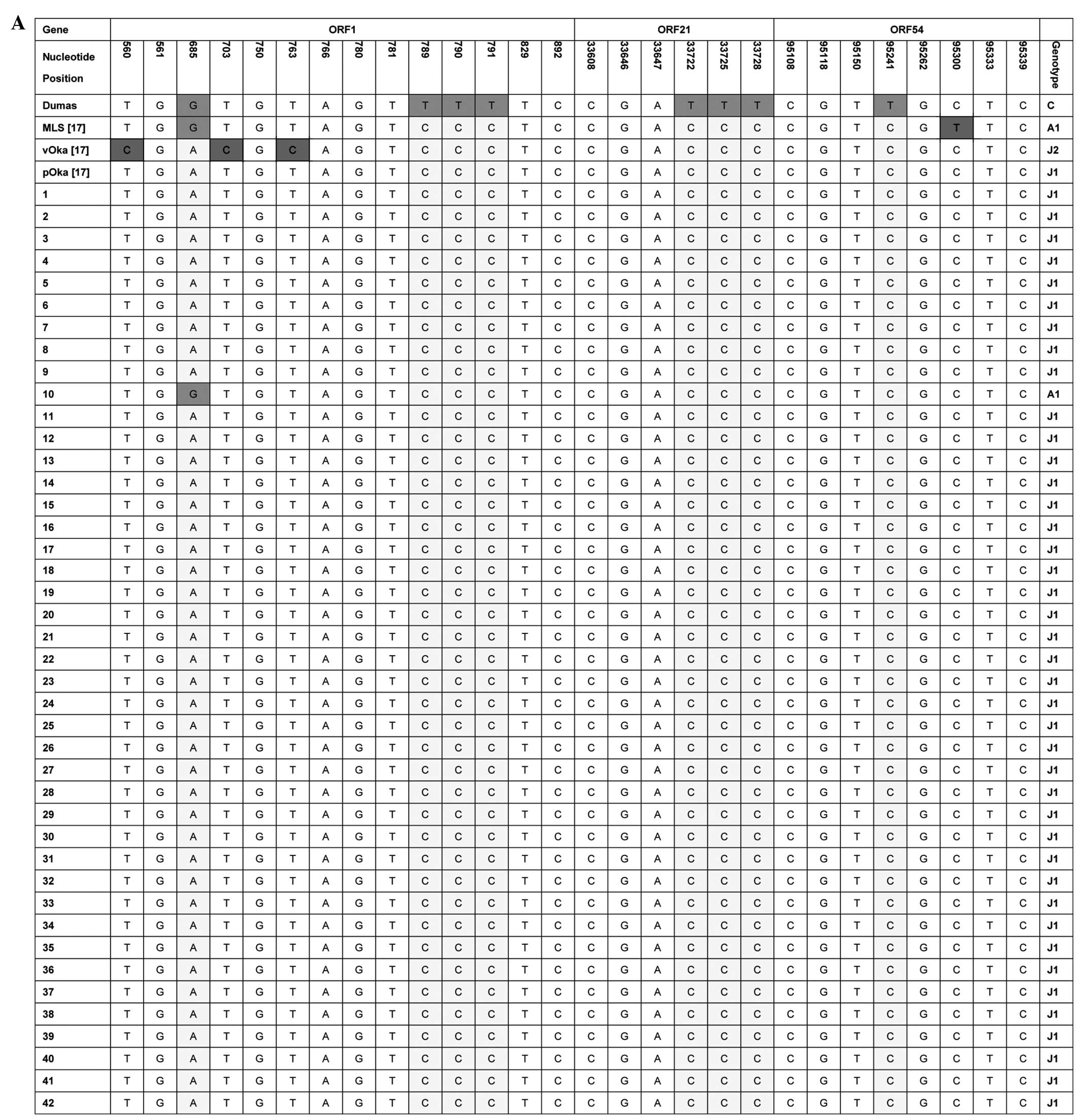
Nucleotide sequence analysis of ORFs
1, 21, 22 and 54
The sequence of ORFs 1, 21, 22 and 54 in VZV genes
have been sequenced and analyzed in certain areas of the world,
such as the United Kingdom, Brazil and Anhui city of China.
Therefore, a nucleotide sequence analysis was performed on ORFs 1,
21, 22 and 54 in the collected VZV isolates. The informative
polymorphic markers on the regions are shown in Fig. 2. Based on the genotyping scheme of
Barret-Muir et al (6), the VZV
isolates in the study could be grouped into genotype J1 (41/42) or
A1 (1/42), as shown in Fig. 2A. The
VZV isolates could be grouped into genotype J (41/42) or M2 (1/42)
according to the genotyping scheme of Loparev et al
(13), as shown in Fig. 2B.
Discussion
VZV is highly infectious for those with no VZV
infection history, and becomes dormant in the sensory ganglia
following the first infection. The genome of VZV is highly
conserved compared to other pathological viruses, such as human
papilloma virus. This may be due to the low reproduction of VZV in
the infected hosts. This feature limits the frequency of the
introduction of new mutations in VZV genes. As the advancement of
VZV molecular epidemiology occurs, numerous genomic variations have
been identified in VZV strains. Based on these genomic variations,
the VZV strains have been classified into different genotypes. The
VZV genotypes in China are rarely studied. The present study made a
preliminary study on the genotypes of VZV isolates collected from
Yunnan province of Southwestern China. To the best of our
knowledge, this is the first investigation on VZV genotype in this
region of China.
The genotype of VZV strains is associated with
climate. In temperate regions, the VZV strains are often
PstI+ and BglI negative
(BglI−) in ORF38 and ORF54, while the majority of
VZV strains in tropical regions are PstI+ and
BglI+ (13,20–22). In the
present study, the VZV isolates collected from the Yunnan province
of Southwestern China were all PstI+ and
BglI+ in ORF38 and ORF54 (Fig. 1A and Table
II). The present results are consistent with the findings that
the VZV strains in the tropical regions are often
PstI+ and BglI− in ORF38 and
ORF54, as the Yunnan province of China is located in the tropical
regions.
ORF38 (PstI), ORF54 (BglI) and ORF62
(SmaI) in VZV genes are molecular genetic markers for the
genotyping of VZV strains (18,20,21). The VZV isolates in the present study
were all PstI+, BglI+ and
SmaI+ (Fig. 1 and
Table II). The VZV isolates collected
from Anhui city of China and the strain MLS (6) were PstI+,
BglI+ and SmaI− (wild-type
VZV). The strain p-Oka was PstI−,
BglI+ and SmaI− (17). The strain v-Oka was
PstI−, BglI+ and
SmaI+ (17). The
results indicated that the VZV isolates collected from the Yunnan
province of Southwestern China were different from that of Anhui
city of middle eastern China. The results supported the conclusion
that the genotype of VZV strains may vary in different regions of
China. Further studies are required to find out the VZV
distributions in China.
The R5 variable region in the VZV genes has been
shown to be geographically related (10,23). The
type of R5A (359 bp) is mainly found in Europe and North America.
The type of R5B (471 bp) is a major type in Japan. In the present
study, R5A and R5B were observed in the collected VZV isolates
(Fig. 1). The genotype frequency of
R5A and R5B are nearly identical in these samples. The results are
consistent with the findings by Liu et al (17). It is possible that either R5A or R5B is
equally distributed in Chinese VZV strains.
Until now, there was no gold standard VZV genotyping
scheme, although several genotyping schemes have been proposed in
recent years. The genetic variations were identified in VZV ORFs 1,
21, 22, 50 and 54 (17,19,24,25). By referring to the SNPs in VZV ORFs 1,
21, 50 and 54, the VZV strains could be grouped into 4 genotypes:
A, B, C and J. By referring to the VZV ORF22, 7 VZV genotypes are
identified, which were E1, E2, J, M1, M2, M3 and M4. The VZV
strains of genotype J are most common in Asia, particular in Japan.
In the present study, the above genotyping scheme was used to
analyze the VZV samples collected from the Yunnan province of
Southwestern China (Fig. 2). The
collected VZV isolates are mainly genotype J or J1, which is a
major genotype in Asia. The results are consistent with the finding
by Liu et al (17), who
collected the VZV isolates from Anhui city of middle eastern China,
which were genotyped as J or J1. In addition to the previously
reported genetic variations in the VZV genes, no new genetic
variations were identified in the collected VZV isolates by
sequencing the ORFs 1, 21, 22 and 54 in VZV genes. The results
indicated that the VZV strains in Yunnan province may be highly
conserved, as several reports have shown new genetic variations in
these sequenced fragments. Of note, one sample in the collected
isolates was classified as genotype A1 or M2. This may be due to
the population migrations.
In conclusion, a preliminary study was performed on
VZV genotypes in the Yunnan province of Southwestern China. The
results of the present study will aid in the understanding of the
genetics of VZV in China. The limitation of the study is that the
sample size is small, although it is larger than the previous
studies in China. Further studies with a larger sample size are
required to understand the VZV genotype distributions in this
region and China.
Acknowledgements
The present study was supported by the Education
Commission of Yunnan Province (grant no. 2014Z036) and the Kunming
University of Science and Technology (grant no. KKZ3201360025). The
authors would like to thank Dr Ping Cao for collecting the VZV
samples, and the patients who participated.
References
|
1
|
Nagel MA, Cohrs RJ, Mahalingam R, et al:
The varicella zoster virus vasculopathies: Clinical, CSF, imaging
and virologic features. Neurology. 70:853–860. 2008. View Article : Google Scholar : PubMed/NCBI
|
|
2
|
Elkind MS: The varicella zoster virus
vasculopathies: Clinical, CSF, imaging, and virologic features.
Neurology. 72:1028–1030; author reply 129–130. 2009. View Article : Google Scholar : PubMed/NCBI
|
|
3
|
Nagel MA and Gilden DH: The protean
neurologic manifestations of varicella-zoster virus infection.
Cleve Clin J Med. 74:489–494, 496, 498, 499 passim. 2007.
View Article : Google Scholar : PubMed/NCBI
|
|
4
|
Becerra JC, Sieber R, Martinetti G, et al:
Infection of the central nervous system caused by varicella zoster
virus reactivation: A retrospective case series study. Int J Infect
Dis. 17:e529–e534. 2013. View Article : Google Scholar : PubMed/NCBI
|
|
5
|
Peters GA, Tyler SD, Grose C, et al: A
full-genome phylogenetic analysis of varicella-zoster virus reveals
a novel origin of replication-based genotyping scheme and evidence
of recombination between major circulating clades. J Virol.
80:9850–9860. 2006. View Article : Google Scholar : PubMed/NCBI
|
|
6
|
Barrett-Muir W, Scott FT, Aaby P, et al:
Genetic variation of varicella-zoster virus: Evidence for
geographical separation of strains. J Med Virol. 70(Suppl 1):
S42–S47. 2003. View Article : Google Scholar : PubMed/NCBI
|
|
7
|
Carr MJ, McCormack GP and Crowley B:
Genetic variation in clinical varicella-zoster virus isolates
collected in Ireland between 2002 and 2003. J Med Virol.
73:131–136. 2004. View Article : Google Scholar : PubMed/NCBI
|
|
8
|
Muir WB, Nichols R and Breuer J:
Phylogenetic analysis of varicella-zoster virus: Evidence of
intercontinental spread of genotypes and recombination. J Virol.
76:1971–1979. 2002. View Article : Google Scholar : PubMed/NCBI
|
|
9
|
Faga B, Maury W, Bruckner DA, et al:
Identification and mapping of single nucleotide polymorphisms in
the varicella-zoster virus genome. Virology. 280:1–6. 2001.
View Article : Google Scholar : PubMed/NCBI
|
|
10
|
Hawrami K and Breuer J: Analysis of United
Kingdom wild-type strains of varicella-zoster virus:
Differentiation from the Oka vaccine strain. J Med Virol. 53:60–62.
1997. View Article : Google Scholar : PubMed/NCBI
|
|
11
|
Hawrami K, Harper D and Breuer J: Typing
of varicella zoster virus by amplification of DNA polymorphisms. J
Virol Methods. 57:169–174. 1996. View Article : Google Scholar : PubMed/NCBI
|
|
12
|
Loparev VN, Argaw T, Krause PR, et al:
Improved identification and differentiation of varicella-zoster
virus (VZV) wild-type strains and an attenuated varicella vaccine
strain using a VZV open reading frame 62-based PCR. J Clin
Microbiol. 38:3156–3160. 2000.PubMed/NCBI
|
|
13
|
Loparev VN, Gonzalez A, Deleon-Carnes M,
et al: Global identification of three major genotypes of
varicella-zoster virus: Longitudinal clustering and strategies for
genotyping. J Virol. 78:8349–8358. 2004. View Article : Google Scholar : PubMed/NCBI
|
|
14
|
Norberg P, Liljeqvist JA, Bergstrom T, et
al: Complete-genome phylogenetic approach to varicella-zoster virus
evolution: Genetic divergence and evidence for recombination. J
Virol. 80:9569–9576. 2006. View Article : Google Scholar : PubMed/NCBI
|
|
15
|
Sergeev N, Rubtcova E, Chizikov V, et al:
New mosaic subgenotype of varicella-zoster virus in the USA: VZV
detection and genotyping by oligonucleotide-microarray. J Virol
Methods. 136:8–16. 2006. View Article : Google Scholar : PubMed/NCBI
|
|
16
|
Chow VT, Tipples GA and Grose C:
Bioinformatics of varicella-zoster virus: Single nucleotide
polymorphisms define clades and attenuated vaccine genotypes.
Infect Genet Evol. 18:351–356. 2013. View Article : Google Scholar : PubMed/NCBI
|
|
17
|
Liu J, Wang M, Gan L, et al: Genotyping of
clinical varicella-zoster virus isolates collected in China. J Clin
Microbiol. 47:1418–1423. 2009. View Article : Google Scholar : PubMed/NCBI
|
|
18
|
Jiang L, Gan L, Chen J, et al: Genetic
analysis of clinical VZV isolates collected in China reveals a more
homologous profile. Biomed Res Int. 2013:6812342013. View Article : Google Scholar : PubMed/NCBI
|
|
19
|
Weidmann M, Meyer-König U and Hufert FT:
Rapid detection of herpes simplex virus and varicella-zoster virus
infections by real-time PCR. J Clin Microbiol. 41:1565–1568. 2003.
View Article : Google Scholar : PubMed/NCBI
|
|
20
|
LaRussa P, Steinberg S, Arvin A, et al:
Polymerase chain reaction and restriction fragment length
polymorphism analysis of varicella-zoster virus isolates from the
United States and other parts of the world. J Infect Dis. 178(Suppl
1): S64–S66. 1998. View
Article : Google Scholar : PubMed/NCBI
|
|
21
|
LaRussa P, Lungu O, Hardy I, et al:
Restriction fragment length polymorphism of polymerase chain
reaction products from vaccine and wild-type varicella-zoster virus
isolates. J Virol. 66:1016–1020. 1992.PubMed/NCBI
|
|
22
|
Quinlivan M, Gershon AA, Steinberg SP, et
al: An evaluation of single nucleotide polymorphisms used to
differentiate vaccine and wild type strains of varicella-zoster
virus. J Med Virol. 75:174–180. 2005. View Article : Google Scholar : PubMed/NCBI
|
|
23
|
Sauerbrei A, Eichhorn U, Gawellek S, et
al: Molecular characterisation of varicella-zoster virus strains in
Germany and differentiation from the Oka vaccine strain. J Med
Virol. 71:313–319. 2003. View Article : Google Scholar : PubMed/NCBI
|
|
24
|
Quinlivan M, Hawrami K, Barrett-Muir W, et
al: The molecular epidemiology of varicella-zoster virus: Evidence
for geographic segregation. J Infect Dis. 186:888–894. 2002.
View Article : Google Scholar : PubMed/NCBI
|
|
25
|
Bostikova V, Bostik P, Chlibek R, et al:
Molecular epidemiology of varicella zoster virus. Epidemiol
Mikrobiol Imunol. 59:21–24. 2010.PubMed/NCBI
|