Morphological and functional changes of the optic nerve following traumatic optic nerve injuries in rabbits

  • Authors:
    • Fei Xue
    • Kunming Wu
    • Tianyou Wang
    • You Cheng
    • Manjie Jiang
    • Junfeng Ji
  • View Affiliations

  • Published online on: January 7, 2016     https://doi.org/10.3892/br.2016.567
  • Pages: 188-192
Metrics: Total Views: 0 (Spandidos Publications: | PMC Statistics: )
Total PDF Downloads: 0 (Spandidos Publications: | PMC Statistics: )


Abstract

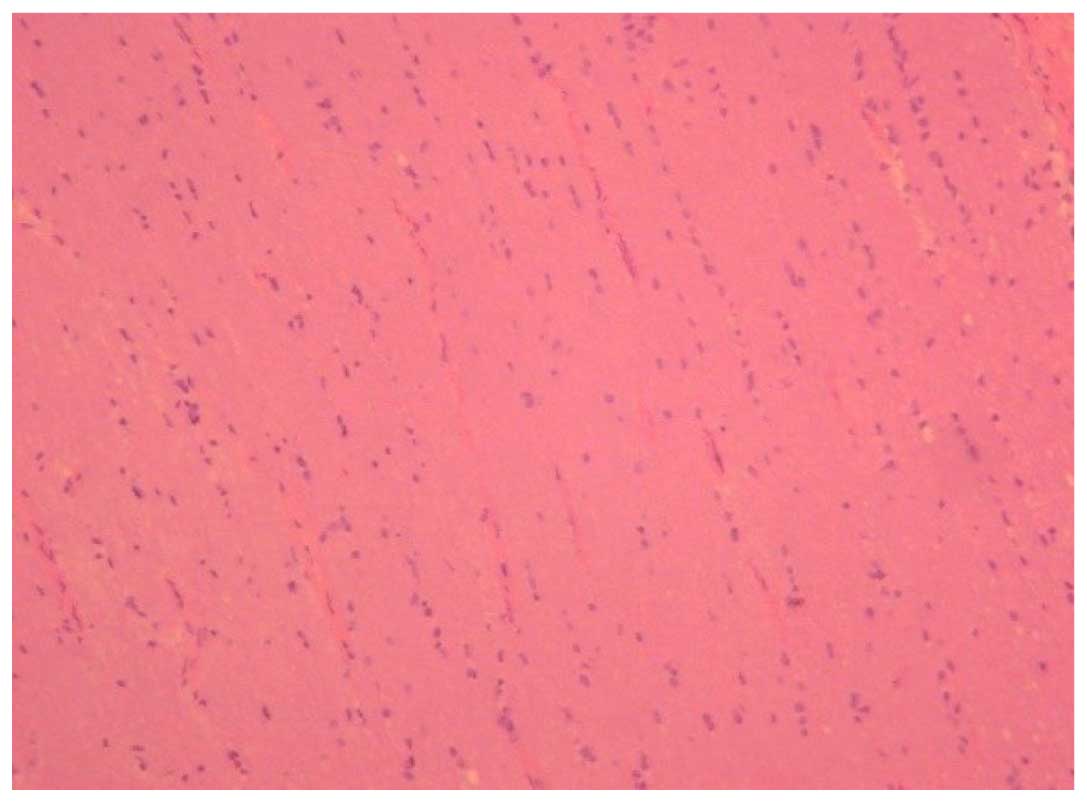
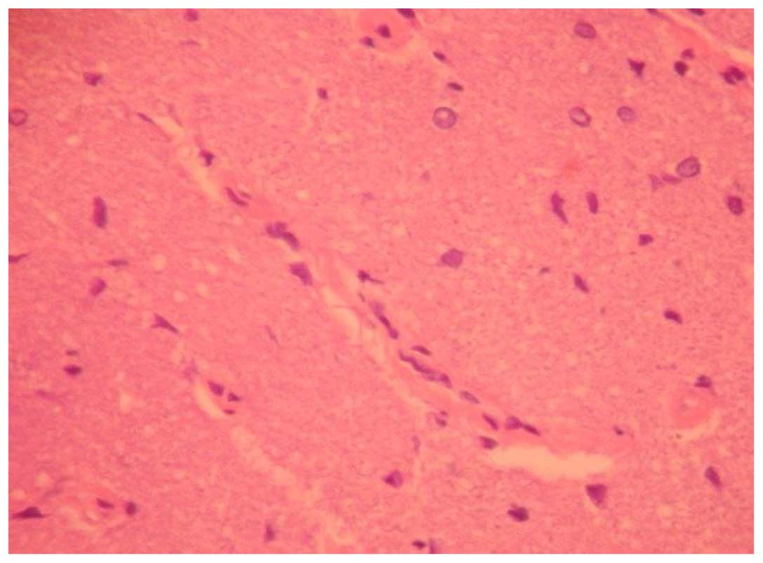
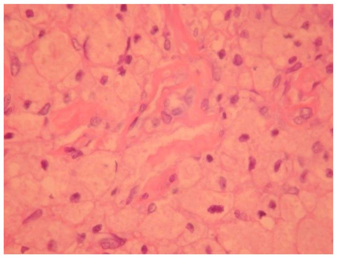
The aim of the present study was to investigate the morphological changes of the optic nerve following traumatic injuries and decompression at different times after injury, and to observe the changes of the visually evoked potentials, to identify the relevant associations between surgical opportunity and the clinical effect of traumatic optic nerve injuries. Rabbits were chosen as the animal model for the study. All the rabbits were randomly divided into five groups (A-E), representing the normal control, decompression in 48 h, in 1 week, in 2 weeks and non-decompression groups, respectively. The pattern reversal visual evoked potentials (P-VEP) and morphological changes of the optic nerve were observed. The P-VEP of each healthy rabbit revealed typical NPN contours, while NPN waves in the injured rabbits were low and flat. The latent period of the P-wave was lengthened and the amplitude was reduced. The differences of the latent period and amplitude pre- and post-trauma were statistically significant. The morphological changes were also assessed. In the normal control group, the astrocytes of the optic nerve exhibited a cylindrical form and were arranged evenly on the vertical section. The neural fibers were arranged neatly, were even following application of a dye, and the cross section exhibited a normal configuration of the blood vessel. For the 48-h decompression group, the arrangement of the astrocytes was even on the vertical section, and vacuoles, slight swelling of the nerve, exudation around the blood vessel and a small amount of astrocytic hyperplasia were observed in the damaged area. In the non-decompression group there were large areas of necrosis, clear nerve demyelination, serious exudation around the blood vessel and astrocytic hyperplasia were observed. In conclusion, the optic nerve decompression is beneficial to protect the visual function in indirect optic nerve injuries. Visual function may be improved by decompression in 48 h compared to 2 weeks. In order to prevent secondary axon injury and to protect visual functions, the decompression should be performed as soon as possible.
View Figures
View References

Related Articles

Journal Cover

February-2016
Volume 4 Issue 2

Print ISSN: 2049-9434
Online ISSN:2049-9442

Sign up for eToc alerts

Recommend to Library

Copy and paste a formatted citation
x
Spandidos Publications style
Xue F, Wu K, Wang T, Cheng Y, Jiang M and Ji J: Morphological and functional changes of the optic nerve following traumatic optic nerve injuries in rabbits. Biomed Rep 4: 188-192, 2016
APA
Xue, F., Wu, K., Wang, T., Cheng, Y., Jiang, M., & Ji, J. (2016). Morphological and functional changes of the optic nerve following traumatic optic nerve injuries in rabbits. Biomedical Reports, 4, 188-192. https://doi.org/10.3892/br.2016.567
MLA
Xue, F., Wu, K., Wang, T., Cheng, Y., Jiang, M., Ji, J."Morphological and functional changes of the optic nerve following traumatic optic nerve injuries in rabbits". Biomedical Reports 4.2 (2016): 188-192.
Chicago
Xue, F., Wu, K., Wang, T., Cheng, Y., Jiang, M., Ji, J."Morphological and functional changes of the optic nerve following traumatic optic nerve injuries in rabbits". Biomedical Reports 4, no. 2 (2016): 188-192. https://doi.org/10.3892/br.2016.567